Chapter 9-Visualizing Cells
 Call Kai
 Learn
 Practice Test
 Spaced Repetition
 Match
 Flashcards
 Knowt Play
1/48

There's no tags or description
Looks like no tags are added yet.
49 Terms
typical animal cell
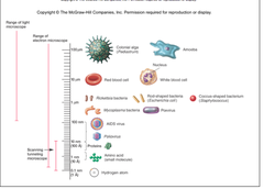
10-20um in diameter
colorless
translucent

naked eye can see
only ~380 to 700 um

light microscopy
allows us to manipulate light with multiple lenses to magnify an image

limit of super resolution (light microscopy)
20 um

limit of conventional resolution (light microscopy)
50 um

light microscopy is non-destructive
will not harm specimen

two light waves in phase
additive effect
bright

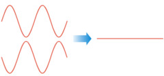
two light waves out of phase
interfere
dull
limitations of light microscopy
optical diffraction
limit of resolution

optical diffraction
phenomena in which light hits an object and bounces off (esp at the edge) and interfere with each other to produce a banding effect

limit of resolution
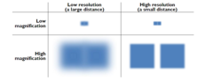
loss of detail; resolution distinguishes the differences between two objects
low resolution
loss of distinction at certain microscopic scales
resolution
minimum distance distinguishable between two objects

objective lens and condenser lens
resolution dependent on

objective lens
lens collects a cone of light rays to create on image

condenser lens
lens focuses a cone of light rays onto each point of the specimen

resolution equation
0.612λ/nsinθ

smaller for better resolving power
resolution equation should be

λ
wavelength of light (0.53um for white light)

1/2 the angular width of the cone of ray collected
θ

refractive index
n

θ & n
should be a bigger value/wider angle

refractive index
ratio of the speed of light in a vacuum to the speed of light in a particular medium

refractive index of air
1.00

refractive index of glass
1.52

refractive index of water
1.33

dry lens
air obscures some light rays

oil immersion lens
100x magnification; oil has simular refractive index to glass

light contrast
color contrast
fluorescence
types of light microscopy
light contrast
directly through cell; an effect in which the intensity of large background regions can modify the lightness of smaller enclosed areas

bright field (light contrast)
Direct light passes straight through the specimen into the objective lens. Contrast comes from how much light the sample absorbs.

dark field (light contrast)
A special opaque disk or stop blocks direct light from hitting the specimen. Only scattered, reflected, or refracted light enters the objective lens
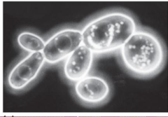
phase contrast (light contrast)
light is further deviated and differentiated to create stronger contrast

differential interference contrast (DIC) (light contrast)
Accentuates diffraction of the light that passes through a specimen; uses two beams of light

color contrast
common for tissue samples; sample is preserved chemically and embedded in wax/resin, where it is sliced into ribbons, stuck on a slide, stained, and mounted under a glass cover slip
when light is passed through, colored image is produced

common stains
hemotoxin and eosin

fluorescence microscopy
uses a fluorescent dye that emits fluorescence when illuminated with ultraviolet radiation

primary antibody
directed against an antigen on the specimen

secondary antibody
attached to a marker directed against primary antibodies

fluorescent marker
Molecule that emits light upon excitation.

excited state
absorption at photon
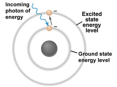
ground state
emission of photon at longer wavelength

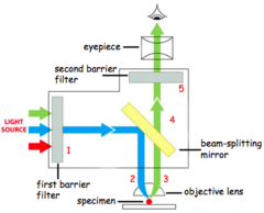
first barrier filter
lets through only blue light with a wavelength between 450 and 490 nm

beam splitting mirror
reflects light below 510 nm but transmits light above 510 nm

second barrier filter
cuts out unwanted fluorescent signals, passing the specific green fluorescein emission between 520 and 560 nm
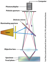
confocal microscope
microscope that can perceive very detailed images due to additional pieces that limit stray light (not from antibody) to get a clean image
- two confocal pin holes
electron microscope
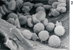
uses a beam of accelerated electrons as a radiation source instead of light photons to capture extremely detailed images of tiny objects

Transmission Electron Microscope (TEM)
A microscope that uses an electron beam to study the internal structure of thinly sectioned specimens.

Scanning Electron Microscope (SEM)
A microscope that uses an electron beam to scan the surface of a sample, coated with metal atoms, to study details of its topography.