Anatomy Lecture Exam 2
 Call Kai
 Learn
Learn Practice Test
 Spaced Repetition
 Match
Match Flashcards
 Knowt Play
1/439

There's no tags or description
Looks like no tags are added yet.
Name | Mastery | Learn | Test | Matching | Spaced | Call with Kai | Chat |
|---|
No analytics yet
Send a link to your students to track their progress
440 Terms
What are the functions of the muscular system?
perform body actions (movement, posture, visceral activities), thermoregulation, electric voltage
What are the muscle classifications?
color, histology, embryonic origin, morphologic appearance, location, control mechanism, actions
What are the muscle colors?
red or white
What are the muscle histologies?
smooth, cardiac, skeletal
What is the embryonic origin of muscle?
mesoderm
What are the muscle locations?
somatic or visceral

Defining traits of skeletal muscle
multinucleated and striated muscle fibers

Defining traits of smooth muscle
one central nucleus and non-striated

Defining traits of cardiac muscle
one central nucleus and striated

What is this muscle?
skeletal

What is this muscle?
cardiac

What is this muscle?
smooth
What make striations?
actin, myosin, troponin, tropomyosin
What is the epimysium?
cover entire muscles
What is the perimysium?
surrounds a muscle fascicle
What is the endomysium?
surrounds individual muscle cells

What part of the muscle connective tissue is in the small red circle (the top)?
endomysium

What part of the muscle connective tissue is the left blue arrow pointing to?
epimysium

What part of the muscle connective tissue is the blue arrow on the right pointing to next to the large red circle?
perimysium
What is the contraction principle (muscle stimulation)?
many motor neurons supply a muscle, each neuron innervates only a few fibers, selective recruitment of additional motor units can increase force

What is this muscle shape and what is its function?
parallel; abdominal muscles

What is this muscle shape and what is its function?
fusiform; biceps brachii, sartorius

What are these muscles shapes and what are their functions?
pennate (unipennate, bipennate, multipennate); digital extensor, rectus femoris, deltoideus

What is this muscle shape and what is its function?
circular; orbicularis oculi (eye)
Muscle extensors
increase joint angle
Muscle flexors
decrease joint angle
Abductors
move away from midline axis
Adductors
move towards midline axis
Protractors
push a body part away from its base
Retractors
pull a body part back
Levators
raise body parts
Depressor
lower body parts
Supinators
turns the palms upward
Pronators
turn the palms downward
Sphincters
constrict an opening
Constrictors
restrict a space
Rotators
turn part on its long axis
What attaches muscle to bone?
tendons
What attaches bone to bone?
ligaments
What is the aponeurosis?
flat sheet of connective tissue
What is the fascia?
thin casing of fibrous connective tissue around muscles, wrap and bind
Define synarthrotic (fibrous) joints
immovable, fibrous connective tissue
Define amphiarthrotic (cartilaginous) joints
limited movement, hyaline or fibrocartilage
Define diarthrotic (synovial) joints
freely moveable, hyaline cartilage
What is neurulation?
neural tube formation (dorsal ectoderm)
What forms from the axial mesoderm?
notochord, somites
What forms from the paraxial mesoderm?
somites (muscle, vertebrae, dermis)
Where do most axial skeletal muscles orginate from?
paraxial membrane
Somites become what?
trunk
Somitomeres become what?
head muscles
What does the hypomere become?
visceral smooth muscle, cardiac muscle
What does the mesenchyme become?
blood vessel smooth muscle, appedicular muscles
Where do axial muscles and fin muscles come from?
somites to myotome to axial muscles/fin muscles
Where do hypobranchial muscles (gill arches) come from?
somites to cervical myotomes to hypobranchial muscles (gill arches)
Where do cranial musculatures come from?
somitomeres to cranial musculatures

What species has the axial muscle in the picture?
teleosts, mostly undifferentiated epaxial and hypaxial (fish)

What species has the axial muscle in the picture?
salamanders, mostly undifferentiated epaxial; differentiated hypaxial (amphibians)

What species has the axial muscle in the picture?
lizards, differentiated epaxial and hypaxial
What appendicular muscles do teleosts have?
dorsal/ventral fin muscle is minor
What appendicular muscles do tetrapods have?
dorsal/ventral muscles prominent, pectoral girdle/forelimb, pelvic girdle/forelimb
What are the body cavities?
thoracic (pleural), abdominopelvic (peritoneal), pericardial
What are the sections of the thoracic cavity?
left, right
What are the sections of the abdominopelvic cavity?
abdominal, pelvic
What are serosal (serous) surfaces?
smooth membrane
What are the functions of the serosal surfaces?
secrete watery fluid that lubricates serosal surfaces; outer surface of organs, lines body cavities
What are the serosal membranes in the thorax/pleural cavity?
parietal pleura, visceral pluera
What are the serosal membranes in the abdomen/peritoneal cavity?
parietal peritoneum, visceral peritoneum
What are the serosal membranes in the pericardial cavity?
parietal pericardium, visceral pericardium

What is label 1?
visceral pleura

What is label 2?
parietal pleura
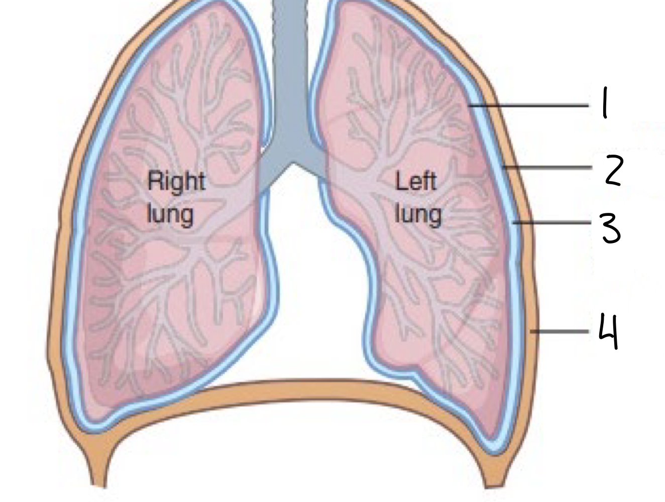
What is label 3?
intrapleural space (pleural cavity)
What is label 4?
thoracic wall
What is the pleural cavity made of?
connective tissue, simple squamous cells (mesothelium)
Where do two pleura meet?
hilum of lung (center)
What is the thin lining covering lungs?
visceral pleura
What is the lining covering internal surface of the thoracic cavity?
parietal pleura
What is the function of pleural fluid?
lubrication, surface tension (pneumothorax)
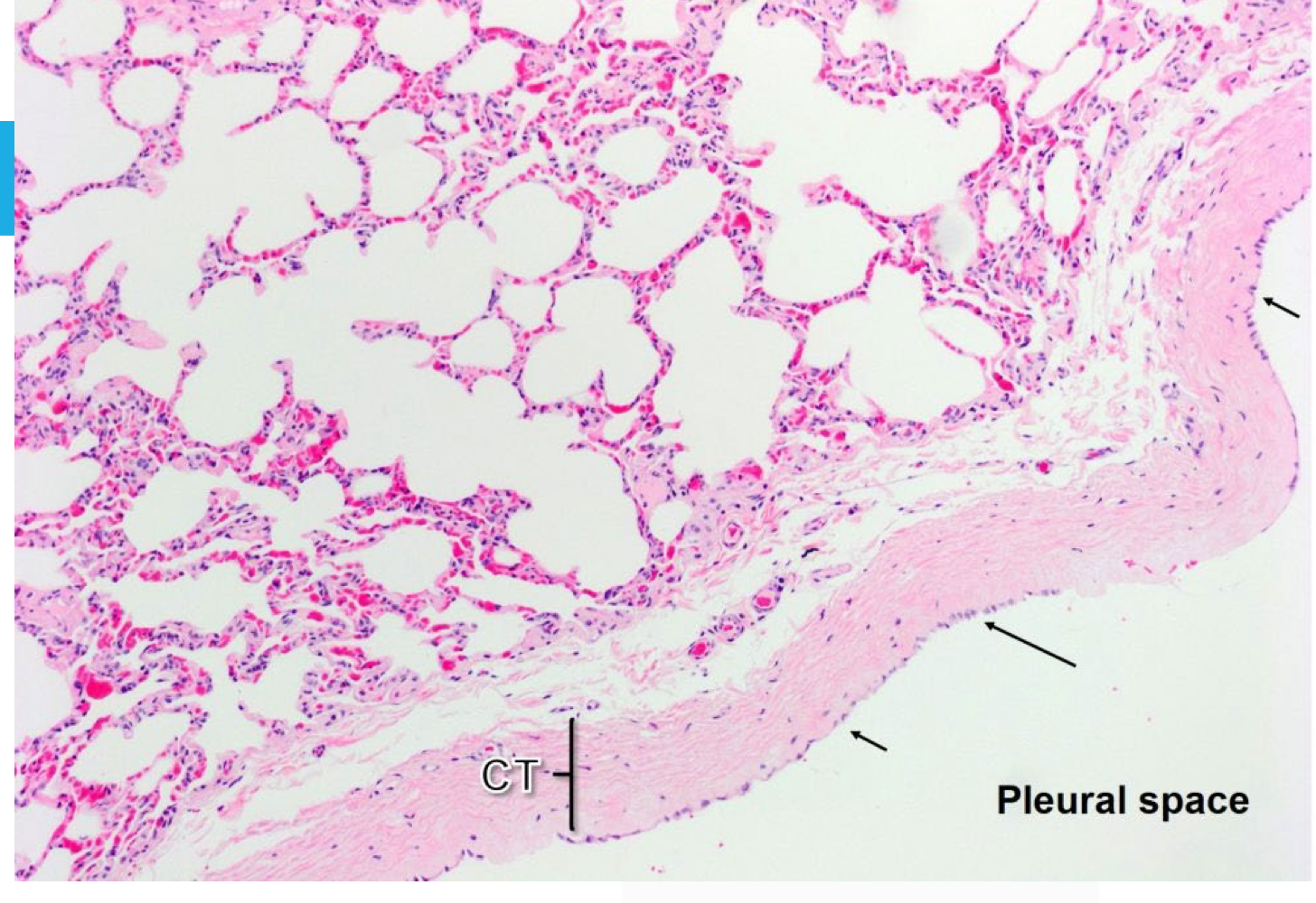
What are the arrows pointing to on this lung histology?
mesothelium (single squamous epithelial cell)
What is emphysema?
air under the visceral pleura
What is pneumothorax?
air between visceral and parietal pleura
What is the mesentary?
formed by double layers of serous membrane
What is the function of the mesentary?
suspends and holds organs loosely in place; contains blood vessels, nerves, lymphatics
What is the embryonic origin of the mesentary?
mesoderm

What is this image depicting?
embryonic mesentery development

What is the bottom arrow pointing to?
ventral mesenstary

What is the top arrow pointing to?
dorsal mesentary

What is the top arrow pointing to?
kidney - nephric ridge (retroperitoneal=behind)

What is the bottom arrow pointing to?
reproductive organs - genital ridge

What is the top arrow pointing to?
dorsal mesentery

What is the middle arrow pointing to?
visceral peritoneum

What is the bottom arrow pointing to?
parietal peritoneum
What happens to the ventral mesentaries?
usually lost
What are the remaining ventral mesenteries?
falciform ligament, lesser omentum
Where is the falciform ligament?
umbilicus to liver
Where is the lesser omentum?
liver to stomach/duodenum, ventral part of mediastinum
What supports the viscera?
dorsal mesenteries

What is 3?
parietal pleura

What is 2?
mediastinum

What is 1?
visceral pleura
What is the function of the mediastinum mesentery?
divides thoracic cavity into right and left cavities; contains heart, nerves, great vessels, lymph nodes