Pos elbow humerus shldr scap
 Call Kai
 Learn
 Practice Test
 Spaced Repetition
 Match
 Flashcards
 Knowt Play
1/29

There's no tags or description
Looks like no tags are added yet.
30 Terms
(Baystate) Elbow protocol (3 views)
8x10 at 40 SID TT
Technique: 1-1.8 mAs @ 60-64 kVp
Pt instructions = dont’ move
AP: arm fully extended with epicondyles parallel to IR + upper extremity (humerus) on same plane CR @ mid-elbow joint
Eval. criteria: radial head, neck, tuberosity slightly superimposed over proximal ulna, elbow joint open, no rotation of humeral epicondyles
External OBL: lateral oblique with elbow rotated 45 degrees laterally (thumb side)
Eval. criteria: radial head, neck, tuberosity projected free of ulna + open elbow joint
Lateral: elbow flexed 90 degrees with epicondyles perpendicular to IR and humerus/forearm parallel to IR wrist in lateral position CR @ mid-elbow joint
Eval. criteria: 3 concentrics arcs visible, olecron process in profile, epicondyles superimposed
(not Baystate Protocol) Internal oblique elbow: forearm pronated 45 degrees
Eval. criteria: demonstrates coronoid process, trochlea notch, medial trochlea; also shows fractures of medial epicondyle/coronoid process
Trauma Elbow (2)
two partially flexed AP projections (when elbow cannot be fully extended)
Humerus parallel = CR perpendicular to mid elbow
Forearm parallel = CR perpendicular to mid elbow
Elbow joint
synovial (diarthrodial hinge joint), 3 joints enclosed in common capsule 2 articulations
Proximal radioulnar joint
Humeroulnar joint
Humeroradial joint
Trochlea of humerus articulates with trochlear notch of ulna
Capitulum of humerus articulates with head of radius
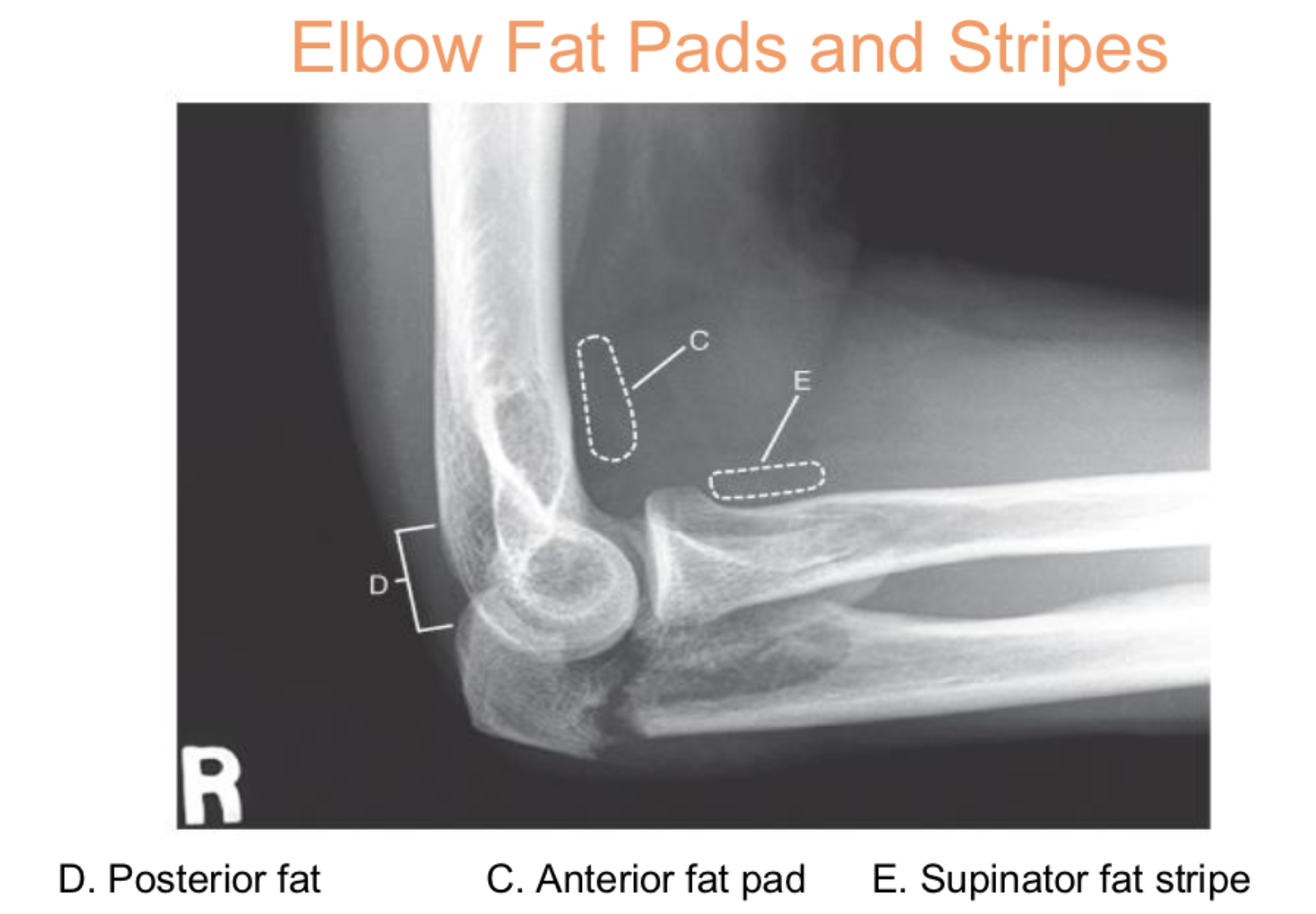
Elbow fat pads/stripes (3)
c. anterior fat pad
d. posterior fat
e. supinator fat stripe
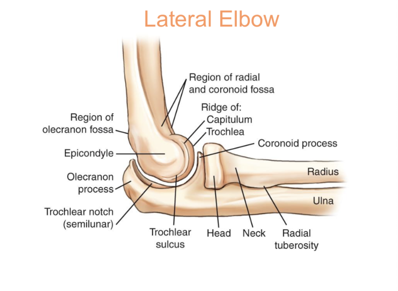
Three concentric arcs
Trochlear sulcus
Outer ridges of capitulum and trochlear
Trochlear notch of ulna

Proximal ulna (2)
olecranon process proximal + posterior
coronoid process proximal + anterior
AP elbow

Lateral elbow
Monteggia fracture
fracture of proximal third ulna with dislocation of radial head
supracondylar fracture
fracture of distal humerus above humeral condyles; most common in children
Humerus (Baystate) protocol
2 views
14x17 lengthwise 40 SID wall bucky
Pt instructions: don’t move don’t breathe
Technique (depends whether done TT/Bucky) = 1.5-5 mAs @68-70
AP (wall bucky) = CR @ mid humerus, include both shoulder/elbow joints no AEC, turn collimator as needed to help collimation, do not allow soft tissue to overlap onto the ribs, ensure epicondyles parallel to IR
Eval. criteria = entire humerus demonstrated with greater tubercle, medial and lateral epicondyles in profile
Lateral (lateromedial/mediolateral humerus project.) = CR @ mid humerus with epicondyles perpendicular to IR (lateral elbow)
Lateral humerus recumbent rotational lateral (supine pos.) = epicondyles perpendicular to IR, CR @ midhumerus using table bucky, suspend respiration
Eval. criteria = entire humerus demonstrated with lesser tubercle in profile and epicondyles superimposed
distal fracture (humerus)
crosstable lateral medial position
lateral medial pos.

Transthoracic Humerus (for proximal fracture)
place affected arm against bucky + raise unaffected arm; use breathing technique to blur our rids (longer secs exposure)
used for fractures of upper 1/3rd of humerus; 63 mAs @ 80 kVp
Shoulder (non trauma) protocol (2 views)
10x12 crosswise 40 SID wall bucky
Technique: 4-8 mAs @ 66-70 kVp
Pt instructions: don’t move don't breathe (suspend respiration)
External rotation shoulder = CR @ 1 inch inferior to coracoid process (soft divot), epicondyles parallel, include SC joint over in light field, palm up
Eval. criteria = greater tubercle (best shown) profiled laterally, scapulohumeral joint centered, proximal humerus, upper scapula + clavicle visualized
Internal rotation shoulder = CR @ 1 inch inferior to coracoid process, epicondyles perpendicular, back of hand against upper leg (medial rotation)
Eval. criteria = lesser tubercle (best shown) profiled medially, scapulohumeral joint centered,
Trauma shoulder routine
AP grashey = 4-8 mAs @68-72 kVp
Y-view = 8-14 mAs @78 kVp
Axillary = 2.5-4 mAs @70 kVp
Grashey (posterior oblique)
Demonstrates glenoid cavity
CR = 2 inches inferior and medial from superolateral border of humerus (coracoid process)
30-45 degree oblique toward affected side
CR perpendicular
10 x 12 crosswise or lengthwise
Suspend respiration
6-8 mAs @70-72 kVp
Evaluation criteria (Grashey; posterior oblique)
Glenoid cavity profiled
Scapulohumeral joint centered
Scapular Y lateral
45-60 degree oblique, affected side farther from IR (kyphotic patient = less oblique)
Can be done AP or PA, but most done AP at clinical
CR proximal to humerus; 2 inches below top of shoulder
10 x 12 lengthwise
Suspend respiration
Looking for dislocation
8-14 mAs @ 78 kVp
Scapular Y lateral evaluation criteria
Body of scapula superimposed
Acromion and coracoid processes in profile
Humeral head and glenoid cavity superimposed
Axillary
CR 25-30 degrees medial to axilla
2.5-4 mAs @70 kVp
best shows humeral head and neck in lateral position
8 × 10 crosswise
Arm supinated abducted 90 degrees (or as near 90 degree as possible)
Can be done supine or sitting down
in supine projections, an angle is needed
When patient is sitting down, a 5–15-degree angle is optional based on body habitus
Supraspinatus outlet view (Neer method)
Specialty view (non routine)
PA y-view position
45-60 degree oblique
10-15 caudal angle
Shoulder impingement
Scapula routine views
Ap and lateral (y-view)
Ap = 4-8 mAs @68-70 kVp
Y-view = 8-14 mAs @78
AP scapula
CR midscapula (2in inferior to coracoid and medial from axillary border)
10 x 12 lengthwise
At clinical = no breathing
Textbook = minimal 3 sec exposure orthostatic breathing technique to blur ribs and lung markings
Abduct arm 90 degrees, with arm in “muscle position,” or resting on head
AP scapula evaluation criteria
Entire scapula demonstrated
Lateral border of scapula free of ribs and lungs
Lateral scapula
CR to proximal humerus (upper ⅓ of humerus)
45-60 degree oblique
10 x 12 lengthwise
Most often done with AP arm reaching across to move humerus away from scapula
Suspend respiration
Non-trauma shoulder
AP external and AP internal views
4-8mAs @66-70 kVp
External rotation nontrauma shoulder
Epicondyles parallel to IR
CR is 1 inch inferior to coracoid process (find divot)
10 x 12 crosswise (include from SC joint over in light field)
Suspend respiration
“Extra” (external) = Greater tubercle
Evaluation criteria (external shoulder)
Greater tubercle profiled laterally
Scapulohumeral joint centered
Proximal humerus, upper scapula, and clavicle visualized
Internal rotation shoulder
Epicondyles perpendicular to IR
CR 1 inch inferior to coracoid process
10 x 12 crosswise
Suspend respiration
Evaluation criteria (shldr internal rotation)
Lesser tubercle profiled medially
Scapulohumeral joint centered