Unit 2: Mycology
 Call Kai
 Learn
Learn Practice Test
 Spaced Repetition
 Match
Match Flashcards
 Knowt Play
1/31

There's no tags or description
Looks like no tags are added yet.
Name | Mastery | Learn | Test | Matching | Spaced | Call with Kai | Chat |
|---|
No analytics yet
Send a link to your students to track their progress
32 Terms
Hortaea werneckii
Causative agent of Tinea nigra

Malassezia furfur
Causative agent of Tinea versicolor. "Spaghetti & Meatballs". Will not grow on routine culture media unless a lipid such as olive oil is layered over it

Piedra hortae
Causative agent of Black piedra - Firmly attached, hard irregular black nodules around the outside of scalp hair (hair shaft)

Trichosporon beigelii
Causative agent of White piedra - Light brown, white, soft nodules around beard and mustache hairs. Colonies appear cream colored and wrinkled.

Microsporum spp.
Invades skin and hair -> Macroconidia (spindle-shaped) are very common; microconidia are less common. Causes dermatophytosis of the body and scalp

Microsporum canis
Macroconidia are thick-walled, spindle shaped (tapered ends) containing 6 to 15 cells. Causes dermatophytosis of the body and scalp. Usually contracted from infected dogs and cats.

Epidermophyton floccosum
Numerous "snowshoe" club-shaped 2-4 celled macroconidia; Microconidia
are absent. Causes athletes foot, jock itch, and onychomycosis
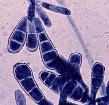
Trichophyton spp.
Numerous Microconidia are common borne in grape-like cluster or singly along hyphae "birds on a fence". Causes dermatophytosis including athlete's foot and tinea barbae.
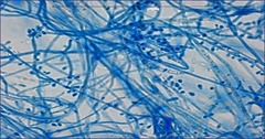
Trichophyton mentagrophytes
The most common cause of athlete's foot. Positive hair perforation test. Microscopic shows numerous round microconidia are found in grape-like clusters.

Cladosporium carrionii
Dematiaceous mold. Microscopy shows branched chains of conidia. Causes Chromoblastomycosis - which presents as "cauliflower-like lesions".

Fonsecaea pedrosoi
Dematiaceous mold that appears as black colonies with dark olive to black aerial mycelium. Most common worldwide cause of chromoblastomycosis.

Phialophora verrucosa
Dematiaceous mold. Microscopy shows balls of conidia. Causes Chromoblastomycosis, mycetoma, mycotic keratitis, intraocular infections and conjunctivitis

Scedosporium apiospermum
Hyaline mold. White, fluffy, colonies develop rapidly and later turn gray with a gray reverse. Causes mycetoma in the U.S.

Sporothrix schenckii
Hyaline mold. Rose gardener's disease.

Blastomyces dermatitidis
Yeast Phase. Thick walled, single budding yeast cells with broad isthmus at mother-daughter attachment. Causes Blastomycosis

Blastomyces dermatitidis
Mold Phase. Single conidia on short conidiophore "lollipop". Causes Blastomycosis
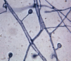
Coccidiodes immitis
Tissue Phase. Round, thick-walled spherules (sporangia) filled with endospores. Causes San Joaquin Valley Fever.

Coccidiodes immitis
Mold Phase. Thick walled, barrel-shaped arthroconidia alternating with empty disjunctor cells. Causes San Joaquin Valley Fever.

Histoplasma capsulatum
Yeast Phase. Appears as small, single-budding yeast cells. Causes histoplasmosis

Histoplasma capsulatum
Mold Phase. Causes histoplasmosis which is associated with bat droppings and caves. Large round macroconidia with smooth spiny or fin-like tuberculate edges.

Paracoccidiodes brasiliensis
Yeast Phase. Large, thick-walled multiple budding yeast cells with narrow necks where the daughter cells attach. Resembles a Mariner's "Ships" Wheel

Absidia
Zygomycotina. Macroscopic shows woolly gray colony that rapidly matures at RT. Microscopic shows branching sporangiophores between the Rhizoids. Causes Mycotic keratitis and Zygomycosis.

Mucor
Zygomycotina. Macroscopic shows white, fluffy mycelium that quickly forms at RT. No Rhizoids or Stolons seen microscopically. Causes otomycosis, allergies, and Zygomycosis.

Rhizopus
Zygomycotina. Microscopic shows unbranched sporangiophores arising opposite of Rhizoids at the nodes. "Bread mold".

Aspergillus fumigatus
Hyaline opportunist. Most common opportunistic pathogenic of genus. Causes disseminated Aspergillosis and Farmer's lung.

Aspergillus niger
Hyaline opportunist. Usually considered a contaminant. Important cause of otomycosis. Macroscopic shows white to yellow colonies at first that turn black.

Fusarium
Hyaline opportunist. Macrophialoconidia are two to five-celled, banana or cylindrical-shaped with a distinctive foot cell at the point of attachment. Most common cause of mycotic keratitis (infection of the cornea).

Paecilomyces
Hyaline opportunist. "ET fingers"

Penicillium
Hyaline opportunist. A genus of fungus used as a source of a chemical used to make penicillin. Flask-shaped phialides which in turn support chains of round phialoconidia

Pneumocystis jiroveci
Atypical fungi. Occurs frequently in AIDS patients.

Candida albicans
Most common cause of yeast infection in the world. Causes thrush. Microscopic shows budding yeast and pseudohyphae. Germ tube positive (+). "Star-shaped" colonies on BAP.

Cryptococcus neoformans
India ink staining of encapsulated yeast in CSF is diagnostic for this pathogen. Urease positive. Can be fatal in immunocompromised patients.