Module 12 - the System of Internal Membranes (Secretion)
 Call Kai
 Learn
 Practice Test
 Spaced Repetition
 Match
 Flashcards
 Knowt Play
1/38

Earn XP
Description and Tags
included on exam 3
39 Terms
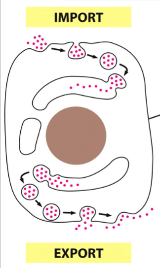
endocytosis
import mediated by the formation of endocytic vesicles
the taking in of other things into the cell (endo, into)
exocytosis
export (secretion) out of a cell
exchange of materials between cell and environment
endocytosis and exocytosis
involves the plasma membrane, small vesicles, endoplasmic reticulum, Golgi apparatus, and lysosomes
peroxisomes
one of the digestive organelles of a eukaryotic cell
contain enzymes that break down lipids and destroy toxic molecules through oxidation reactions
(perOXIsomes, OXIdative breakdown)
eukaryotic nucleus
the center of a eukaryotic cell
site of DNA storage and replication
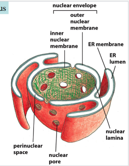
consists of nuclear envelope (and ER?), perinuclear space, and nuclear pores
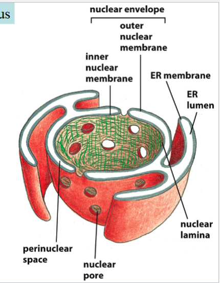
nuclear envelope
a double membrane surrounding the nucleus
(made of outer and inner nuclear membranes)
outer membrane is continuous with the ER
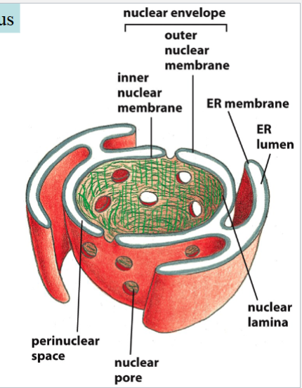
perinuclear space
the cavity in between the two membranes of the nuclear envelope
nuclear pores
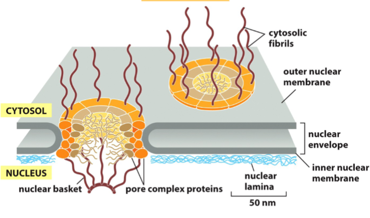
holes in the nuclear envelope that allow both passive and active transport
contain protein complexes that facilitate transport through the nuclear envelope (e.g. cytosolic fibrils, nuclear basket)
nuclear lamina
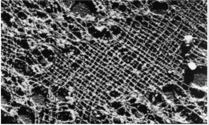
a network of lamin protein fibers that lines the inner wall of the nuclear envelope
provides structural support for nucleus
provides attachment sites for chromatin
when destroyed, the entire nucleus collapses
a component of cytoskeleton (categorized as intermediate filaments)
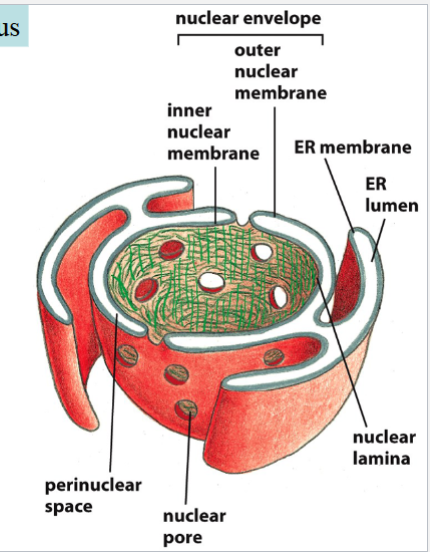
(green in the picture)
passive diffusion through nuclear pores
for small molecules and small proteins (<50 kDalton)
no energy required
left side of the picture
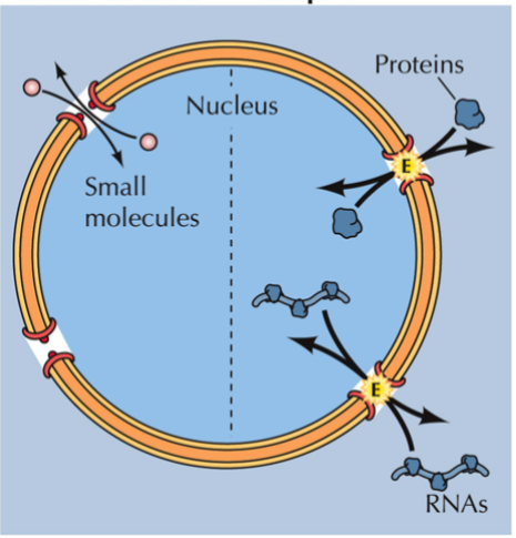
active transport through nuclear pores
required for larger proteins and all RNAs
energy-dependent
right side of the picture
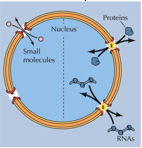
nuclear localization signal (NLS)
an amino acid sequence that is included in proteins that are transported through the nuclear envelope
allows a protein to enter the nuclei of cells
(e.g. included in DNA polymerases, RNA polymerases, transcription factors, histones, other chromatin assembly proteins, lamins, SV40 T-antigen)
the SV40 T-antigen
contains an NLS: 7 amino acids in the middle of the protein
oncogenic; capable of altering nuclear transcription factor activity of cells → transforming those cells
NLS allows it to enter the nuclei of infected cells, cause malignancy
if a protein (e.g. pyruvate kinase) lacking an NLS is given the SV40 T-antigen NLS as an expression vector, pyruvate kinase still fails to translocate to the nucleus (? not sure on this one)
process of active transport through a nuclear pore
transport complex forms, containing cargo protein and nuclear transport receptor protein
“cargo” protein: nuclear protein with an NLS
nuclear transport receptor protein: recognizes and binds to NLSs in cargo protein
translocation cycle:
entire transport complex is translocated through pore by interactions with pore proteins (e.g. cytosolic fibrils)
Ran-GTP binds to the nuclear transport receptor protein → causes dissociation and release of cargo protein inside nucleus
Ran-GTP with nuclear transport receptor protein goes to cytoplasm
ran GTPase activating protein (Ran-GAP) removes a Pi from the GTP on Ran
nuclear transport receptor protein dissociates from Ran-GDP, returns to the cycle
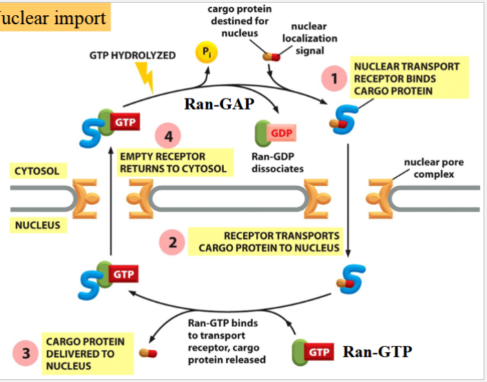
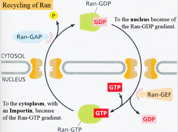
the Ran-GTP gradient
important for active transport through a nuclear pore
Ran-GTP is more abundant inside nucleus → will tend to go to cytoplasm
in cytoplasm, GTP is hydrolyzed to GDP by Ran GAP
→ Ran-GTP gradient is maintained across nuclear envelope → its diffusion drives the nuclear import cycle
energy-dependent: requires GTP hydrolysis
Ran-GDP produced in cytoplasm goes back to nucleus with its own concentration gradient
GDp on ran is exchanged for GTP (catalyzed by Ran-GEF)
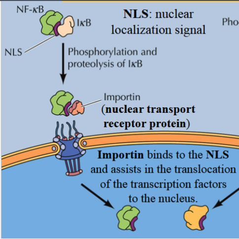
regulation of import of NF-KB
NF-KB is dormant in cytoplasm with IKB bound to it
IKB masks the NF-KB’s NLS → prevents importin from binding to it
if IKB is phosphorylated and destroyed, NF-KB is released → importin can bind, translocate it to the nucleus so it can activate transcription
NF-KB is a transcription factor with a key role in regulating immune response to infections
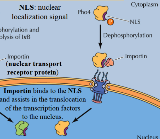
regulation of import of Pho4
Pho4: a transcription factor, exists in cytoplasm in an inactivated phosphorylated state
phosphate groups block NLS
if dephosphorylated, importin can bind to it and take it to the nucleus
involved in regulation of the cell division cycle
smooth endoplasmic reticulum (ER)
the site of lipid synthesis and processing
builds membranes and membrane systems for the cell
contains no ribosomes
rough ER
site of protein synthesis and processing
site of protein folding, addition of cofactors, lipids, and carbohydrate groups
association of ribosomes → rough appearance
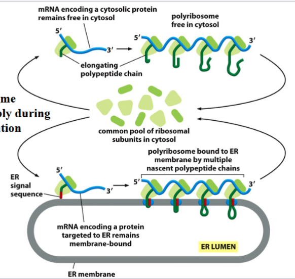
translation
polypeptide synthesis by ribosomes in the cytoplasm
occurs in a free ribosome if a synthesized protein is meant to stay in the cytoplasm, go into the nucleus, chloroplast, mitochondria, or peroxisome
occurs in rough ER ribosomes if the synthesized protein is a membrane protein, lysosomal, or secreted
^translocated into the lumen of the ER right after synthesis
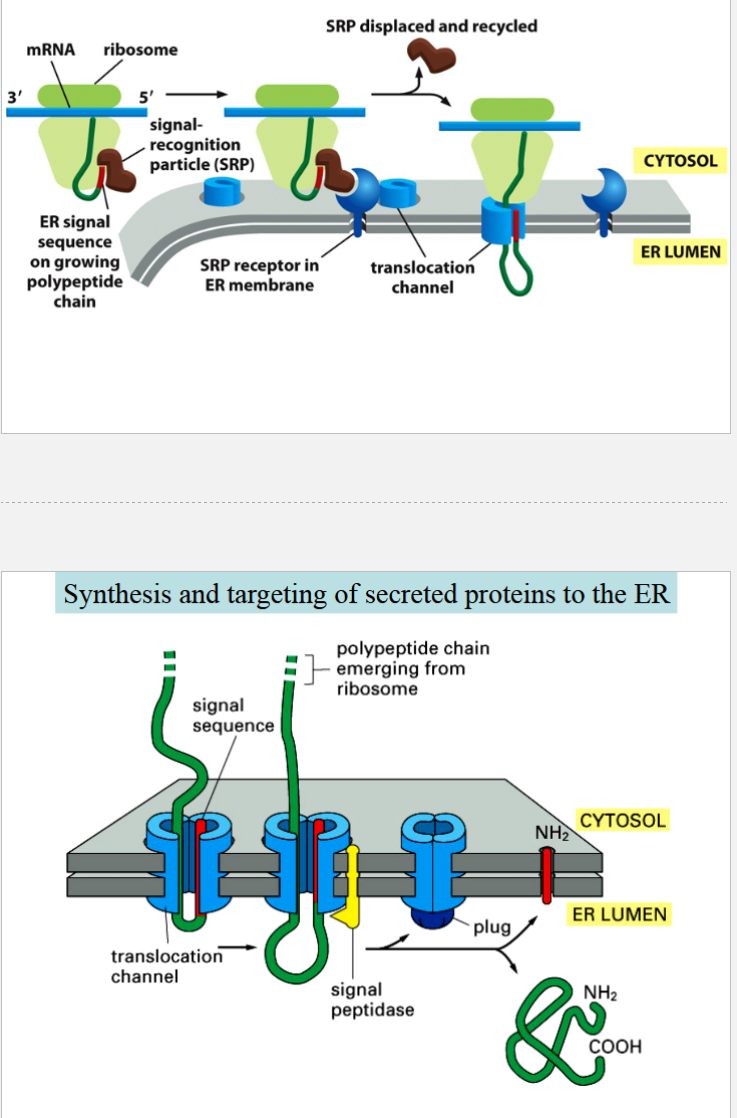
synthesis of secreted proteins
all secreted proteins contain an ER signal sequence in their amino terminal
signal is necessary for translocation into ER; gets removed after translocation
ER signal sequence is recognized by SRP (signal recognition particle)
SRP docks on SRP receptor on ER membrane
growing polypeptide moves through translocation channel
signal peptidase removes translocation signal from the new polypeptide
secretion pathway
path taken by proteins synthesized on the rough ER
ER for protein processing → ERGIC for retrieval of ER-resident components → Golgi apparatus for protein sorting
from Golgi, can go to:
out of cell through secretory vesicles (secreted proteins), of
to plasma membrane (membrane proteins), or
to lysosomes (lysosomal proteins)
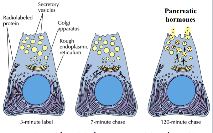
the pulse-chase experiment
a way to track the secretion pathway
steps:
1) incubate cultured pancreatic cells with 35S-labeled amino acids for 3 minutes (pulse: all proteins synthesized during that time will be radioactive)
2) remove radioactive medium (no more radioactive protein synthesis)
3) follow movement of radioactive proteins by periodic autoradiography (chase: migration of radiolabeled proteins is tracced following radioactive signal)
ERGIC
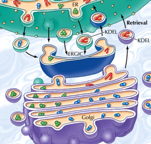
ER-Golgi intermediate complex
a system of membrane stacks located between the ER and the Golgi
function: a checkpoint to ensure that ER-resident proteins do not reach the Golgi
reason: traffic from ER to Golgi is non-selective
ER-resident proteins contain a KDEL-sequence retention signal
if they reach the ERGIC, their are recognized, retrieved, sent back to ER
(blue in the picture)
Golgi apparatus
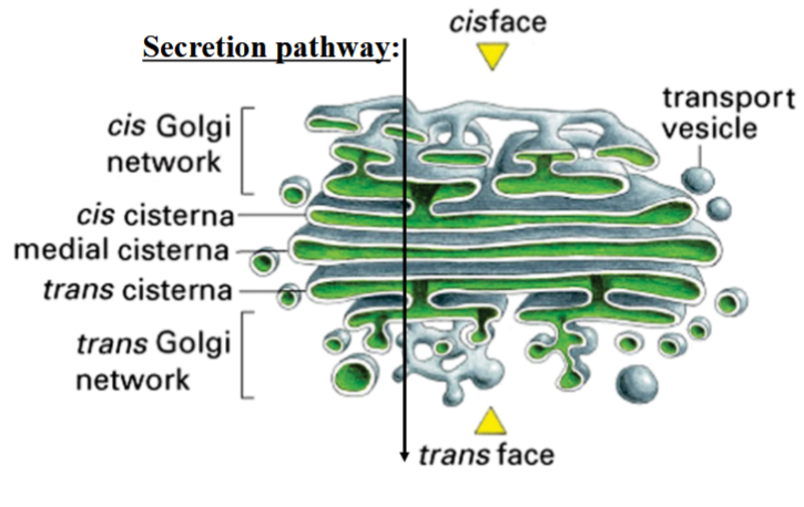
the protein sorting center of the cell (post office)
functions: protein modification, sorting, and packaging for secretion or delivery to other organelles8
protein-containing vesicles coming from ER enter at the cis face of the Golgi
proteins are moved through medial and trans Golgi stacks and released in vesicles from the trans face
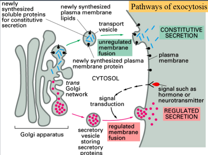
3 types of transport from the Golgi
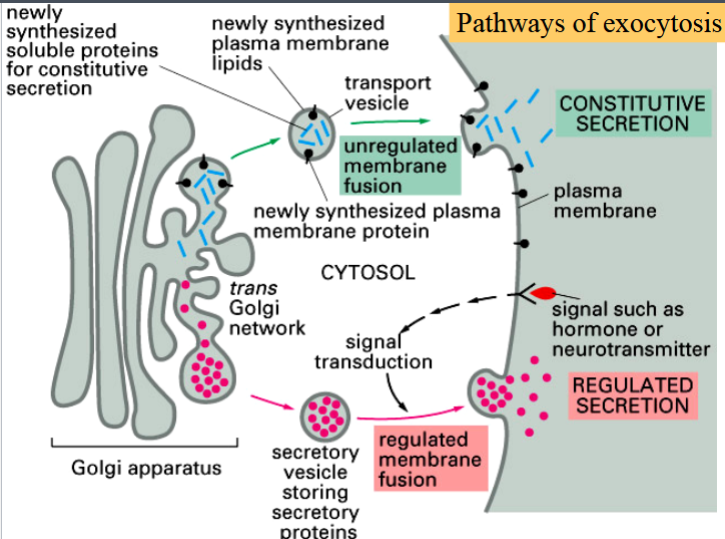
constitutive
regulated
lysosomal
constitutive transport from the Golgi
operates in all cells
unregulated secretion
also includes delivery of membrane proteins and lipids
(blue in the picture)
regulated transport from the Golgi
operates only in specialized cells
cells release their secreted products only when stimulated by a signal
signal is usually external, sometimes internal
products to be secreted are sorted into special, large vesicles that wait in the cell until the signal is received
exocytosis is calcium-dependent
e.g. endocrine cells secreting hormones, neurons secreting neurotransmitters
(red in the picture)
calcium-dependence of regulated secretion
fusion of secretory vesicles to cell membrane for release of products is induced by Ca2+
in simple endocrine cells: external signals cause opening of Ca2+ stores in ER
in neurons: extracellular Ca2+ inflow is observed in axon terminals (uncommon in simple endocrine cells)
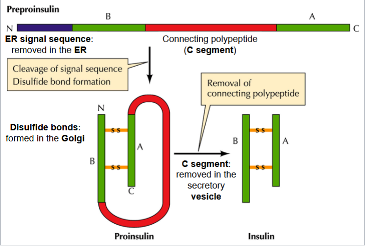
synthesis and regulated secretion of insulin
preproinsulin starts with signal sequence, B segment, connecting C segment, and A segment
ER signal sequence is removed in the ER, disulfide bond forms
proinsulin: B and A disulfide-linked with C still attached
C segment is removed in secretory vesicle → insulin has just B and A
regulated by blood glucose: when high, insulin is released into extracellular space
lysosomal transport from the Golgi
transport of digestive proteins into the lysosome
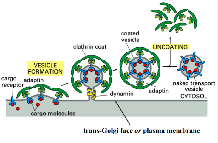
all vesicles that target the lysosome are clathrin coated (either from Golgi or from the plasma membrane)
clathrin-coated vesicles
originate from trans-golgi (if carrying lysosomal enzymes) and plasma membrane (from endocytosis)
headed toward lysosomes
when lysosomal protein contacts inner surface of trans-Golgi, clathrin coat is assembled on other side
formation of coat mechanically induces vesicle formation
lysosomal proteins are recognized by receptors on the Golgi inner surface, contact of lysosomal cargo protein with receptor induces clathrin coat assembly)
dynamins assemble around the budding vesicle’s neck and pinch it off
vesicle is released, coat is shed (because clathrin is only needed for mechanically bending the membrane to form the vesicle)
lysosome
one of the digestive organelles of the eukaryotic cell
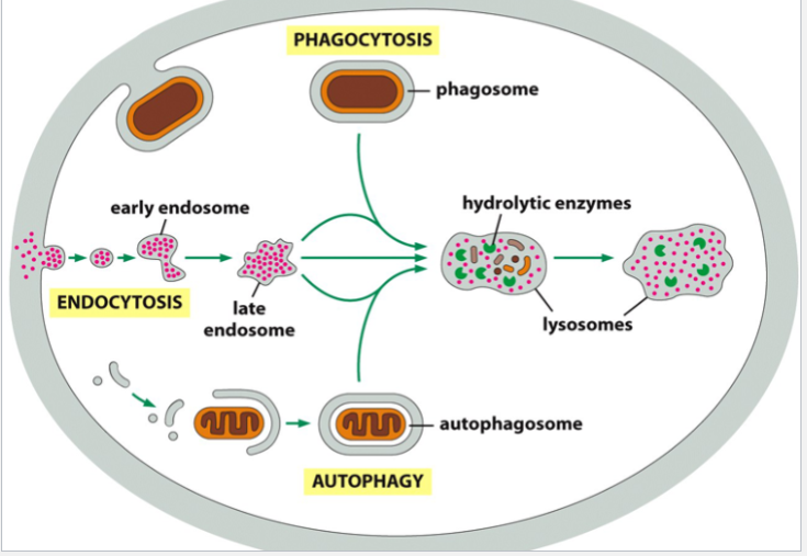
receives molecules/particles taken in by endocytosis, or cells taken in by phagocytosis, or obsolete/damaged cell components by autophagy for enzymatic degradation
contains acid hydrolases (digestive enzymes)
contains hydrolytic enzymes (not oxidative enzymes like peroxisomes)
needs a highly acidic environment inside to function (pH lowered by a H+ gradient) (pH~5 instead of pH~7.2 like in the cytoplasm)
components: early endosome, late endosome, maturation into functional lysosome
only forms as a result of its function
formation of the lysosome
early endosome forms from fusion of endocytic/phagocytic/autophagocytic vesicle
late endosome forms when hydrolase-containing vesicles from Golgi fuse with early endosome
late endosome matures into functional lysosome as pH decreases and hydrolytic enzymes become active
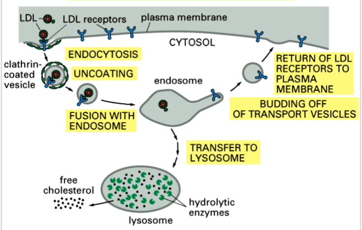
receptor-mediated endocytosis
taking in of specific molecules or particles recognized by cell surface receptors
e.g. endocytosis of cholesterol
endocytosis of cholesterol
an example of receptor-mediated endocytosis
cholesterol is blood-insoluble
transported from liver to bloodstream and around body bound to proteins → LDLs (low density lipoproteins)
LDL is recognized by surface LDL receptors, taken in by cell, delivered to lysosome
LDL is dissociated by digestive enzymes in lysosome, cholesterol is released for membrane synthesis
LDL receptors return to surface and get recycled
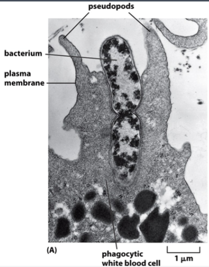
phagocytosis
the ingestion of other cells
done by specialized cells
e.g. a neutrophil ingesting a dividing bacterium
e.g. a macrophage ingesting damaged red blood cells
phagocytic cells have pseudopods: extensions of the membrane that extend over the ingested cell
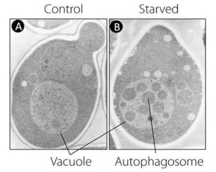
autophagy
self-digestion
the process by which cells eliminate damaged or obsolete cell components
also occurs when cells are starved; cells “eat” their own components for food
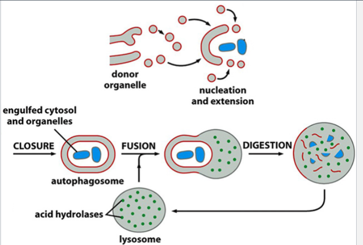
4 steps: nucleation, closure, fusion, digestion
its existence was demonstrated in yeast cells by Yoshinori Ohsumi: mutant cells lacking degradation enzymes were starved, autophagosomes accumulated in the vacuole (see picture)
4 steps of autophagy, expanded
1) nucleation (recruitment of membrane and necessary molecules), extension of delimiting membrane
2) closure of double membrane autophagosome with its components
4) fusion of outer membrane with a lysosome
4) digestion of inner membrane and its contents