Bone development
 Call Kai
 Learn
 Practice Test
 Spaced Repetition
 Match
 Flashcards
 Knowt Play
1/23

There's no tags or description
Looks like no tags are added yet.
24 Terms
Osteogenesis/Ossification
The process of forming bone tissue
2 types of osteogenesis/ossification
Intramembranous ossification and Endochondral ossification
What is intramembranous ossification replacing
Forms bone directly within a fibrous connective tissue membrane formed by mesenchymal cells
Step 1 of intramembranous ossification
ossification centers appear in the fibrous connective tissue

Step 1 of intramembranous ossification cellularly
Centrally located mesenchymal cells differentiate into osteoblasts and form trabeculae of spongey bone

Step 2 of intramembranous ossification
Osteoid secreted within fibrous membrane calcifies

Step 2 of intramembranous ossification cellularly
Osteoblasts secrete osteoid which then calcifies. Some osteoblasts become trapped and convert to osteocytes

Step 3 of intramembranous ossification
Spongy bone and periosteum form

Step 3 of intramembranous ossification cellularly
Blood vessels enter the tissue. Mesenchyme condenses into periosteum. Osteoid is secreted around the blood vessels trabecular/woven bone formation.

Step 4 of intramembranous ossification
Compact bone replaced woven bone and red marrow appears
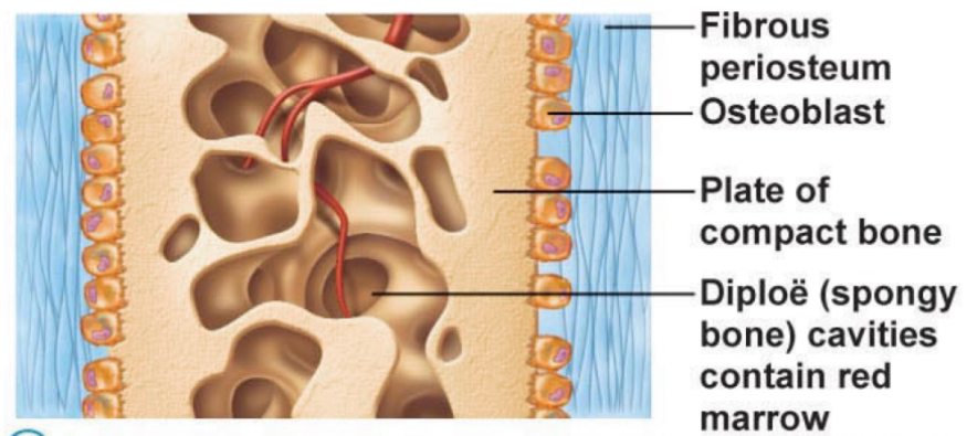
Step 4 of intramembranous ossification cellularly
Spongy bone just deep to periosteum thickens and forms compact bone. Spongy bone internally and vascular tissue produces red marrow that persists into adulthood.
What is endochondral ossification replacing
Forms bone by replacing an existing hyaline cartilage model
Examples for intramembranous ossification
Flat bones of cranium and clavicles
Examples for endochondral ossification
All bones of the post cranial skeleton except the clavicle

Step 1 of endochondral ossification
Bone collar forms around the diaphysis of the hyaline cartilage model

Step 1 of endochondral ossification cellularly
Osteoblasts in the osteogenic layer of the periosteum make Osteoid surrounding the hyaline model (primary ossification center)
Step 2 of endochondral ossification
Cartilage in center of diaphysis calcifies and develops cavities

Step 2 of endochondral ossification cellularly
Chondrocytes in diaphysis start to hypertrophy which signals surrounding cartilage to calcify. Chondrocytes become isolated from nutrients and die- leaving cavities. Ends of model continue to elongate.

Step 3 of endochondral ossification
Periosteum bud invades internal cavity- begins to form spongy bone

Step 3 of endochondral ossification cellularly
Periosteal bud- nutrient artery, vein, nerve fibers, red marrow elements, osteogenic cells, and osteoclasts. Osteoblasts form Osteoid around remaining cartilage forming spongy bone

Step 4 of endochondral ossification
Diaphysis elongates and medullary cavity forms

Step 4 of endochondral ossification cellularly
Osteoclast break down newly formed spongy bone and clear medullary cavity. Hyaline cartilage model at epiphysis continues to grow and diaphysis continues to elongate. Ossification chases the cartilage. Some secondary ossification centers start to form

Step 5 of endochondral ossification
Epiphyses ossify

Step 5 of endochondral ossification cellularly
At birth most epiphyses are still cartilage models- shortly after birth they become secondary ossification centers. Process is similar to primary ossification centers except no medullary cavity forms. Eventually the epiphyseal plate ossifies leaving behind the epiphyseal line