Lesson 62: Stomach Function, Emesis, Mucosal Barrier and GI Motility
 Call Kai
 Learn
 Practice Test
 Spaced Repetition
 Match
 Flashcards
 Knowt Play
1/40

There's no tags or description
Looks like no tags are added yet.
Name | Mastery | Learn | Test | Matching | Spaced | Call with Kai | Chat |
|---|
No analytics yet
Send a link to your students to track their progress
41 Terms
ENTERIC NS = Brain of the Gut
Contains as many neurons as the spinal cord
Between esophagus and anus
Consists of a myenteric and a mucosal plexus
Both are local (short) reflex systems with sensory receptors, sensory & motor fibers, cell bodies and effectors
Myenteric Plexus
between circular and longitudinal muscle layer
its mechanoreceptors respond to stretch = distension of the gut wall = presence of food
its effectors are smooth muscle cells ➔ controls GI motility
Anatomy of the myenteric plexus
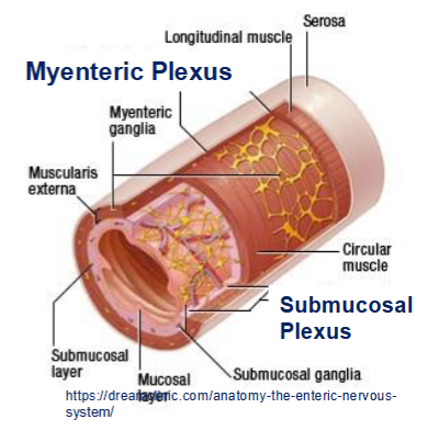
Submucosal Plexus:
between mucosa and muscle layer
its chemoreceptors respond to ingesta composition: pH, fat content, protein content
effectors are mucus cells, secretory cells ➔ Controls secretion rates of fluids, bicarb and mucus
The ENS uses a multitude of neurotransmitters
Excitatory, such as serotonin, substance P, motilin, acetylcholine
Inhibitory, such as vasoactive intestinal peptide (VIP), nitric oxide, dopamine, opioids, NE etc
very responsive system to many endogenous compounds, bacterial products and drugs
Sympathetic GIT main effects
Decreases GIT motility (beta-2, alpha-2)
Decreases GIT secretion rates (alpha-2)
Decreases blood flow to the GIT (alpha-1)
Stimulates GIT sphincters (alpha-1)
Enteric NS (local reflexes) and the Autonomic NS (central reflexes) are stimulated by and share the same?
chemoreceptors and mechanoreceptors and work in tandem
The strongest stimulator for both is GI wall distension
Enteric Immune System:
Composed of mesenteric lymphnodes, gut-associated lymphoid tissue (GALT), Peyers patches, mucosal WBCs
Function of the enteric immune system
If challenged, release pro-inflammatory cytokines
Cytokines act as local stimulators of the ENS and greatly stimulates GI motility and secretions
PERISTALTIC RUSH to flush out pathogens
GI smooth muscle: Peristaltic Contractions
travel long distances via coordinated local (ENS) reflexes: contractions cranial to a bolus with relaxation caudal to it → push ingesta slowly along the GI tube
GI smooth muscle: Mixing / Segmentation Contractions
travel short distances; circular muscle contractions mostly ENS generated → squeeze ingesta and move it back and forth
Interstitial cells of Cajal
Upon stimulation by a priming factor such as stretch, a motor ENS or ANS neuron
releases acetylcholine at a motor junction, which depolarizes the muscle membrane
further and pushes the top of slow waves above threshold potential → initiates action
potentials → muscle contraction.
Storage occurs in the proximal stomach = Fundus, facilitated via the Accommodation Reflex:
Food enters stomach ➔ distention stimulates stretch receptors ➔ sensory signals ascend via vagus nerve to medulla / vagal nuclei ➔ motor signals descend via vagal motor fibers to stomach muscles ➔ release of inhibitory NTs(nitric oxide) ➔ relaxation of muscles in proximal stomach ➔ enables expansion without pressure increase
Accommodation champions: Carnivores
can accommodate up to 25% of their body weight in the proximal stomach.
Purpose: “Feast-fasting” feeding pattern, regurgitation feeding of young
MIXING, GRINDING & RELEASE (Body & Antrum): Induction
Pacemaker area induces rhythmic slow waves traveling towards the pylorus, e.g. 3 waves/min
Stomach wall distension during feeding stimulates the ENS, ANS and endocrine cells, pushing slow waves above threshold and causing up to 3 strong peristaltic waves/min
Mixing, grinding, & release: Process
Contractions towards antrum → small volumes of fluid chyme pass into duodenum before the pylorus contracts ahead of a wave → most of the content is squirted back(=retropulsion) → repeat cycle
Content is kneaded for several hours, involving more of the stored ingesta as fluid chyme is released into the small intestines.
Stomach retention time of solid food: 4-12 hours /dog.
Motility pattern during fasting
Involves a powerful persitaltic wave action(5-10 minutes) sweeping from the stomach, along the small intestines into the large intestines („ stomach rumbling“).
Clears stomach and SI of residual ingesta, Avoids bacterial overgrowth in the SI.
Stimulated largely by the hormone Motilin, released from endocrine M-cells in the antrum and duodenum in the absence of food
Parietal cells produce hydrochloric acid
Pure HCl solution has a pH of <1
Mixed with stomach content: pH 1-2 in carnivores; pH 3-4 in omnivores
Production of HCL
active H+ / K+ pump = Proton Pump
it pumps H+ into the stomach lumen in exchange against K+ → Cl- follows down an electrical gradient
Functions of HCL
Bactericidal
Denatures dietary proteins
Activates gastric pepsinogen to pepsin
Emulsifies dietary fat into smaller globules
Triple control of HCL secretion
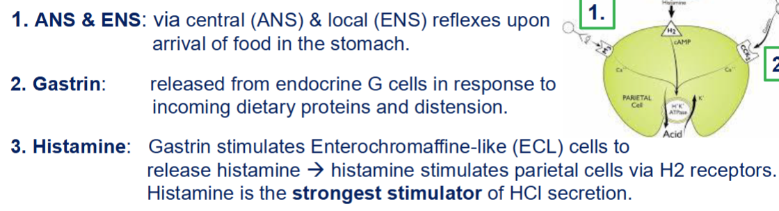
Somatostatin
released from endocrine D cells when the stomach pH is too low = negative feedback → inhibits proton pump, gastrin and histamin release
Intrinsic Factor is co-secreted with HCl
Binds to Vit B12(cobalamin) and protects it from being digested by pancreatic proteases
Both are absorbed in the ileum via a specific transporter.
Vit B12 is a necessary cofactor for DNA replication / mitosis.
Lack of Intrinsic Factor / Vit B12 deficiency affects:
RBC production / anemia
Nerve function / demyelination
Normal body growth
Most animals produce Intrinsic Factor mostly in the pancreas
pancreatic dysfunction can lead to Cobalamin deficiency in dogs, cats, horses
A genetic lack of IF-absorption mechanism exists in Giant Schnauzers
Chief cells produce Pepsinogens
HCL activates the proenzyme pepsinogens to the active pepsins
Pepsins’ pH optimum lies between pH 1-3.
Pepsins are proteolytic enzymes and hydrolyze large polypeptides into smaller polypeptides and oligopeptides
Chief cells co-produce gastric lipases
Hydrolyse some triglycerides; minor overall importance
Control of pepsinogen secretion:
Central & local reflexes (acetylcholine)
Gastrin
Carbohydrate digestion
salivary amylase hydrolyzes polysaccharides (typically 1000s of Glc) into a mixture of smaller polysaccharides and oligosaccharides (3-20 Glc).
10-30% of carbohydrate digestion occurs in the stomach
Amylase is destroyed once exposed to HCl (works only in the accommodation phase)
Protein digestion
HCl: denatures proteins = breaks up 2-3-4 structures into primary polypeptide chains.
Pepsin: Enzymatic hydrolysis of primary polypeptide chains to a mixture of smaller
polypeptides, oligopeptides and some amino acids
10-20% of protein digestion occurs in the stomach
Fat digestion
HCl: Begins the emulsification process = dividing larger into smaller fat globules.
Lingual & Gastric Lipases: Some hydrolysis of triglycerides into monoglycerides and free fatty acids (less than 5% of fat digestion) → More important in neonates for milk fat digestion.
Causes of vomiting
Peripheral signals from the GIT, pancreas, liver, peritoneum: Distension!! Inflammation, Irritation, Toxins, Foreign bodies
Chemical signals: endotoxins, exotoxins, waste products (ammonia, urea, ketone bodies, lactic acid, CO2), some hormones (hCG)
Vestibular Signals: motion sickness, vestibular disease
Higher CNS: pain, fear, stress, olfactory & gustatory signals
Prodromal Phase of vomiting(Nausea)
Drooling, lip licking, frequent swallowing, isolation
Retching (Dry Heaves)
Rhythmic abdominal contractions; SI antiperistaltic waves push ingesta back into the stomach; pylorus closes & cardia opens; expulsion of ingesta into esophagus
Expulsion (Vomiting)
Epiglottis closes; diaphragm and abdominal muscles contract powerfully; ingesta is pushed out via mouth, sometimes nose
Blood-borne signals activate the Chemoreceptor Trigger Zone (CTZ)
Its chemoreceptors detect and bind to a multitude of chemicals: toxins, waste products →
stimulating the CTZ neurons → transmit signals to the Vomiting Center to induce vomiting
Location of the CTZ
sits in the 4th ventricle above the medulla, outside of the blood-brain-barrier(BBB) → has direct access to circulating emetogenic substances
The Vomiting Center and the CTZ can be activated via many different causes and carry multiple receptors → drug targets:

Consequences of vomiting
Dehydration
Hypokalemia → hyperpolarization of excitable cells → nerve & muscle weakness
Alkalosis (or acidosis if small intestinal content is expulsed)
Esophagitis / aspiration pneumonia
Regurgitation
passive expulsion of ingesta from the esophagus or forestomach
No nausea , no abdominal contractions → Controlled by local reflexes in the esophagus
Reverse peristalsis pushes undigested food back into the mouth
Causes of regurgitation
Esophagitis
Megaesophagus (myesthenia gravis)
Esophageal stricure (RPAA; parasites)