Functional Neuroanatomy Knowt 1
 Call Kai
 Learn
Learn Practice Test
 Spaced Repetition
 Match
Match Flashcards
 Knowt Play
1/160

There's no tags or description
Looks like no tags are added yet.
Name | Mastery | Learn | Test | Matching | Spaced | Call with Kai | Chat |
|---|
No analytics yet
Send a link to your students to track their progress
161 Terms
Are dorsal/ventral classifications the same as superior/inferior?
Not necessarily. If you’re referring to the BRAIN, yes dorsal/ventral are the same as superior and inferior. But if you’re referring to the spinal chord, dorsal is posterior and ventral is anterior.
Now how would you describe the rostral/caudal classifcations?
In a human brain, rostral is towards the front and caudal to the back. In relation to the spinal chord, rostral is up the spine and caudal is down the spine.
In relation to the BRAIN, what is a classifcation that is the same was ventral/dorsal and what is one that is the same as rostral/caudal?
Inferior/Superior: Ventral/Dorsal, Anterior/Posterior: Rostral/Caudal
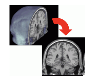
What is this view of the brain?
Coronal
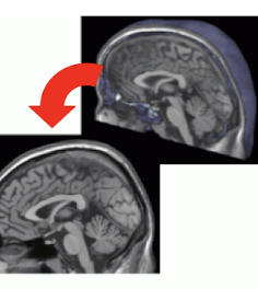
What is this view of the brain?
Saggital
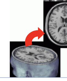
What is this view of the brain?
Transverse or axial
What are slices called that are not parallel to an orthogonal plane?
Oblique, neither coronal nor axial.
What are the ridges on the surface of the cerebrum and cerebellum?
gyrus
What are the grooves on the surface of the cerebrum?
Sulci, deep grooves are fissures.
What is Gray matter?
Generic term for neurons in the CNS
What is the Ganglion?
Collection of neuron cell bodies in the PNS
What is a substantia?
Less distinct borders of cell bodies than nuclei
What is Locus?
Small but well-defined mass of neuron cell bodies (Smaller than a nucleus)
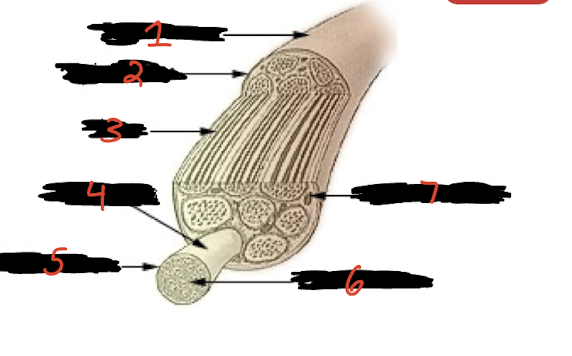
First, what is this? Second, label each number.
This is a nerve. 1) Spinal nerve 2) Epineurium 3) Axon 4) Fasciculus 5) Perineurium 6) Endoneurium 7) Blood vessels
What is a chiasm?
An x shaped crossing of nerve fibers between the cerebral hemispheres
What is a commissure?
A group of nerve fibers crossing the sagittal plane without forming a cross shape
What is neural induction?
Commitment of ectodermal tissue to neural tissue
What is neurulation?
The process of neural tube formation
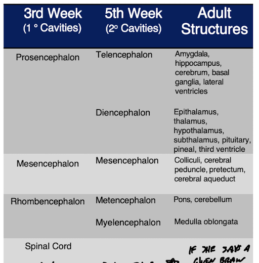
What are the 4 major vesicles that arise from the neural tube by the 3rd week?
Prosencephalon, Mesencephalon, Rhombencephalon, Spinal chord
By the 5th week, what changes with the 4 major vesicles?
Mesencephalon and spinal chord stay the same. Prosencephalon splits between Telencephalon and Diencephalon, while Rhombencephalon splits between Metencephalon and Myelencephalon.
What does the ectoderm give rise to?
Skin and neural tissues arise from the ectoderm after receiving signals provided by the mesoderm. A number of molecules interact to determine whether the ectoderm becomes neural tissue or develops in another way to become skin.
Describe the first appearance of neural areas
Budding of neural areas occurs in the rostral part of the neural tube which will eventually become the brain, regions called neuromeres. Occurs by 4 weeks.
What are the 8 phases of embryonic and fetal development at the cellular level?
1) Mitosis/Proliferation - creation of new cells 2) Migration - cells move to new areas 3) Differentiation - specialization of cell type 4) Aggregation - cluster together with like cell types 5) Synaptogenesis - create new synapses 6) Neuron death - “pruning” of unsuccessful nerve cells 7) Synapse rearrangement - modifying synapses 8) Myelination - formation of myelin sheath around axons

1) Neural plate 2) Neural Groove 3) Neural Crest 4) Neural Tube 5) Ectoderm 6) Gastrula
Is it fair to say that all cells that underwent neuralation also underwent neural induction?
Ja
What are the prosenphealon, diencephalon, and mesencephalon examples of?
Neuromeres
Describe neural induction
Once the ectoderm is formed, molecules within interact to determine whether the ectoderm becomes neural tissue or develops in another way to become skin.
Is it true that if a cell has undergone neurulation, it has also undergone neural induction? Why?
Yes, because neural induction occurs before neuralation. Neural induction is the commitment of ectodermal tissue to neural tissue. Neurulation is the process of neural tube formation from that neural tissue.
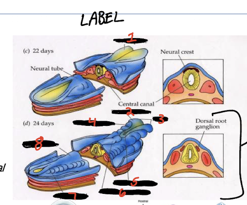
1) Brain plate 2) Telencephalon 3) Brain 4) Diencephalon 5) Mesencephalon 6) Rhombencephalon 7) Spinal chord 8) Neural tube
What happens immediately after neurulation… explain?
Immediately after neurulation, neuromere formation occurs in the rostral part of the newly formed neural tube. Usually by 4 weeks
Write the neuromere graph
What is the function of the choroid plexus
It is a complex of blood vessels that filters out blood cells to produce CSF in each of the 4 ventricles after they have developed.
After neuromere formation, what occurrs?
Within the ventricles, the choroid plexus forms and produces CSF within the ventricular spaces. Then, mitosis occurs in the ventricular layer, vertical cleavage to increase the number of precursor cells and stay within the ventricular zone, horizontal cleavage to result in one cell migrating to the developing cortex.

First, what is this picturing? Then label.
This is a coronal view of the developing brain, after the ventricles have formed. 1) Cortical plate 2) Ventricular/subventricular layers 3) 3rd ventricle 4) Choroid plexus 5) Telencephalon 6) Diencephalon 7) Mesencephalon
What occurs after the divided neuroblasts in the ventricular layer have migrated to the cortical plate? When in the timeline do neuroblasts begin to differentiate?
They differentiate, change appearance to take characteristics of a specific cell. Neurons differentiate first, followed by astrocytes and oligodendrocytes. Neuroblasts begin to differentiate during 2nd-3rd month of development.
Explain what occurs after migrated neuroblasts have differentiated.
Aggregation, meaning similar cell types cluster together into layers and regions based on common properties.
What occurs after aggregation?
Synaptogenesis, as the nerve cell develops, it sprouts a handlike distal end called the growth cone. Filopodia probe the environment, and integrins allow filopodia to grasp and move. It navigates the developing nervous system using complex guidance cues.
In synaptogenesis, how do axons locate their target tissues?
By using chemical attractants and repellants located around or on the surface of guide cells
By what month have all neurons been produced? And when synaptogenesis is over? What then occurs after this?
By the 6th month. After this, apoptosis (neuronal death) occurs in neurons with faulty connections.
What occurs after neuronal death?
Stage 7, synaptic rearrangement.
What is the stage after synaptic rearrangement?
Myelination
What is myelin
supportive sheathing that wraps around axons during and after development
What types of cells are responsible for myelination?
Glial cells. Oligodendrocytes wrap around numerous axons in the CNS, and Schwann cells can wrap around a singular axon in the PNS.
Give all info about the ventricles
Set of structures that hold CSF in the brain
Cushions the CNS within the bone
4 total ventricles: 1 lateral ventricle in each cerebral hemisphere, 3rd ventricle at the midline, and the 4th in the hindbrain
Where is most of the choroid plexus located?
The lateral ventricles
Telencaphlon has the appearance:
Telencephalon has the appearance of the third ventricle, but it eventually goes down into the diencephalon
Describe the cranium
22 immovable bones, plus a movable mandible and 3 pairs of auditory ossicles. Neurocranium are the bones that form the cranial cavity, while the viscerocranium makes up the features of the face.
What are sutures?
Where your skull is connecting together over your brain
Describe the 3 main sutures
Metopic suture - where the 2 frontal bones connect. Coronal suture - where the frontal bone is paired with the 2 parietal bones. Saggital suture - where the 2 parietal bones meet
What is the anterior fontanelle
The intersection of the coronal and sagittal suture
Describe the vertebral column
Consists of 33 separate bones (vertebrate) that stack to form this structure
Vertebrae are separated by vertebral discs that help provide cushion during movement
Each vertebrae has the central foramen, which when stacked create the vertebral canal, which houses the spinal chord
What part of the spinal nerve transmits info to move, and which transmits sensory info?
To move is ventral root, whereas sensory info is the dorsal root. Dorsal root ganglion is where the spinal nerve splits into the two
Why is the Shh gene important
Involved with retinal ganglion cell pathfinding
Attracts those axons at low concentrations of Shh, and repels them at high concentrations
Attracts commiseral axons at the central midline of the spinal chord, which means it also affects motor movement
Mutation in shh gene can develop tiny malformed eyes and failure to separate the left and right hemispheres of the brain/facial dismorphia
What are the 2 main neural tube defects (NTDs) that we covered in class?
Spina biffida and anencephaly
Describe anencephaly
Brain initially protrudes through a defect in the cranial vault
Eventually all that is left is a small vascular mass of disorganized neural tissue
Eyes evaginate from the forebrain before it is destroyed and are preserved
Describe spina bifida
Latin for split stone
Affects brian, spinal chord, and muscles surrounding it
Loss of movement/sensation in the legs/feet, as well as bowel movement and bladder dysfunction ]
Arises from abnormalities during 3 migrations of cell groups during embryogenesis: neural tube, notochord, and mesenchymal tissue
What are the 3 types of spina bifida
Oculta (mild)
Meningocele (minor cyst, can be removed surgically)
Myelomeningocele (cyst blocking sensory info from getting in or out)
Describe the pattern of brain maturation/postnatal development
Vision, sensory, motor, in that order.

1) Ventral root 2) Pedicle 3) Dorsal root ganglion 4) Spinal nerve 5) Vertebral body 6) Intervertebral disc 7) Spinous process 8) Neural foramen 9) Dorsal root
What is the brainstem composed of? What is its function?
Medulla, the pons, and the midbrain. Provides main motor and sensory innervation to face and neck via cranial nerves.
What 2 cranial nerves are not part of the brainstem?
Olfactory and optic

Label the places of all cranial nerves
1) Olfactory 2) Oculomotor 3) Trigeminal 4) Facial 5) Glossopharyngeal 6) Accessory 7) Optic 8) Trochlear 9) Abducens 10) Vestibulocochlear 11) Vagus 12) Hypoglossal
Draw cranial nerve graph


Label the sensory cranial nerve nuclei
1) Mesencephalic of CN 5 2) Principal of CN 5 3) Spinal descending CN5 4) Solitary of CNs7,9,10 5) Superior vestibular CN8 6) Lateral vestibular CN8 7) Cochlear CN8 8) Inferior vestibular CN8 9) Medial vestibular CN8
Draw CN7 Faciial motor nerves diagram.

How do the olfactory and optic nerves enter the brain?
Through the inferior surface of the cerebrum
What is the neglected cranial nerve?
Nervus Terminalis, has relations to the olfactory nerve and may detect pheromones (ongoing research)…
What is the Trigeminal Nerve mainly governing?
Sensory in the head; touch, proprioception, pain/temperature. CNV and its nuclei are to the head what dorsal roots and spinal chord are to the body
What are the 3 fibers of the Trigeminal Nerve?
V1 - Opthalmic (sensory only) V2 - Maxillary (Sensory only) V3 - Mandibular (Both)
What are the characteristics of the Facial Nerve?
muscles of facial expression via the motor nucleus in the pons
eye closure
To salivary and lacrimal glands from superior salivatory nucleus
Palate and taste buds on anterior 2/3 of tongue via solitary nucleus of medulla
What does a stroke model and show in terms of Facial Nerve anatomy?
It would show a lesion in the corticobulbar/corticonuclear tract, which would result in contralateral lower face weakness. That is why weakness on one lower side of the face is an early sign of a stroke.
What does Bell’s Palsy model and show in terms of Facial Nerve anatomy?
It would show a lesion in the motor nucleus of VII or the peripheral nerve, which would result in ipsilateral upper AND lower face weakness
What does the Vestibulocochlear nerve govern?
Cochlear (auditory) and Vestibular (balance/motion)
Glossopharyngeal means
“tongue and throat”
Where does the glossopharyngeal nerve receive afferents from?
Carotid sinus, walls of the pharynx, mucous membranes, taste bud from posterior 1/3 of tongue
What are the 2 important muscles that the Spinal Accessory nerve governs?
Sternocleidomastoid - turn and nod head, part of trapezius - move head and shoulder blade.
Hypoglossal nerve governs…
motor for muscles of the tongue via its Hypoglossal nucleus
What are the 3 important pathways the Pons houses?
Corticospinal tract - connects brain and descends down spinal chord. Lemniscal pathways - transmit sensory info. Spinothalamic tract - originates in brainstem and regulates pain, temp, touch, itch. Pons is an input/output point (nuclei) for cranial nerves V - VIII
What are the 2 main parts of the pons?
1) Dorsal pons (pontine tegmentum), important for sleep. 2) Ventral pons, with orderly arranged transverse/longitudinal fiber bundles with pontine neurons (pontine nuclei) between them. Some longitudinal bundles include 1) Corticobulbar (corticonuclear) fibers, 2) corticospinal fibers, 3) corticopontine fibers. Trasverse fibers are connections from the cerebellum that wrap around the brainstem
Tell me about the Medulla
Most caudal part of the brainstem, continuous with the spinal chord
Site of attachment of CN VII - XII on the ventral side
How do you know you are looking at a ventral view of the medulla?
You can see prominent pyramids, underlying the pyramidal tracts from the cortex down to the spinal column. You should be able to see the decussation of the pyramidal tracts as they cross to the opposite side here before continuing down the spinal chord
How do you know you are looking at a dorsal view of the medulla?
Has an inferior (closed part) and a superior (open part). Open part is the floor of the 4th ventricle. Part where opening begins is called the obex, and the closed part is where the central canal begins
What is the Inferior Olive?
Part of the medulla. Receives afferent fibers from the muscle and joint receptors via spino-olivary pathways, connects to cerebellum

Label these motor cranial nerve nuclei
1) Edinger-Westphal CN III 2) Superior CN VII and Inferior CN IX 3) Dorsal motor CN X 4) Spinal Accessory CN XI 5) Hypoglossal CN XII 6) Ambiguous CN IX and X 7) Facial CN VII 8) Abducens CN VI 9) Motor CN V 10) Trochlear CN IV 11) Oculomotor CN III
Where is the hypoglossal nucleus?
The medulla
What is the GENERAL part of the brain that the Prosencephalon becomes?
Forebrain
What are the exterior structures of the Forebrain?
Superior Frontal Sulcus, Inferior frontal gyrus, Sylvian fissure, temporal lobe, superior and inferior parietal cortex divdied by the intraparietal sulcus
Describe the Sylvian Fissure
Divides temporal lobes from the frontal lobes and anterior parietal
What is the function of the vagus nerve?
Back and forth signals between brain and major organs, swallowing
What are the interior structures of the forebrain?
Cingulate gyrus, cingulate sulcus, Paracentral lobule, lingual gyrus, calcarine fissure, cuneus

Label each
1) Paracentral lobule 2) Lingual gyrus or lingula 3) Central sulcus 4) Cingulate sulcus 5) Cingulate gyrus 6) Cingulate sulcus
Anatomically describe the cingulate gyrus
Like a belt surrounding the corpus callosum
Anatomically describe the cingulate sulcus
Its marginal branch runs up to the superior surface, allowing you to easily find the central sulcus directly anterior to it
Anatomically describe the paracentral lobule
Surrounds the central sulcus and extends to medial surface
Anatomically describe the lingual gyrus or lingula
Medial occipital lobe structure below the calcarine fissure
Anatomically describe the calcarine fissure
Fissure at the very bottom right of the occipital lobe
Anatomically describe the Cuneus
Directly above the calcarine fissure