SBMS1431- Anatomy of Muskoskeletal System II
 Call Kai
 Learn
Learn Practice Test
 Spaced Repetition
 Match
Match Flashcards
 Knowt Play
1/49

There's no tags or description
Looks like no tags are added yet.
Name | Mastery | Learn | Test | Matching | Spaced | Call with Kai | Chat |
|---|
No analytics yet
Send a link to your students to track their progress
50 Terms
Muscoskeletal system - Key Components
Bones: Rigid framework providing structure and protection
Muscles: Contractile tissues enabling movement
Joints: Connections between bones allowing mobility
Tendons: Connects muscles to bones
Ligaments: Connects bones to other bones
Muscoskeletal system - Primary Functions
Support and movement of the body
Protection of vital organs
Blood cell formation (hematopoieses)
Mineral Stirage and homeostasis
Overview: Joints
Forms connections between bones that allow various types of movement
Overview: Tendons
Transfer force from muscles to bones to create movement
Overview: Ligaments
Connect bones to other bones, stabilizing joints and guiding movement
Overview: Cartilage
Cushions joints and reduces friction between moving parts
Integration of the muscoskeletal system
The system works through linked roles: support (bones), movement (joints), force, (tendons), stability (ligaments), low-friction (cartilage), and generate (muscles)
Movement Pathways
When you decide to move, your brain sends signals yjtough nerves to skeletal muscles, which contract and pull-on tendons → movement occurs at a joint
Stability + Protection
Ligaments stabilise and guide joint motion. Cartilage cushions, distributes load and reduces friction.
Cartillage: Nature’s shock absorber
Hyaline Cartilage
Elastic Cartilage
Fibrocartilage
Hyaline Cartilage
Found in joints, ribs, and respiratory tract
Smooth, glassy appearance with high collagen content
Primary type in articulation surfaces of synovial joints
Elastic Cartilage
Present in ear, epiglottis, and parts of the larynx
Contains elastic fibers allowing flexibility
Supports structures that bend repeatedly
Fibrocartilage
Located in menisci (of the knee), intervertebral discs (spine), and pubic symphysis (pelvis)
Contains abundant collagen fibers for strength
Strongest cartilage type
Designed to resist compression and tension
The Muscular System: Types of muscle
Cardiac Mucle
Smooth Muscle
Skeletal Muscle
Cardiac Muscle
Involuntary, controlled by the autonomic nervous system
Forms the muscular layer of the heart (myocardium)
Striated, involuntary control
Has unique properties allowing it to contract rhythmically
Smooth Muscle
Comprises walls of blood vessels and hollow organs
Non-striated, involuntary control (autonomic nervous system)
Example: Muscles in blood vessels, digestive tract, bladder
Skeletal Muscle
Attaches to bones and provides voluntary movement
Striated, voluntary control (somatic nervous system)
Example: Biceps, quadriceps, hamstrings
Anterior View
Deltoid (flex, abduct, and extend arm)
Biceps (flex forearm)
Abdominal muscles (flex and rotate trunk, hip)
Wrist flexors (flex wrist and fingers)
Pectoralis major (adduct arm)
Triceps (extend forearm)
Rectus abdominis ( flex trunk, hip)
Quadriceps (flex hip, extend leg)
Calf Muscles (plantarflexion & invert foot, flex toes)

Posterior View
Trapezius- (extend head, elevate shoulder)
Deltoid- ( posterior fibers extend arm)
Triceps- (extend forearm)
Latissimus dorsi- (extend, adduct arm)
Gluteus maximus- (extend, abduct hip/thigh)
Hamstring- biceps femoris ( extend hip, flex knee)
Calf muscles- (olantarflexion foot & invert foot, flex toes)

Skeletal Muscle Attachments: Original, Insertion, & Belly
Origin- the relatively fixed attachment of a muscle; often proximal (closer to the center of the body/ trunk), but this depends on the action being performed.
Insertion- The attachment that usually moves toward the origin during contraction. It is often the end farther from the body’s center (distal)
Muscle belly- The thick, contractile middle region of the muscle, made up of fascicles and muscle fibers. When the region shortens, it generates tension that is passed on to the bone through tendons.
Tendon- A connective tissue structure that attaches muscle to bone and carries the force produced by the muscle belly to the bone.
In short: A muscle anchors at its origin (stays put), pulls at its insertion (moves), generates force in its belly (the fleshy contractile part), and delivers that force to bone through its tendon.

Skeletal Muscle: Organization
Muscle → Fascicle → Muscle Fiber → Myofibril → Sarcomere
Muscle
Organ composed of fascicles, blood vessels, nerves and connective tissue coverings.
Fascicle
Bundle of muscle fibers wrapped by perimysium
Muscle Fiber
Long multinucleated muscle cell surrounded by sarcolemma.
Myofibril
Cylindrical bundle of repeating contractile units
Sarcomere
Functional contractile unit containing actin and myosin. Actin and myosin are arranged in repeating sarcomeres. Their orderly pattern produces skeletal-muscle striations.
Skeletal Muscle: Overview
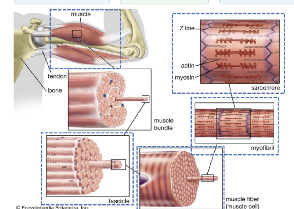
Connective Tissue coverings organize force transmission
Epimysium: wraps whole muscle
Perimysium: wraps each fascicle
Endomysium: wraps each fibe
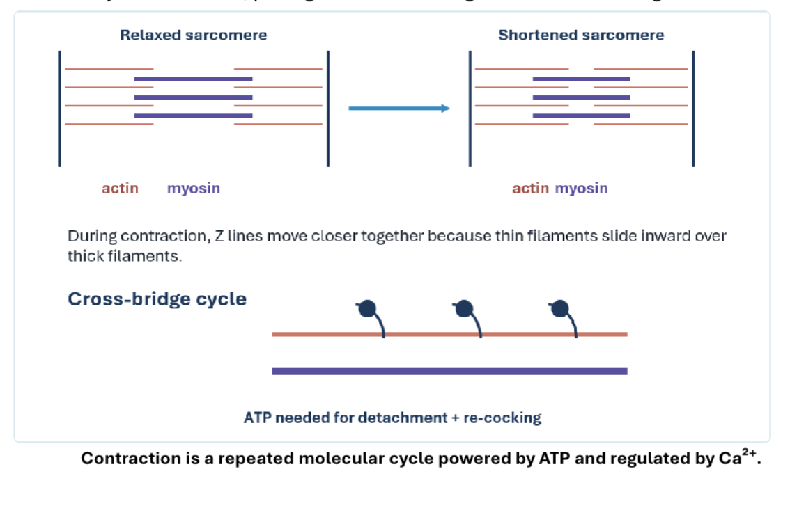
Sliding Filament Theory
Calcium exposes binding sites
A nerve signal triggers the release of calcium ions (Ca2+) inside the muscle Fiber. The calcium binds to troponin (a protein on the Acton filament), which causes tropomyosin to shift out of the way. This uncovers the myosin- binding sites on actin that were previously blocked.
Sliding Filament Theory
Cross-Bridge Formation
The myosin head (already energized by ATP) now attaches to the exposed binding sites on actin. This connection is called a cross-bridge
Sliding Filament Theory
Power stroke pulls actin
Once attached, the myosin head pivots (tilts), dragging the actin filament toward the centre of the Sarcomere. This is the power stroke- the actual pulling motion that generates force.
Sliding Filament Theory
ATP resets myosin
For the cycle to continue, myosin must let go of actin. ATP binds to the myosin head, causing it to detach from actin. Then the ATP is broken down (hydrolysis), which re-cocks the myosin head back into its energized, ready position.
Sliding Filament Theory
Cycle Repeats
As long as calcium and ATP are present, the cycle repeats: bind → pull → detach → reset → bind again. Each cycle pulls the actin a little farther inward.
Sliding Filament Theory: Overview
Actin and myosin filaments themselves don’t get shorter- thin actin filaments just slide past the thick myosin filaments, pulling Z discs closer together and causing the sarcomere to shorten.
Muscle Actions
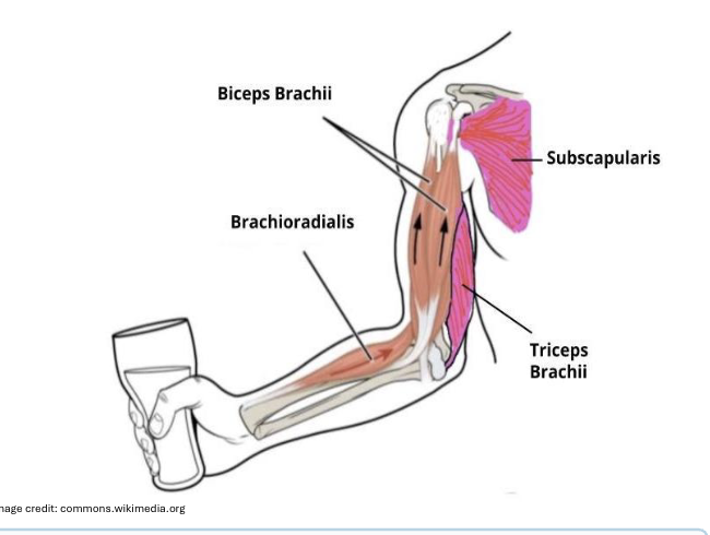
Agonist: Prime mover that generates the desired movement (e.g, biceps, in elbow flexion)
Antagonist: Opposes the action of the agonist and relaxes during movement (e.g., triceps during elbow flexion)
Synergist: Assists the agonist in producing movement.
Fixator: Stabilizes the origin of the agonist (e.g., deltoid stabilizing shoulder during bicep curl)
Muscle Actions: Example
A person lifting a class uses biceps brachial as the prime mover, brachioradialis acts as the synergist, subcapularis as the fixator and triceps brachial as the antagonist
Four key properties of muscle
Excitability
Contractibility
Extensibility
Elasticity
These four physiological properties enable muscles to respond to stimuli, generate force, adapt to movement demands, and maintain their integrity during physical activity.
Excitability
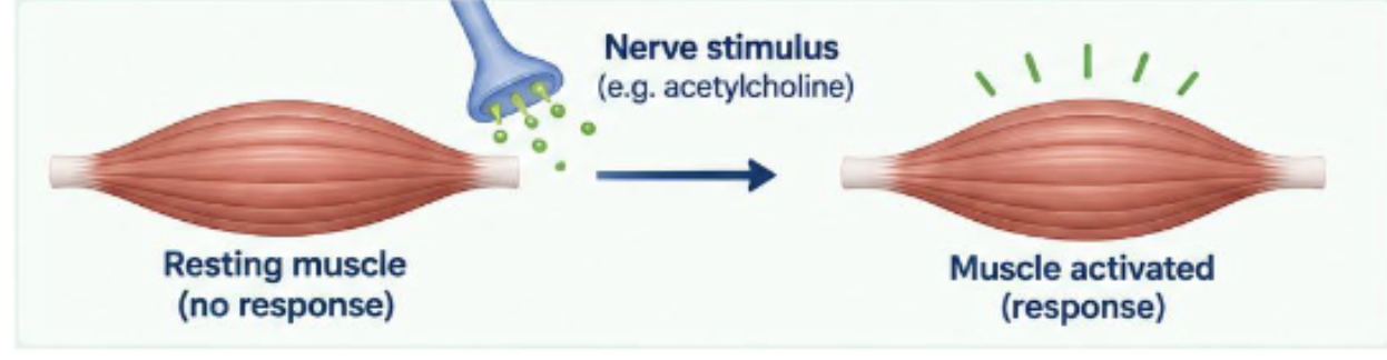
The ability to detect neural stimuli
Muscle Fibers can respond to an appropriate stimulus (such as a nerve signal) by generating an electrical and mechanical response
Contractibility
The ability to contract in response to a neural stimulus
When stimulated, muscle fibers develop tension and shorten, producing force and movement

Extensibility
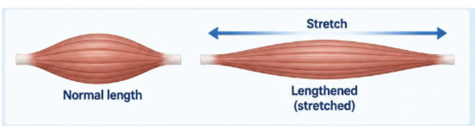
The ability of a muscle to be stretched without tearing
Muscle fibers can be stretched to a longer length and return to their original length, within physiological limits, without being damaged.
Elasticity
The ability to return to normal shape after being extended.
After being stretched or contracted, muscle fibers recoil and return to their original resting length,

Types of Contraction
Isometric: Muscle length does not change during contraction
Isotonic: Tension remains unchanged while length changes
Concentric: Muscle shortens (lifting a weight)
Eccentric: Muscle lengthens while maintaining tension (lowering a weight)
Contraction Mechanism
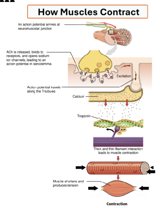
Nervous system generates action potential
Signal travels through motor neurons to neuromuscular junction
Acetylcholine released into the synaptic cleft
Calcium ions released into synaptic cleft
Actin and myosin slide over each other using ATP
Muscle contracts as signal diminishes
The neuromuscular Junction (NMJ)
Presynaptic Nerve Terminal: Contains vesicles filled with acetylcholine (ACh) neurotransmitter
Synaptic Cleft: Space where ACh diffuses from nerve to muscle
Postsynaptic Endplate: Contains nicotinic ACh receptors that trigger muscle contraction.
Acetylcholineesterase (AChE) enzyme rapidly degrades ACh in the synaptic cleft, terminating the signal. This junction is essential for precise muscle contraction control.
Muscoskeletal Pathologies- Osteoporosis
Decreased bone density increasing fracture risk
Osteoclasts: Break down (resort) old bone
Osteoblasts: Build new bone
In a healthy bone, build up and break down stay balanced. In osteoporosis, osteoclasts break down bone faster than osteoblasts can rebuild it, so bone mass steadily declines.
Result: Bones become porous and fragile → higher fracture risk (especially hip, spine, and wrist)
The pharmacology: Goal is to tip the balance back toward bone preservation- mainly by slowing down osteoclast activity.
Bisphosphonates, SERMs, calcitonin
Target: Osteoclast activity and bone remodelling
Muscoskeletal Pathologies- Neuromuscular Junction (NMJ) Disorders
Myasthenia gravis: autoimmune disease that blocks or destroys nicotinic acetylcholine receptors (AChRs) at the neuromuscular junction
Result: acetylcholine can’t bind properly → muscles receive weak signals → muscle weakness and fatigue (especially with repeated use)
Pharmacology: Neostigmine, pyridostigmine (acetylcholinesterase inhibition)
Target: Inhibit the breakdown of acetylcholine in the NMJ by blocking AchEs- allowing ACh to stat longer in the synaptic cleft.
Osteoarthritis
A degenerative joint disease caused by the breakdown of articular cartilage. As cartilage wears away, bones rub together, causing pain, stiffness, and inflammation.
Pharmacology: NSAIDs, corticosteroids, hyaluronic acid (these treat the symptoms by reducing pain/ inflammation to improve joint movement, but not the underlying cartilage loss)
Target: Inflammation and joint lubrication
Gout
Crystal-induced arthritis caused by deposition of urate (uric acid) crystals in joints. The crystals trigger intense inflammation and sudden, severe joint pain.
Pharmacology: allopurinol, febuxostat
Xanthine oxidase is the enzyme that produces uric acid. Allupurinol and febuxostat inhibit xanthine oxidase, lowering uric acid production. Less uric acid in the blood- fewer crystals forming in joints → fewer gout attacks.
Long term prevention drugs as they lower uric acid levels rather than addressing an acute attack
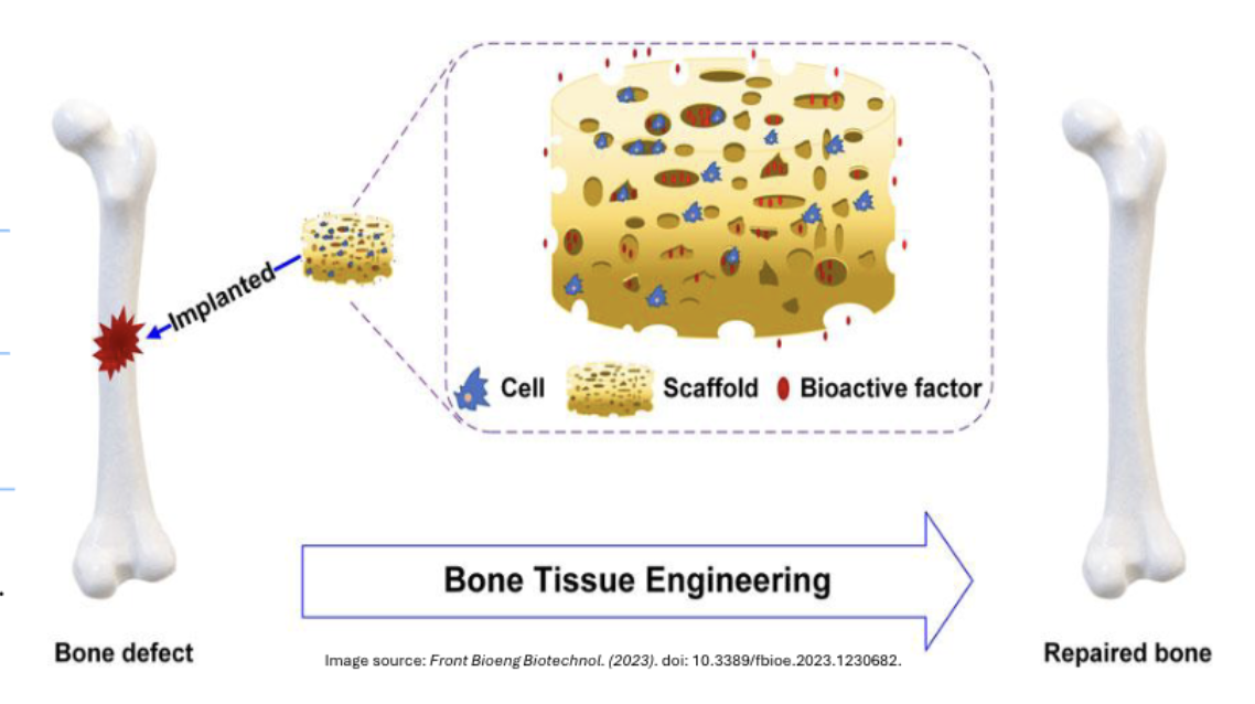
Bone Tissue Engineering Applications
Mesenchymal Stem Cells: Multipotent stem Cells that can differentiate into osteoblasts, chondrocytes, and other connective tissue cells.
Bioactive scaffolds: Three dimensional frameworks, made from hydroxyapatite or other biocompatible materials.
Tissue Regeneration: Cells proliferate, differentiate, and organise into functional bone tissue.
Key Engineering Processes
Cell Adhesion: Cells attach to scaffold surfaces through specific protein interactions
Proliferation: Rapid cell division increases cell population within the scaffold
Differentiation: Stem cells transform into specialized bone-forming osteoblasts
Matrix Formation: New bone tissue develops with proper mineral composition and structure
Modern tissue engineering harnesses the natural power of bone cells, combining them with advanced biomaterials to regenerate lost bone tissue and restore function where traditional treatments fall short.