Cardiac Action Potential and Arrhythmias
 Call Kai
 Learn
 Practice Test
 Spaced Repetition
 Match
 Flashcards
 Knowt Play
1/32

There's no tags or description
Looks like no tags are added yet.
33 Terms
In what order does electrical conduction travel through the heart during sinus rhythm?
The SAN initiates the electrical impulse
The impulse spreads across the atria, causing atrial depolarisation and contraction
The impulse reaches the AVN, where conduction is delayed by ~0.12s
The impulse travels rapidly down the Bundle of His and its right and left branches
The impulse spreads through the Purkinje fibres, causing ventricular depolarisation and contraction.
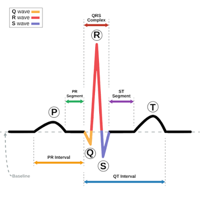
How does sinus rhythm present on an ECG?
P wave: Atrial depolarisation
QRS complex: Ventricular depolarisation
T wave: Ventricular repolarisation
PR interval: Atrial depolarisation and atrioventricular delay
QT interval: Ventricular depolarisation & repolarisation
PR segment: Atrioventricular delay
ST segment: Ventricular plateau

What are the two types of action potentials in the heart?
Cardiac action potential in atrial and ventricular myocytes, bundle of His, and in the Purkinje fibres.
Pacemaker action potential in the SAN and AVN

What is the cardiac action potential responsible for?
Defines heart rhythm, regulates heart rate, and controls the contractility
What is the pacemaker action potential responsible for?
Sets the heart rate (SAN), controls the rate of the heartbeat (AVN)

What are the ion mechanisms in the cardiac action potential?
Phase 0: Rapid Na+ influx (depolarisation) = Fast voltage gated Na+ channels open
Phase 1: Transient K+ efflux and Cl- influx (initial repolarisation)
Phase 2: Ca2+ influx balanced by K+ efflux (plateau)
Phase 3: Increased K+ efflux (repolarisation) = Several voltage gated K+ channels open
Phase 4: K+ efflux through IK1 channels (stable resting potential).
What determines the refractory period of the heart?
The duration of the cardiac action potential determines how long the cardiac cells remain in refractory.
Effective refractory period: A 2nd action potential cannot be generated, even with the strongest stimulus
Relative refractory period: A 2nd action potential can be generated but requires a stronger stimulus.


On an ECG, where is each phase of the cardiac action potential shown?
Rapid depolarisation Phase 0: The P wave and the QRS complex
Plateau Phase 2: The ST segment
Repolarisation Phase 3: The QT interval


What are the pharmacological features of the cardiac action potential?
Rapid depolarisation Phase 0: Target for Class I anti-arrhythmic drugs
Plateau Phase 2: Target for Class IV anti-arrhythmic drugs
Repolarisation Phase 3: Target for Class III anti-arrhythmic drugs

What are the ion mechanisms of the pacemaker action potential?
Phase 4: Na+ influx (pacemaker potential that is unstable) = Na+ influx via the funny current (If)
Early Phase 0: T-type calcium channels allow Ca2+ influx
Phase 0: Ca2+ influx (slow depolarisation) = Voltage gated L-type Ca2+ channels open
Phase 3: K+ efflux (repolarisation) = Voltage gated K+ channels open


What are the pharmacological features of the pacemaker action potential?
Slow depolarisation Phase 0: Target for Class IV anti-arrhythmic drugs
Phase 4: Target for Class II anti-arrhythmic drugs

What is the pacemaker current (If)?
The inward movement of mainly Na+ through HCN channels causing slow spontaneous depolarisation during Phase 4 of the pacemaker action potential.
What are the key features of the pacemaker current (If)?
The pacemaker current is the movement of ions across the membrane of pacemaker cells in the SAN.
Membrane repolarisation and increased cAMP causes HCN channels in the pacemaker cells to open
Na+ and K+ flow through HCN channels
More positive charge enters the cell than leaves it due to Na+ influx
This gradually makes the inside of the cell less negative, causing slow depolarisation
Slow depolarisation helps the SAN reach threshold and control heart rate.
How is the pacemaker current regulated by the ANS?
Sympathetic stimulation via β1 and β2 receptors increases cAMP and If, increasing heart rate
Parasympathetic stimulation via M2 receptors decreases cAMP and If, reducing heart rate
How do medicines affect the pacemaker current?
Ivabradine directly blocks HCN channels, reducing If
β-blockers indirectly reduce If by decreasing sympathetic stimulation
Digoxin indirectly reduces If by increasing parasympathetic input
Adenosine reduces If via A1 receptors and Gi/o proteins
What is the role of the pacemaker current in the regulation of heart rate?
It controls the rate of spontaneous depolarisation in the SAN.
Increased If increases heart rate.
Decreased If reduces heart rate.
What is the difference between rate control and rhythm control drugs?
Rate control drugs slow the heart rate without necessarily restoring normal sinus rhythm. Examples: Class IV CCBs, β-blockers (ivabradine and adenosine) and digoxin (indirectly).
Rhythm control drugs aim to restore or maintain normal sinus rhythm (sodium channel blockers, CCBs, drugs that prolong potassium channels and prolong the AP duration).
What are the 4 different ways arrhythmias are classified?
By the effect on the heart rate
By the effect on the heart rhythm (whether the heartbeat follows a consistent pattern; irregular or regular)
By the site of origin in the heart
By the type of QRS complex
What are the arrhythmias classified by the effect on the heart rate?
Bradycardia
Tachycardia (tachyarrhythmia)
How are arrhythmias classified by the site of origin in the heart?
Supraventricular – This means the arrhythmia originates above the ventricles, such as in the SAN, atria, or AVN
Ventricular – This means the arrhythmia originates in the bundle of His, Purkinje fibres, or ventricles
How are arrhythmias classified by the type of QRS complex?
Narrow complex means the QRS duration is less than 120ms
Broad complex means the QRS duration is greater than 120ms.
QRS complex represents ventricular depolarisation, so its width gives information about how electrical activity is travelling through the ventricles.
What are the signs and symptoms of arrhythmias?
Feeling heartbeats (palpitations)
Shortness of breath and fatigue
Chest pain
Dizziness
Blackouts or syncope
Cardiac arrest
What is bradycardia?
An abnormally slow heart rate, usually below 60bpm.
Sinus bradycardia is when the SAN generates electrical impulses more slowly than normal. What are the physiological causes of sinus bradycardia?
The vagal tone (the level of parasympathetic activity through the Vagus nerve) increases, slowing the heart rate through increased parasympathetic activity. Trained athletes may have physiological sinus bradycardia.
What are the pathological, extrinsic (non-cardiac) causes of bradycardia?
Endocrine disorders (e.g. hypothyroidism)
An electrolyte imbalance: Severe hyperkalaemia, and hypo/hypercalcaemia can disrupt the heart’s electrical activity
Drugs: anti-arrhythmic drugs can slow the heart rate. This includes beta-blockers, CCBs, digoxin, and clonidine (an antihypertensive)
Hypothermia: slows heart rate
What are the pathological, intrinsic causes of bradycardia?
Sick sinus syndrome: Disease or damage of the SAN affects its ability to generate electrical impulses. SSS includes ischaemia and infarction of the SAN
Atrioventricular blockage or heart block: Impaired conduction of electrical impulses through the AVN slows heart rate. Causes include:
Ischaemia and fibrosis
Congenital heart defects
Infections and inflammations such as myocarditis, diphtheria, and rheumatic fever
What is sick sinus syndrome?
Sick sinus syndrome is a group of heart rhythm disorders due to malfunction of the SAN that may present on the ECG as:
Sinus pause
Sinus arrest (pause for more than 3 seconds)
Bradycardia-tachycardia syndrome
How do we treat sick sinus syndrome?
Implantable transcutaneous pacemaker (TCP)
What is AV block?
AV block occurs when electrical impulses are slowed or blocked as they travel from the atria to the ventricles through the AVN.
There is a 1st degree, 2nd degree, and 3rd degree AV block.
1st degree AV block
AV conduction is slower than normal
The PR interval is prolonged to more than 0.2s
2nd degree AV block
Some electrical impulses fail to reach the ventricles
This causes some ventricular beats to be missed
There are 2 subtypes of 2nd degree AV blocks: Mobitz I (progressive prolongation of PR intervals) and Mobitz II.
3rd degree AV block
No electrical impulses are conducted from the atria to the ventricles
P waves and QRS complexes occur independently of each other.
How do you treat AV block?
In emergencies, give atropine or isoprenaline intravenously.
For a long term solution, give an implantable TCP in cases of the 2nd and 3rd degree block.