neurology II
 Call Kai
 Learn
 Practice Test
 Spaced Repetition
 Match
 Flashcards
 Knowt Play
1/66

There's no tags or description
Looks like no tags are added yet.
67 Terms
multiple sclerosis
Epidemiology: Young women (20-40 yrs).
Course: Relapsing-remitting (most common) → neurologic deficits separated in time and space.
Optic neuritis (painful vision loss, ↓ color vision).
Internuclear ophthalmoplegia (diplopia).
Sensory changes: paresthesias, numbness.
Motor: weakness, spasticity.
Lhermitte sign: electric shock sensation down spine with neck flexion.
Fatigue, heat sensitivity (Uhthoff phenomenon).
symptoms associated with multiple sclerosis
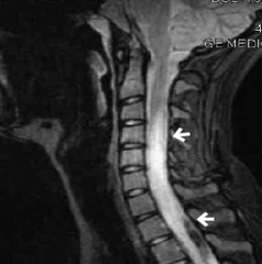
transverse myelitis
inflammation of the spinal cord
-can occur post-infection, post-vaccination, or with MS/NMO
Acute/subacute motor, sensory, and autonomic dysfunction localized to a spinal cord level.
Bilateral weakness, sensory level, sphincter dysfunction (urinary retention, bowel issues).
symptoms of transverse myelitis
transverse myelitis
Key history clue: rapid onset of bilateral deficits with a clear sensory level.
progressive multifocal leukoencephalopathy
Reactivation of JC virus in immunocompromised (HIV/AIDS, natalizumab, rituximab).
Subacute, progressive neurologic decline (weeks-months).
Cognitive impairment, behavioral changes.
Visual field deficits, ataxia, motor weakness.
symptoms of PM
PML
Key history clue: immunosuppression + progressive multifocal neuro deficits without fever.
Acute disseminated encephalomyelitis (ADEM)
encephalitis in children/young adults often post viral or post-vaccinaation
Monophasic illness (one-time event).
Encephalopathy (confusion, decreased consciousness) + multifocal neuro deficits (weakness, ataxia, vision changes).
symptom of ADEM
Acute disseminated encephalomyelitis (ADEM)
Key history clue: acute, widespread CNS demyelination following infection/vaccine, with encephalopathy.
Campylobacter jejuni, CMV, EBV, Mycoplasma
most common triggers of guillain barre syndrome
Rapidly progressive, symmetric ascending weakness (legs → arms).
Areflexia.
Possible cranial nerve involvement (facial weakness, dysphagia).
Autonomic instability (arrhythmias, BP fluctuations).
symptoms of guillaiin barre syndrome
Guillain-Barre syndrome
Key history clue: acute ascending paralysis after infection.
myasthenia gravis
Epidemiology: Young women, older men; often associated with thymoma or thymic hyperplasia.
Fluctuating weakness, worse with activity, better with rest ("fatigability").
Ocular: ptosis, diplopia (most common presenting).
Bulbar: dysphagia, dysarthria.
Respiratory weakness (myasthenic crisis).
symptoms of myasthenia gravis
myasthenia gravis
Key history clue: weakness that worsens with use, improves with rest.
Etiology: Often paraneoplastic (esp. small cell lung carcinoma).
etiology of lambert eaton myasthenic syndrome
Proximal muscle weakness (legs > arms).
Improves with repeated use (opposite of MG).
Autonomic symptoms: dry mouth, impotence, constipation.
Hyporeflexia.
symptoms of LEMS
Lambert-Eaton myasthenic syndrome
Key history clue: paraneoplastic + weakness improves with activity + autonomic symptoms.
No specific antibody test for MS.
Diagnosis = clinical + MRI + CSF findings.
CSF: Oligoclonal IgG bands (present in >85% of MS patients).
testing diagnostic of MS
JC virus DNA in CSF by PCR.
Antibody testing not used directly for diagnosis (JC virus antibody serology sometimes used to estimate risk in immunosuppressed patients, e.g., natalizumab therapy).
diagnostic testing for PML
Anti-GM1, GD1a, GD1b, GQ1b ganglioside antibodies in certain variants.
antibodies associated with GBS
Anti-acetylcholine receptor (AChR) antibodies (most common).
Anti-MuSK antibodies (muscle-specific kinase) in some seronegative MG patients.
antibodies diagnostic of myasthenia gravis
Anti-VGCC (voltage-gated calcium channel) antibodies
antibodies associated with LEMS
-oligoclonal IgG bands
-elevated IgG index
-normal/mildly elevated protein, normal glucose
CSF findings diagnostic of multiple sclerosis
MRI findings of multiple sclerosis
-white matter lesions hyperintense on T2/FLAIR
-Both enhancing (active, new) and non-enhancing (older) lesions on the same MRI.

-Dissemination in space (lesioins in >2 CNS regions)
-dissemination in time (evidence that lesions occurred at different points in time)
McDonald diagnostic criteria of multiple sclerosis
1st line: IV methylprednisolone
2nd line: plasmapehresis (used for severe rellapses not responding to steroids)
1st and 2nd line management for relapsing MS:
amantadine or modafinil
Rx treatment for the fatigue associataed with MS
gabapentin
tx for neuropathic pain associated with MS
Multiple sclerosis
a chronic, immune-mediated demyelinating disease of the CNS.
The pathology involves autoimmune attack against myelin and, secondarily, axonal injury.
Both innate and adaptive immunity play roles
CD4+ Th1 and Th17 cells cross blood-brain barrier → release cytokines (IFN-γ, IL-17, TNF-α) → activate macrophages & microglia → demyelination.
describe the pathophys of the immune mediated demyeliniation in MS
B cells
which immune cells in MS Produce oligoclonal IgG in CSF, act as antigen-presenting cells, form ectopic follicles in meninges?
Myelin Basic Protein (MBP)
Myelin Oligodendrocyte Glycoprotein (MOG)
Proteolipid Protein (PLP)
what are some of the antibodies that target myellin proteins in MS?
Often triggered by preceding infection (molecular mimicry):
Campylobacter jejuni (most common)
CMV, EBV, Zika, Mycoplasma
what triggers GBS?
Immune response → antibodies cross-react with gangliosides (GM1, GD1a, GQ1b) on peripheral nerves → demyelination ± axonal injury
describe the pathophys of GBS
-ascending symmetric weakness
-hyporeflexia
-mild paresthesias
-facial palsy, dysphagia
symptoms of GBS
Albuminocytologic dissociation → high protein, normal WBC count.
CSF findings associated with GBS
Weakness of diaphragm & intercostal muscles → ventilatory failure.
most common cause of death due to GBS
anti-acetylcholine receptor antibodies aainst post-synaptic NMJ
most common Ab associated with myasthenia gravis
myasthenia gravis
Antibodies block or destroy receptors → impaired neuromuscular transmission → weakness.
Strongly associated with thymic abnormalities (thymoma or thymic hyperplasia)
-fluctuating skeletal m. weakness (worsens with activity)
-ptosis, diplopia
-dysphagia, dysarthriaa
main symptoms of myasthenia gravis
Repetitive nerve stimulation → decremental response
EP findings associated with myasthenia gravis
Ice pack test → improvement of ptosis after application of ice.
Bedside test to aid in diagnosis of myasthenia Gravis
Chest CT/MRI to evaluate for thymoma.
should I get imaging in assessment of MG?
Caused by antibodies against presynaptic P/Q-type voltage-gated calcium channels (VGCCs)
antibodies associated with LEMS
-antibodies against VGCC
Result: ↓ calcium influx into presynaptic terminal → ↓ acetylcholine (ACh) release.
MOA of lambert eaton
-proximal muscle weakness
-autonomic dysfunction: dry mouth, constipation, impotence
-ptosis, diplopia
-hyporeflexia, areflexia
strength improves with repeated activity ("facilitation"), unlike MG.
symptoms of LEMS
Chest CT or PET scan to evaluate for small-cell lung carcinoma
imaging indicated for pts wit LEMS
transverse myelitis
Inflammation across one spinal cord segment → immune-mediated demyelination and axonal injury → sensory, motor, and autonomic dysfunction below the lesion.
CNS white matter
site of PML
JC virus infects and lyses oligodendrocytes → multifocal CNS demyelination. Not antibody-driven, but opportunistic viral-mediated.
pathopys of PMLL
CNS --diffuse white matter
site of ADEM
Widespread perivenular inflammation & demyelination → monophasic encephalopathy with multifocal deficits. Involves T-cell cross-reactivity (molecular mimicry).
pathophys of ADEM
dawson's fingers
In multiple sclerosis, perivascular demyelination creating the appearance of finger projections oriented transversely on an axial scan.

Test for AQP4-IgG, MOG-IgG
antibodies associated with transverse myeliitis
MRI: multifocal asymmetric white matter lesions, no enhancement or mass effect
MRI findings associated with PML

oligodendrocytes
JC virus infects what cells?
HIV work-up, immunosuppression history
lab workup indicatedi n pt diagnosed with PML
ADEM MRI
bilateral symmetric inflammatory changes of the same age
fluffy white spots on T2 weighted

Internuclear ophthalmoplegia
a lesion of the medial longitudinal fasciculus (MLF); disorder of conjugate lateral gaze in which the affected eye fails to adduct when the contralateral eye abducts (w/ nystagmus); most commonly due to MS plaques when presenting in a young person

upper motor neuron signs
spastic paralysis with hyperreflexia, increased muscle tone, positive Babinski sign
upper
is MS associated with upper or lower motor neuron signs?
Bilateral weakness, sensory level, sphincter dysfunction
physical exam findings of transverse myellitis
PML
History: Immunocompromised patient (HIV, natalizumab, transplant), progressive neurologic deficits (aphasia, hemiparesis, vision loss).
ADEM
History: Child/young adult, recent viral illness or vaccination, monophasic acute neurologic deficits + encephalopathy.
Exam: Multifocal deficits, altered mental status.