RAD 112-Humerus & Shoulder Girdle
 Call Kai
 Learn
Learn Practice Test
 Spaced Repetition
 Match
Match Flashcards
 Knowt Play
1/170

There's no tags or description
Looks like no tags are added yet.
Name | Mastery | Learn | Test | Matching | Spaced | Call with Kai | Chat |
|---|
No analytics yet
Send a link to your students to track their progress
171 Terms
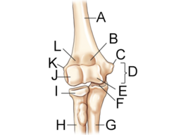
label the anatomy (A-F) of the elbow
A. Body/Shaft of humerus
B. Coronoid Fossa
C. Medial Epicondyle
D. Humeral Epicondyles
E. Trochlea
F. Trochlear notch

label the anatomy (G-L) of the elbow
G. Ulna
H. Radius
I. Radial Head
J. Capitulum
K. Lateral Epicondyle
L. Radial Fossa
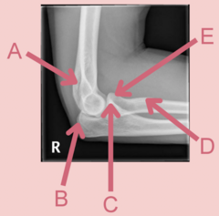
What view of the elbow is this?
lateral

label the anatomy
A. area of olecranon fossa
B. olecranon process
C. coronoid process
D. radial tuberosity
E. radial head

label the anatomy of the humerus
1. Greater tubercle (tuberosity)
2. Intertubercular groove (bicipital groove)
3. Surgical neck
4. Head
5. Anatomical neck
6. Lesser tubercle (tuberosity)
7. Deltoid tuberosity
8. Body

label the anatomy of the humerus
1. Greater tubercle
2. Intertubercular (bicipital) groove
3. Surgical neck
4. Head
5. Anatomical neck
6. Lesser tubercle
7. Body

label the anatomy of the shoulder girdle
1. Scapulohumeral (glenohumeral) joint
2. Lateral (axillary) border
3. Axilla
4. Superior border
5. Medial (vertebral) border

label the anatomy of the scapula
1. Acromion
2. Glenoid cavity (fossa)
3. Lateral angle (head)
4. Neck
5. Costal surface/subscapular fossa
6. Coracoid process
7. Scapular notch
8. Superior angle
9. Body (blade/wing)
10. Inferior angle

label the anatomy of the scapula
1. Infraspinous fossa
2. Supraspinous fossa
3. Crest of spine
4. Acromion
5. Dorsal surface (posterior)
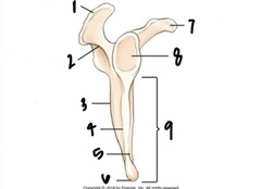
label the anatomy of the scapula
1. Acromion
2. Spine of scapula
3. Dorsal surface
4. Lateral border
5. Ventral (costal) surface
6. Inferior angle
7. Coracoid process
8. Glenoid cavity (fossa)
9. Body

label the shoulder muscles listed here
1. Subscapularis
2. Supraspinatus
3. Infraspinatus

What projection is this?
AP Humerus (supine)

What anatomy is circled in this?
Greater Tuberosity

What projection is this?
AP Humerus (erect)

label the anatomy in this image
1. Acromion
2. Coracoid profile
3. Greater tubercle
4. Body
5. Lateral Epicondyle
6. Medial Epicondyle

What projection is this?
Lateral Humerus

label the anatomy in this image
1. Acromion process
2. Lesser tubercle
3. Body
4. Capitulum
5. Olecranon process

What projection is this?
Neutral Rotation Humerus
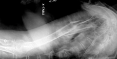
What projection is this?
Transthoracic lateral

label the anatomy of the clavicle
1. Lateral aspect
2. Acromial Extremity
3. Body
4. Sternal Extremity
5. Medial aspect
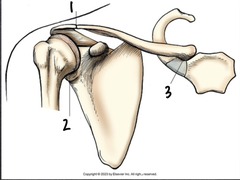
Label the joints of the shoulder
1. Acromioclavicular joint
2. Scapulohumeral joint
3. Sternoclavicular joint

What projection is this?
AP Internal Rotation Shoulder

What projection is this?
AP External Rotation Shoulder

label the anatomy in this image
1. Clavicle
2. Acromion
3. Coracoid process
4. Scapulohumeral joint
5. Scapula
6. Humerus

What projection is this?
AP clavicle

What projection is this?
AP axial clavicle

What projection is this?
AP scapula

label the anatomy in this image
1. Acromion
2. Clavicle
3. Coracoid process
4. Glenoid cavity
5. Lateral border of scapula
6. Medial border of scapula
7. Inferior angle of scapula

What projection is this?
Lateral Scapula to demonstrate acromion and coracoid processes

What is the line pointing to?
the coraco-acromial arch

What projection is this?
Lateral Scapula to demonstrate body of scapula

What clinical indication does this image show?
Dislocation (posteriorly) under the acromion process

What clinical indication does this image show?
Dislocation (anteriorly) under the coracoid process

What projection is this?
AP Neutral Rotation Shoulder

What projection is this?
Scapular Y view

label the anatomy of this image
1. Acromion
2. Coracoid process
3. Body of Scapula
4. Inferior Angle
5. Humerus

What projection is this?
Supraspinatus Outlet: Neer Method

What projection is this?
AP oblique: Grashey

label the anatomy of this image
1. Acromion
2. Humeral head
3. Glenoid cavity
4. Clavicle
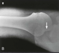
What projection is this?
Inferosuperior Axial: Lawrence method

label the anatomy of this image
1. Lesser tubercle
2. Humerus
3. Coracoid process
4. Acromioclavicular joint
5. Clavicle
6. Scapulohumeral joint
7. Acromion

What projection is this?
PA Axial Clavicle
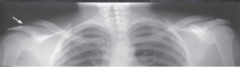
What projection is this?
AP Bilateral AC joints
What is another name for the intertubercular groove?
Bicipital groove.
What is another name for the body of the humerus?
Shaft.
What is another name for the capitulum?
Capitellum.
What three bones make up the elbow joint?
Humerus, radius, and ulna.
Where does the head of the humerus sit?
Within the glenoid fossa.
What joint is formed by the articulation of the humeral head and glenoid fossa?
Glenohumeral joint.
Which neck of the humerus is more likely to experience a fracture?
Surgical neck.
What are the routine projections for the humerus?
AP and lateral.
What are the special projections for the humerus?
Neutral rotation and transthoracic lateral.
What are the clinical indications for humerus radiographs?
Fracture, dislocation, and pathologic processes including osteoporosis.
What should you NEVER do if a humeral fracture or dislocation is suspected?
Do NOT attempt to rotate the arm.
What projection should be performed instead of rotating the arm when a fracture or dislocation is suspected?
Neutral rotation.
What is the kVp range for the humerus?
70–85 kVp.
What patient positions can be used for an AP humerus?
Erect or supine.
Which patient position is usually more comfortable for an AP humerus, if possible?
Erect.
How are the hand and forearm positioned for an AP humerus?
Extend the hand and forearm and supinate the hand.
How is the arm positioned for an AP humerus?
Abduct the arm.
How should the humeral epicondyles be positioned for an AP humerus?
Parallel to the IR.
What size IR is used for an AP humerus?
14 × 17 inch lengthwise IR.
Where is the IR placed for an AP humerus?
Within the bucky, not tabletop.
Where is the CR directed for an AP humerus?
Perpendicular to the mid-humerus.
How should the tube head be oriented for an AP humerus?
Rotate the tube head to follow the orientation of the arm.
What should be included in the collimation for an AP humerus?
Shoulder joint, elbow joint, and soft tissue.
Where should the marker be placed for an AP humerus?
In the light field but not over the anatomy.
What breathing instructions are given for an AP humerus?
Suspend respiration.
What anatomy should an AP humerus demonstrate?
Entire humerus, shoulder joint, elbow joint, and soft tissue.
What indicates a true AP humerus?
The greater tubercle is in profile laterally and the humeral epicondyles are parallel to the IR.
How can you tell that exposure is adequate on an AP humerus?
Bony trabeculae and soft-tissue detail are visible.
What is the lateral humerus projection called in the class slides?
Lateromedial lateral humerus.
What patient positions can be used for a lateral humerus?
Erect or supine.
Which patient position is usually more comfortable for a lateral humerus if possible?
Erect.
How is the arm positioned for a lateral humerus?
Abduct and internally rotate the arm.
How much should the elbow be flexed for a lateral humerus?
90°.
Where should the patient's hand be placed for a lateral humerus?
Place the dorsal aspect, or back, of the hand against the hip.
How should the humeral epicondyles be positioned for a lateral humerus?
Perpendicular to the IR.
What size IR is used for a lateral humerus?
14 × 17 inch lengthwise IR.
Where is the IR placed for a lateral humerus?
Within the bucky, not tabletop.
Where is the CR directed for a lateral humerus?
Perpendicular to the mid-humerus.
How should the tube head be oriented for a lateral humerus?
Rotate the tube head to follow the orientation of the arm.
What should be included in the collimation for a lateral humerus?
Shoulder joint, elbow joint, and soft tissue.
Where should the marker be placed for a lateral humerus?
In the light field but not over the anatomy.
What breathing instructions are given for a lateral humerus?
Suspend respiration.
What anatomy should a lateral humerus demonstrate?
Entire humerus, shoulder joint, elbow joint, and soft-tissue structures.
Which tubercle should be seen in profile on a true lateral humerus?
Lesser tubercle in profile medially.
How should the humeral epicondyles appear on a true lateral humerus?
Superimposed and perpendicular to the IR.
What positioning features indicate a true lateral humerus?
Lesser tubercle in profile medially and humeral epicondyles superimposed and perpendicular to the IR.
How can you tell that exposure is adequate on a lateral humerus?
Bony trabeculae and soft-tissue detail are visible.
What is a quick way to distinguish an AP from a lateral humerus?
AP = greater tubercle in profile laterally and epicondyles parallel to IR. Lateral = lesser tubercle in profile medially and epicondyles perpendicular to IR/superimposed.
What two bones make up the shoulder girdle?
The clavicle and scapula.
Which scapular process projects anteriorly?
The coracoid process.
Which scapular process projects posteriorly?
The acromion process.
What is the shallow socket that articulates with the humeral head?
The glenoid cavity (glenoid fossa).
What border of the scapula is closest to the vertebral column?
The medial (vertebral) border.
What border of the scapula is closest to the axilla?
The lateral (axillary) border.
What structure separates the supraspinous and infraspinous fossae?
The scapular spine.
Which fossa lies above the scapular spine?
The supraspinous fossa.
Which fossa lies below the scapular spine?
The infraspinous fossa.