L16 - Lymphocyte development: T-cell development
 Call Kai
 Learn
 Practice Test
 Spaced Repetition
 Match
 Flashcards
 Knowt Play
1/56

There's no tags or description
Looks like no tags are added yet.
57 Terms
What Structural Features do BCRs and TCRs share?
Antigen-binding sites
Variable regions
Constant regions
Transmembrane regions

How do BCRs and TCRs differ in Antigen Binding and Secretion?
BCR: Bivalent (two identical antigen-binding sites)
Immunoglobulins can be secreted as antibodies
TCR: Monovalent (one antigen-binding site)
Never secreted and does not cross-link antigens

What is the Basic Structure of an αβ TCR?
A heterodimer of alpha (α) and beta (β) chains, each containing a variable and constant region
The TCR resembles a membrane-bound Fab fragment
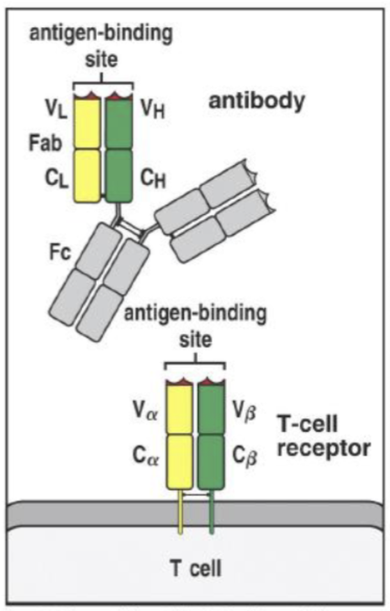
What are the Two Classes of TCRs, and Which predominates?
αβ TCR: Predominant type (over 95%)
γδ TCR: Comparatively enriched in ruminants, where it can account for up to 35% of peripheral T cells

Where does TCR Recombination occur, and What Mechanisms generate Diversity?
In the thymus
Through
Random V(D)J gene-segment recombination
Random nucleotide addition at the joining sites
How is TCR Recombination similar to BCR Recombination?
BOTH use DNA-level rearrangement of V, D, and J gene segments to generate millions of antigen receptor specificities
BCR rearrangement occurs in the bone marrow, while TCR rearrangement occurs in the thymus
What Two Properties are Evaluated during Thymic Selection?
The goal is to retain functional T cells that are tolerant to self
Functionality: Can the TCR recognize antigen presented by self-MHC?
Self-reactivity: Does the TCR react too strongly to the body's own antigens?
QUESTION: What Types of Antigens and Epitopes do TCRs recognize?
Most conventional TCRs recognize processed, fragmented peptide epitopes presented by MHC molecules

How does Antigen Recognition differ between BCRs and TCRs?
BCR: Directly recognizes native antigens, including linear and conformational epitopes
TCR: Recognizes processed peptide fragments presented by MHC molecules

What Types of Molecules can BCRs recognize?
Proteins
Carbohydrates
Lipids
Nucleic acids
What are Linear and Conformational Epitopes?
Linear: Continuous amino acid sequences
Do not require the original three-dimensional protein structure
Conformational: Discontinuous amino acids brought together by protein folding
Depend on tertiary or quaternary structure
What are the Two Universal Rules of Immunology?
Nothing happens without a receptor–ligand interaction
Immune responses are usually mediated by cytokines and chemokines

What is an Antigen-Presenting Cell (APC), and Which Three Cells are Professional APCs?
An APC processes antigens and displays peptide fragments on MHC molecules for T-cell recognition
Professional APCs
Dendritic cells
Macrophages
B cells
How do Dendritic Cells connect Innate and Adaptive Immunity?
They present processed antigens to T cells, connecting innate immune detection with adaptive immune responses

What is MHC, and What is Its Main Function?
Major histocompatibility complex (MHC) consists of molecules that display peptide fragments for recognition by T-cell receptors
Where are MHC Class I and MHC Class II Expressed?
MHC I: Nearly all nucleated cells
MHC II: Primarily professional APCs (dendritic cells, macrophages, and B cells)
How do NK Cells use MHC Class I to Monitor Cells?
MHC I provides inhibitory signals to NK cells
Reduced MHC I expression, as may occur in infected or cancerous cells, can remove inhibition and permit NK-cell killing
What are the Main Steps of Antigen Processing and Presentation by an APC?
Phagocytosis of antigen
Lysosomal digestion
Peptide loading onto MHC
Presentation on the cell surface for TCR recognition

What Structural Feature do MHC I and MHC II share for Antigen Presentation?
BOTH contain a peptide-binding cleft that holds peptide fragments for TCR recognition

What is MHC Polymorphism, and Why is It Beneficial?
MHC polymorphism is extensive genetic variation in MHC molecules among individuals
Different MHC variants present different pathogen peptides → increasing the diversity of immune responses across a population
How does MHC Diversity differ from BCR/TCR Diversity?
BCR/TCR: Generated through V(D)J recombination
Producing millions of receptor specificities
MHC: Inherited genetic variation
With thousands of variants across populations; an individual's MHC genotype is fixed for life
QUESTION: Which MHC Class does CD4 recognize, and What Type of T Cell expresses CD4?
CD4 recognizes MHC class II and is expressed by helper T cells

QUESTION: Which MHC Class does CD8 recognize, and What Type of T Cell expresses CD8?
CD8 recognizes MHC class I and is expressed by cytotoxic T cells

What are the Main Functional Differences between CD4+ and CD8+ T Cells?
Memory trick: CD4 × MHC II = 8; CD8 × MHC I = 8.
CD4+ helper T cells: Coordinate immune responses
CD8+ cytotoxic T cells: Kill infected or abnormal cells

What are the Primary and Secondary Lymphoid Organs, and What are Their Functions?
Primary lymphoid organs: Bone marrow and Thymus
Sites of lymphocyte development and maturation
Secondary lymphoid organs: Lymph nodes, Spleen, and Mucosa-associated lymphoid tissues
Sites where lymphocytes encounter antigens and initiate adaptive immune responses
What is the Main Function of the Thymus in T-Cell Development?
The thymus is the primary lymphoid organ where T-cell precursors undergo
TCR rearrangement
CD4/CD8 lineage commitment
Selection for functional, self-tolerant receptors
What are the Three Main Stages of T-Cell Development based on CD4/CD8 Expression?
Sequence: DN → DP → SP
Double negative (DN): CD4−, CD8−
Double positive (DP): CD4+, CD8+
Single positive (SP): CD4+ or CD8+
What happens to T Cells during Co-Receptor Selection?
Developing T cells progress from expressing neither CD4 nor CD8 to expressing both
Finally to expressing only one → becoming either CD4+ helper or CD8+ cytotoxic T cells when leaving the thymus

What is Positive Selection, and Why is It Necessary?
Positive selection preserves developing T cells capable of recognizing the body's own MHC molecules
This ensures that mature T cells can later recognize peptides presented by self-MHC

What happens to T Cells during Positive Selection based on Their Recognition of Self-MHC?
Weak-to-moderate recognition: Receives a survival signal
Insufficient recognition: Dies by neglect
Strong recognition of self-peptide–MHC: Can trigger negative selection
What is Negative Selection, and Why is It Necessary?
Negative selection eliminates strongly self-reactive T cells before they leave the thymus, helping prevent autoimmunity

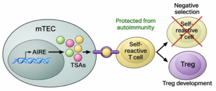
What are Tissue-Specific Antigens (TSAs) and Medullary Thymic Epithelial Cells (mTECs)?
TSAs: Antigens normally associated with particular tissues, such as insulin from the pancreas
mTECs: Specialized thymic epithelial cells that express tissue-specific antigens to help identify self-reactive T cells

What is Aire, and What is Its Role in Negative Selection?
Aire is a transcription factor that allows mTECs to express tissue-specific antigens normally found outside the thymus
Helping identify T cells that react strongly to self

What are the Possible Outcomes of Negative Selection?
Strong self-reactivity: T cell undergoes apoptosis or may develop into a regulatory T cell (Treg)
No strong self-reactivity: T cell can survive and mature
What is the Main Function of Regulatory T Cells (Tregs)?
Suppress immune responses
Help maintain self-tolerance, reducing the risk of autoimmunity
What is Central Tolerance, and How does It differ from Peripheral Tolerance?
Central tolerance: Elimination or control of self-reactive lymphocytes during development in primary lymphoid organs
Peripheral tolerance: Additional mechanisms that control self-reactive lymphocytes outside primary lymphoid organs
How can Failure of Thymic Selection contribute to Autoimmunity?
If strongly self-reactive T cells escape thymic selection and are not adequately controlled by peripheral tolerance, they may attack the body's own tissues
What are the Four Main Outcomes of T-Cell Maturation in the Thymus?
Mature T cells:
Have a functional rearranged TCR
Are committed to a CD4+ or CD8+ lineage
Can recognize self-MHC through positive selection
Have undergone selection against strong self-reactivity through negative selection
What is a Naïve T Cell, and Where does It travel after Leaving the Thymus?
A naïve T cell is a mature T cell that has NOT yet encountered its specific antigen
It circulates through secondary lymphoid organs, including lymph nodes, spleen, and mucosa-associated lymphoid tissue
QUESTION: How many epitopes can an BCR bind?
ANSWER → Two
An immunoglobulin is bivalent, meaning it has two antigen-binding sites that recognize the same epitope
TCR recognition is monovalent, unlike the bivalent immunoglobulin
QUESTION: Which Types of Epitopes can BCRs recognize?
ANSWER → Linear and conformational epitopes
Why: BCRs can directly bind native antigens, including structures created by protein folding
QUESTION: What are the Two Types of Epitopes based on Their Structure?
ANSWER → Linear (continuous) & conformational (discontinuous)
Why: Linear epitopes consist of consecutive amino acids, while conformational epitopes depend on protein folding
QUESTION: Which MHC Class does CD4 or CD8 recognize?
ANSWER →
CD4 → MHC II
CD8 → MHC I
Why: These coreceptors help T cells recognize the appropriate peptide–MHC complex
QUESTION: During T-Cell Development in the Thymus, What is the Main Purpose of Positive Selection?
a) To activate T cells that recognize a foreign pathogen
b) To preserve T cells capable of recognizing self-MHC molecules
c) To eliminate T cells that strongly recognize self-antigens
d) To generate diverse T-cell receptors through gene rearrangement
ANSWER → b) To preserve T cells capable of recognizing self-MHC molecules
Why: Positive selection ensures T cells can recognize self-MHC molecules, which is necessary for future antigen recognition. Choice C describes negative selection
QUESTION: What Structural Feature is shared by Both αβ TCRs and Immunoglobulins (Antibodies)?
a) Antigen-binding sites located in the constant regions
b) Two antigen-binding sites per receptor molecule
c) Two heavy chains and two light chains
d) Antigen is bound by CDR3 of antigen receptor
ANSWER → d) Antigen is bound by CDR3 of the antigen receptor
Why: Both receptors have variable regions containing complementarity-determining regions (CDRs)
CDR3 is particularly important for antigen-binding specificity.
TCR/BCR vs. MHC Diversity
TCR/BCR | MHC | |
How is it created | V(D)J recombination | Inherited |
Number of specificities | Millions | Thousands |
Plasticity | Dependent on exposure history | Fixed for life |
TABLE 1. BCR vs. TCR Comparison
Feature | BCR | TCR |
Found on | B cells | T cells |
Structure | 2 heavy + 2 light chains | αβ or γδ heterodimer |
Binding sites | 2 (bivalent) | 1 (monovalent) |
Variable region | Determines specificity | Determines specificity |
Constant region | Determines antibody class/function | Structural region |
Antigen recognition | Directly binds native antigen | Recognizes processed peptide–MHC |
Antigen types | Proteins, carbohydrates, lipids, nucleic acids | Primarily peptides |
Epitope types | Linear and conformational | Linear peptides |
Secreted form | Antibody | Never secreted |
Receptor diversity | V(D)J recombination | V(D)J recombination |
Development | Bone marrow | Thymus |
Key concept: BCRs directly recognize antigens, while conventional αβ TCRs recognize processed peptides presented by MHC molecules.
TABLE 2. TCR Types and Generation of Diversity
Feature | αβ TCR | γδ TCR |
Chains | Alpha + beta | Gamma + delta |
Relative abundance | Predominant type | Less common in many species |
Veterinary significance | Main focus of lecture | Enriched in ruminants |
Antigen recognition | Primarily peptide–MHC | Can differ from conventional αβ recognition |
TABLE 2a. Mechanisms of Initial TCR Diversity
Mechanism | Description |
Somatic recombination (V(D)J) | Randomly rearranges receptor gene segments |
Combinatorial diversity | Different combinations of V, D, and J segments |
Random nucleotide addition | Adds nucleotides at gene-segment junctions |
Junctional diversity | Additional receptor variation produced at joining sites |
Location | Thymus |
Result | Millions of different TCR specificities |
Remember: Somatic recombination and V(D)J recombination refer to the same process. Random nucleotide addition increases diversity further.
TABLE 3. MHC Class I vs. MHC Class II
Feature | MHC Class I | MHC Class II |
Expression | Nearly all nucleated cells | Primarily professional APCs |
Antigen source | Primarily intracellular proteins | Primarily extracellular proteins |
Antigen processing | Cytosolic pathway | Endosomal/lysosomal pathway |
Recognized by | CD8+ T cells | CD4+ T cells |
T-cell subset | Cytotoxic T cells | Helper T cells |
Additional role | Provides inhibitory signals to NK cells | Presents antigens to helper T cells |
Memory trick: CD4 × MHC II = 8; CD8 × MHC I = 8.
The intracellular/extracellular distinction is a useful general immunology connection; the lecturer primarily emphasized MHC distribution and CD4/CD8 matching.
TABLE 4. CD4+ vs. CD8+ T Cells
Feature | CD4+ T Cell | CD8+ T Cell |
Name | Helper T cell | Cytotoxic T cell |
Coreceptor | CD4 | CD8 |
MHC recognition | MHC II | MHC I |
Main function | Coordinates immune responses | Kills infected or abnormal cells |
Final developmental stage | Single positive | Single positive |
Key concept: T cells become committed to the CD4+ or CD8+ lineage during development in the thymus.
TABLE 5. Stages of T-Cell Development in the Thymus
Stage | CD4 | CD8 | Key Concept |
Double negative | − | − | Immature thymocyte |
Double positive | + | + | Expresses both coreceptors; undergoes selection |
Single positive | + or − | − or + | Committed to helper or cytotoxic lineage |
Remember: DN → DP → SP. The lecturer said you do not need to memorize the exact anatomical location of each stage.

TABLE 6. Positive vs. Negative Selection
Feature | Positive Selection | Negative Selection |
Main purpose | Ensure T cells recognize self-MHC | Prevent strong self-reactivity |
What is tested? | Ability to recognize self-peptide–MHC | Strength of recognition of self-antigens |
Survival outcome | T cells with sufficient recognition survive | Strongly self-reactive T cells are eliminated or diverted |
Failure outcome | Death by neglect | Apoptosis or possible Treg development |
Importance | Produces functional, MHC-restricted T cells | Promotes central tolerance |
TABLE 6a. TCR Recognition Strength and Outcome
TCR Recognition | Outcome | Explanation |
No/insufficient recognition | Death by neglect | Cannot adequately recognize self-MHC |
Weak-to-moderate recognition | Positive selection | Receives survival signal |
Strong recognition | Negative selection | Potentially self-reactive |
Strong recognition with appropriate developmental signals | Possible Treg development | Helps suppress immune responses |
Memory trick: Positive selection = keep the useful T cells. Negative selection = remove dangerous self-reactive T cells.
TABLE 7. Aire, mTECs, and Central Tolerance
Component | Definition | Main Function |
mTECs | Medullary thymic epithelial cells | Express tissue-specific antigens in thymus |
TSAs | Tissue-specific antigens | Represent proteins normally expressed in other organs |
Aire | Autoimmune regulator transcription factor | Promotes expression of tissue-specific antigens by mTECs |
Negative selection | Removal of strongly self-reactive T cells | Prevents potentially harmful T cells from maturing |
Tregs | Regulatory T cells | Suppress immune responses and help maintain tolerance |
TABLE 7a. How Aire Supports Central Tolerance
Aire in mTECs
Promotes tissue-specific antigen expression
Self-Antigen Presentation
Developing T cells encounter self-antigens
Negative Selection
Strongly self-reactive T cells are identified
Central Tolerance
Reduces the risk of autoimmunity
Key concept: Aire allows developing T cells to be tested against antigens from tissues throughout the body, even though development occurs in the thymus.
TABLE 8. Central vs. Peripheral Tolerance
Feature | Central Tolerance | Peripheral Tolerance |
Location | Primary lymphoid organs | Outside primary lymphoid organs |
T-cell location | Thymus | Peripheral tissues and secondary lymphoid organs |
Timing | During lymphocyte development | After lymphocytes leave primary organs |
Main purpose | Eliminate/control self-reactive cells during development | Control self-reactive cells that escape central tolerance |
Mechanisms emphasized | Negative selection; Treg development | Additional control of self-reactive cells |
Failure can lead to | Autoimmunity | Autoimmunity |
Remember: Central tolerance is not perfect, so peripheral tolerance provides another layer of protection.