Unit 1.4 Vocab AP Psych
 Call Kai
 Learn
 Practice Test
 Spaced Repetition
 Match
 Flashcards
 Knowt Play
1/46

There's no tags or description
Looks like no tags are added yet.
47 Terms
contains the midbrain, pons, and medulla oblongata; connects the cerebrum to the spinal cord and cerebellum
Brain stem
emotional center of brain that also plays roles in smell, motivation, and memory
Limbic system:
back part of cerebral cortex specialized for vision
Occipital lobes:
The lowest part of the brainstem that connects the higher levels of the brain to the spinal cord
Medulla
a forebrain structure that processes sensory information for all senses, except smell, and relays it to the cerebral cortex
Thalamus
lower part of cerebral cortex involved in hearing, understanding language, and memory
Temporal lobes:
network of neurons in the brain stem involved in mediation of behavior and arousal
Reticular activating system:
: a structure below the thalamus responsible for maintaining a constant internal state
Hypothalamus
upper middle part of the cerebral cortex lying behind the frontal lobe that is specialized for touch and perception
Parietal lobes (RAS):
makes, stores, and releases several important hormones including those for growth and metabolism
Pituitary gland:
parts of the cerebral cortex responsible for the coordination and interpretation of information, as well as higher mental processing
Association areas:
A group of connected regions that process pleasure and motivation by releasing dopamine
Reward center:
part of the brain that plays a role in spatial memory
Hippocampus:
area at the front of the parietal lobes that registers and processes body touch and movement sensations
Somatosensory cortex:
: a structure located at the base of the brain that controls muscle movement and procedural learning
Cerebellum
: part of limbic system involved in fear, excitement, and arousal; the 4 F’s
Amygdala
a region of the cerebral cortex that has specialized areas for movement, abstract thinking, planning, memory, and judgement
Frontal lobes:
outermost part of forebrain; controls voluntary muscular movements as well as sensation, movement, memory, emotions, and executive function
Cerebral cortex:
a thick band of nerve fibers that connects large areas of the cerebral cortex on each side of the brain and supports communication of information across the hemispheres
Corpus callosum:
when language areas of the brain connect meanings of words, grammar, and rules of language to put together an utterance
Linguistic processing:
includes higher cognitive skills such as problem solving, thinking, reasoning, planning, and organizing
Higher-order thinking:
a set of mental skills that include working memory, flexible thinking, and self-control
Executive functioning
part of frontal lobe responsible for thinking, planning, and language
Prefrontal cortex:
an area at the rear of the frontal lobes that controls voluntary movements
Motor cortex:
study of patients with a severed corpus callosum; demonstrates right and left brain specialization; has been used to treat epilepsy
Split brain research:
each hemisphere of the brain handles specific tasks (left - logic, language; right - creativity, spatial reasoning, art, emotion)
Hemispheric specialization:
the language expression area in the frontal lobe (usually in the left hemisphere) that directs the muscle movements involved in speech
Broca's area:
part of the temporal lobe involved in understanding speech
Wernicke's area:
impairment of language, usually caused by left hemisphere damage either to Broca's area (impairing speaking) or to Wernicke's area (impairing understanding).
Aphasia (Broca's and Wernicke's):
opposite side communication between the brain and the body (the left side of the brain controls the right side and vice versa)
Contralateral hemispheric organization:
When the brain changes and builds new neural pathways in response to a person’s experiences
Plasticity:
recording of the brain's electrical activity at the surface of the skull
EEG (electroencephalograph):
damage to a part of the brain that results in destruction to the neurons; can be natural (like brain injury or disease) or deliberate (surgery)
Lesioning:
a type of MRI scan that can show which areas of your brain are most active during specific functions
fMRI:
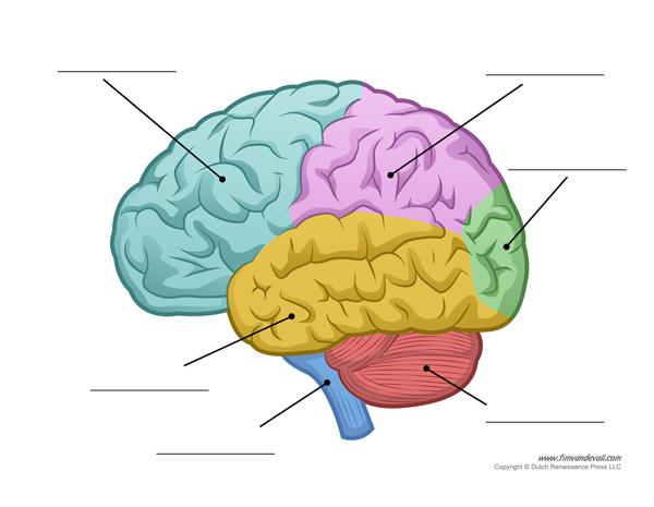
light blue - frontal lobe
Purple - Partiel lobe
Yellow - temporal lobe
Green - Occipital lobe
Red - cerebellum
dark blue - spinal cord
1 -
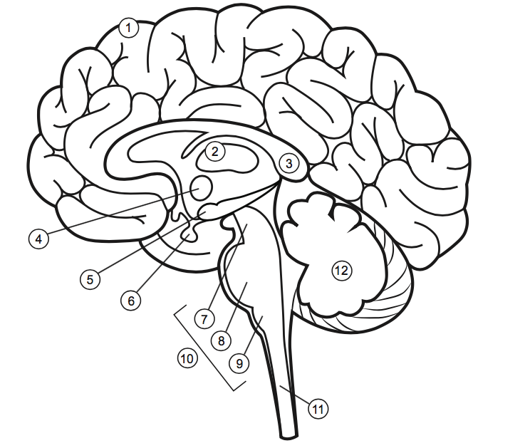
cerebral cortex
2

thalamus
3 -

corps callosum
4-
hypothalamus
5
hippocampus
6 -

pituitary gland
7 -

midbrain
8 -

pons
9 -

medulla
10 -
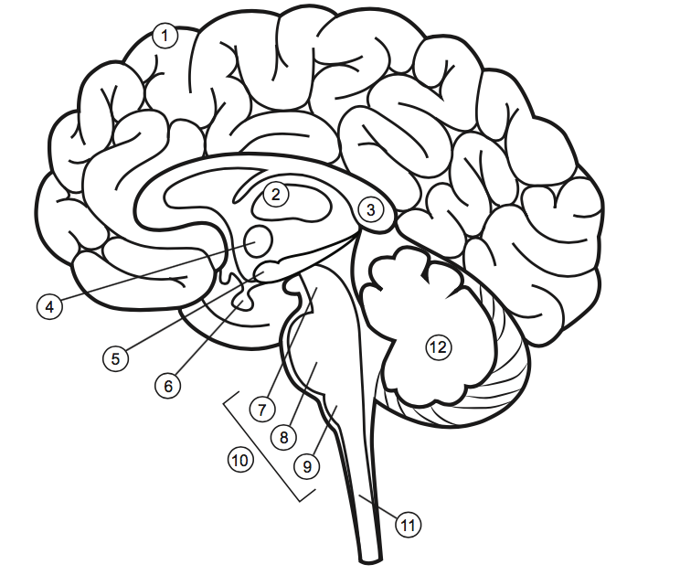
brain stem
11 -
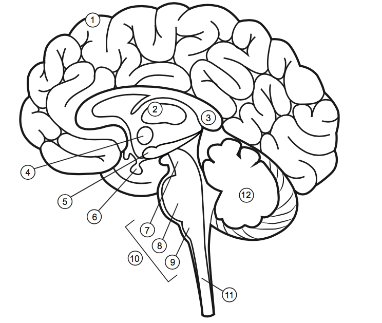
spinal cord
12 -

cerebullum