Cell Bio - Lec 2 (exam 3)
 Call Kai
 Learn
 Practice Test
 Spaced Repetition
 Match
 Flashcards
 Knowt Play
1/40

There's no tags or description
Looks like no tags are added yet.
41 Terms
fab area
variable part, fragment antigen binding, change this area to lock and key with antigen
primary antibody
primary contact with the antigen
secondary antibody
amplify the signal
isolate proteins through...
SDS-page, western plot & RPPA , Localization: sub-cellular fractionation
How to Visualize Protein Localization in Cells?
1. Fix live cells with formaldehyde
for 30 minutes at room
temperature
2. Block with goat serum in
Phosphate buffered Saline (PBS)
and Triton X-100
3. Incubate with antibodies that
target your protein of interest in
PBS and Triton X-100
4. Visualize under Fluorescence
microscope
DAPI
nuclear stain
What is RPPA? Reverse Phase Protein Array
➢ Quantitative high throughput method
➢ Femtogram sensitivity; nanograms of
starting protein
➢ Requires high quality validated antibodies
and robotics
➢ 200 antibodies validated
➢ Up to 1000 samples per slide
You are working with Alveolar Type II cells and would like to determine the role of Rab30 in the secretion of pulmonary surfactant. Your mentor has experimental results showing that Rab30 becomes palmitoylated when the cells secrete surfactant. Which of the following would provide you with some experimental evidence of its contribution to this cellular response?
I would overexpress Rab30 in cells that are deficient in this protein and use alveolar type II cells that are not capable of secreting surfactant. This experiment would tell me whether the cellular secretion of surfactant can be recovered.
Which step in the PCR process ensures that the primers hybridize to the complementary DNA sequences?
Annealing
Basic elements of plasmid
selectable marker? - different cells with and without plasmid
promoter - express the gene (transcription)
restriction sites - molecular cut/paste
origin of replication - multiple copies
antibiotic resistence - grow bacteria to put the plasmid differentiate
Memorize the design
Restriction Nucleases
bacterial enzymes which cut at specific nucleotide sequences in the DNA. Each bacteria expresses a different restriction enzyme and these recognize a different sequence of DNA.
where to they cut always? (restriction enzyme site)
G and A
G/AA
G^A-A
less number of base pairs =
more cuts
asymmetrical
sticky ends (ends generated)
symmetrical
blunt (ends generated)
Restriction Nucleases Cut DNA Molecules at Specific Sites
True
right in the middle of the base pairs
blunt cut (symmetrical)
asymmetrical, there is nothing attaching to it, it can pair up with other bases complementary to it
sticky
Bacteria express specific enzymes that physically "cut" (break) the DNA at specific sites
(4-8bp)
Separates DNA Fragments of Different Sizes
Gel Electrophoresis
component used to create gel
Agarose Gel to identify products
gel 1% or 2% more resistence to travel?
2% gel (higher percentage so higher resistence)
DNA charge
negative
proteins charge
side chain tells use (SDS makes it equal)
load samples which charge
negative
Incubate DNA with Restriction Enzymes temp?
37C for 1 hour.
1 fragment
no site for the enzyme
Ethidium Bromide in Gel
a dye that intercalates into the DNA double helix (nitrogenous bases) and fluoresces under UV light

Example 1: Digestion with EcoR1: How many fragments?
2. Digestion with HindIII: How many fragments?
3. Digestion with EcoR1 & HindIII: How many fragments?
1. 3
2. 3
3. 5
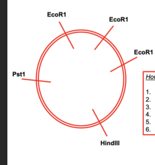
How many fragments if:
1. Cut with EcoR1?
2. Cut with HindIII?
3. Cut with Pst1?
4. Cut with EcoR1 + HindIII?
5. Cut with EcoR1 + Pst1?
6. Cut with EcoR1 + HindIII + Pst1?
1 = 3
2 = 1
3= 1
4= 4
5= 4
6= 5
(SOUTHERN BLOTTING, CGH)
DNA
(NORTHERN BLOTTING, in Situ Hybridization, Microarrays)
RNA
circular
less resistence, faster
travel faster with or with mutation?
with mutation
Process of making many identical copies of a DNA sequence
Begins with the isolation and manipulation of the DNA sequence
How to DNA clone in bacteria?
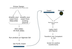
Use restriction nucleases to cut long DNA into pieces
These two pieces of DNA can be joined to other DNA pieces (using DNA Ligase) • Can use a vector/plasmid (as a carrier) to ligate your DNA into & which will allow amplification of your DNA in bacteria
Ligase?
phosphodiester bonds
DNA polymerase
- blunt end then join DNA polymerase
promoter sequence (expression vector)
- create multiple copies of transcript
- more copies of mRNA
- more proteins
Chimeric Protein that is Fluorescent
Fuse the DNA of a target with that of GREEN FLUORESCENT PROTEIN Jellyfish protein
- fusion protein