Normochromic Anemias
 Call Kai
 Learn
 Practice Test
 Spaced Repetition
 Match
 Flashcards
 Knowt Play
1/38

There's no tags or description
Looks like no tags are added yet.
39 Terms
Spleen function
Storage: 1/3 of body’s platelets and granulocytes (by mass)
Filtration: Checks RBCs and culls abnormal cells or pits inclusions
Promotes phagocytosis of capsule bacteria
Fetal hematopoiesis (with liver)
Hereditary Spherocytosis
Membrane shaved off
RBC becomes spherical and more compact
Shortened life span
Mild to moderate hemolysis
HS gene + population
In 1:2000 Caucasians
75% autosomal dominant, 25% autosomal recessive + de novo mutation
HS is a deficiency of…
Ankyrin
Band 3
Band 4.2
This affects the elasticity and flexibility of the RBC

HS RBC elasticity and environment
Able to stretch 117% normally
HS only stretches 3% until Lysis
Spleen environment dangerous for spherocytes
Low pH, low ATP, low glucose
Splenectomy helps with RBC survival
Issues with membrane diffusion
Accumulates Na at higher rate in spleen → swell and lyse


Spherocytes indices + smear
Happens in peripheral circulation, doesn’t exist in bone marrow
Spherocytes present, lacking central pallor
Polychromatic macrocytes
Reticulocytes: 3-10% (normal: 0.5-2.5)
MCHC: >36% in 50% of patients
Moderate anemia
RDW slightly elevated
Increased Bilirubin
Due to continued hemolysis
Cholelithiasis (gall stones) in adolescents/ young adults

Spherocytosis mechanisms
Thermal injury: denatures RBC membrane proteins and lipid bilayer. Forms vesicles without losing cytoplasm (microsphero)
Intrinsic abnormalities: deficiency of spectrin, ankyrin, band 3 (HS)
Immune hemolysis: Antibodies attach to RBC membrane, triggering binding of macrophages (microsphero)

HS lab diagnosis
Screening test:
MCHC, RDW, Spherocytes, bilirubin
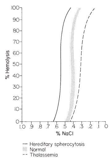
Osmotic fragility test (preferred):
RBC subject to serial dilutions of isotonic water (0.85% NaCl → 0.0%)
At 0.85% NaCl, RBC has little hemolysis
Lysis starts when solution becomes more hypotonic (more diluted)
Data reports level of complete hemolysis
Normal: 0.45%,
HS Spherocyte: 0.65%
Hereditary Elliptocytosis gene + population
Defective/ deficient Spectrin
Decreased thermal stability
ALL ethnic groups
Subtypes:
Common Hereditary Elliptocytosis
Southeast Asian Ovalocytosis
Spherocytic Hereditary elliptocytosis
Hereditary Pyropoikilocytosis

Common Hereditary elliptocytosis
Mild Common HE: 30-100% RBC are elliptic. Most have no clinical S/S
More severe Common HE. Infantile pyropoikilocytosis. Fragmented, bizarre RBCs since birth. Mild hemolysis, jaundice. Converts to mild HE as they age

Southeast Asian Ovalocytosis (SAO) peripheral
Due to Band 3 defect
RBCs spoon-shaped. may have 1-2 slit-like pallor
Shape may protect from malaria
Strongly heat-resistant and rigid. Maintains shape under temperature that would normally cause it to burst

Spherocytic hereditary elliptocytosis (SHE)
Cross between elliptocyte & spherocyte
More common in Northern Europe
Gallbladder disease common, splenectomy indicated
Spherocytic hereditary elliptocytosis (SHE) peripheral
Mild hemolysis, increased osmotic fragility
Spherocytes and oval elliptocyte
Hereditary Pyropoikilocytosis
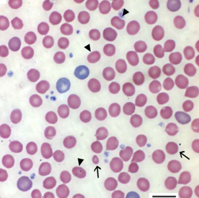
α and β Spectrin have reduced assembly
Mutant Spectrin more easily degradable
More common in African Americans
Hereditary Pyropoikilocytosis (HP) Indices/ Peripheral
Hgb: 6.5 g/dL
RBC budding
MCV 50-75 fL
Rare elliptocytes, spherocytes
Sensitivity to heat
Fragments at 37-46 °C instead of normal 49 °C
Severe hemolysis, poor growth, facial abnormalities

Hereditary Stomatocytosis/ Xerocytosis
Deficiency in Stomatin→ in Na/K permeability
Regulates ions across RBC channels
Overhydrated stomatocytosis:
Net gain of intracellular Na+→ water follows
Dehydrated stomatocytosis/ xerocytosis:
Net loss of intracellular K+→ water moves out
Hereditary Stomatocytosis indices/ smear
10-30% are stomatocytes
Elevated MCV
Decreased MCHC
Slit-like central pallor
Splenectomy can help

Hereditary Xerocytosis
Hereditary autosomal dominant
Unknown etiology/ cause
Hereditary Xerocytosis indices/ smear
mild anemia
decreased osmotic fragility
increased surface:volume ratio
high MCHC
Loss of K+→ dehydrated RBC
Causes xerocytes: RBC where hgb puddled at the end
Glucose 6 Phosphate dehydrogenase deficiency (G6PD) pathway
G6PD keeps glutathione in the reduced state by converting NAD to NAD
Deficiency can’t restore NADPH and GSH which causes oxidative stress
Usually apparent after oxidative chemicals or drugs, or have infection

G6PD genetics
Most common human enzyme deficiency in the world
>400m people affected
X linked recessive disorder
Homozygous females have the disease. Heterozygous are carriers
>400 variants of the G6PD gene from AA substitution
5 genotypes:
2 normal, 3 abnormal. Hemolysis vary
More common in Mediterranean, Africa, China
malarial protection due to oxidative stress

G6PD indices/ smear
In heterozygous women, two populations are present: normal and G6PD cells
Polychromasia
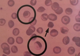
Heinz bodies (supravital stain) from denatured/ precipitated Hgb after RBC exposed to oxidizing agents
Bite cells present due to spleen pitting them

Favism
Ppl with G6PD can develop this after consuming Fava or Broad beans
Due to chemicals isouramil, divicine
Triggers hemolytic episodes on 25% of G6PD patients
Become irritable, lethargic, nausea, fever, ab pain
Hemoglobinuria after 48 hours
Normocytic, normochromic, polychromasia
Decreased haptoglobin, increased bilirubin
Indicates RBC breakdown
Neonatal Jaundice
G6PD related. Occurs within 2-3 days after birth
high bilirubin (kernicterus)
can cause neurological complications
High sensitivity to vitamin K
Substitute triple dye used to treat umbilical cords and camphorated powder.
Treatments
Phototherapy, exchange transfusion (pt blood replaced with donated blood to clear out waste/ toxins)
Congenital Non-Spherocytic Hemolytic anemias
History of neonatal jaundice w/ gallstones, enlarged spleen. Extravascular hemolysis with hyperbilirubinemia
Splenectomy may help
Varies from mild to severe transfusion dependent
Diagnosis of G6PD deficiency
Testing must be done in time. If testing is during an acute hemolytic episode, reticulocyte production increased
Testing should be done after hemolytic episode resolved and counts return to normal
G6PD enzyme assay of older RBCs
Fluorescent Spot Test- routinely
Measures NADP and NADPH reduction
Pyruvate Kinase Deficiency + population + indices
Rare enzyme disorder of Embden Meyerhof pathway
Lack of Pyruvate Kinase= unable to convert ADP to ATP
Build up of 2,3 DPG
Rigid, inflexible cells. Sequestered by spleen and hemolyzed
Occurs in Northern Europe and close Amish populations in Pens
HCT 18-36%
Polychromasia
Few Nucleated RBC

Aplastic Anemia: definition + mechanism
Hypo-proliferative disorder
cellular depletion
Pancytopenia: reduced production of all blood cells
Mechanisms:
Auto-antibody against antigen on stem cells
Immune mechanisms where T lymphocytes suppress stem cell proliferation
Aplastic Anemia: predispositions
These can predispose someone to an aplastic episode
Radiation
Chemotherapy/ chemicals
Benzene exposure
Epstein-Barr virus, HepB, HepC
Aplastic anemia: indices + smear + characteristics
BM has decreased cellularity, pancytopenia, reticulopenia
Slow progress with S/S reflecting pancytopenia
When RBCs significantly depleted:
Fatigue, heart palpitations, dyspnea (shortness of breath)
When platelets depleted:
Ecchymosis (skin discoloration from blood beneath)
Mucosa bleeds
Lymphocytosis (increased lymphs)
Normocytic, normochromic anemia
Managed by transfusions and steroids
Need marrow transplant in severe cases
Fanconi’s Anemia
Autosomal recessive
Affects physical characteristics and BM development
Chromosomal abnormalities, defective DNA repair, chromosomal breaks
Fanconi’s Anemia indices + smear + appearance
BM has macrocytosis, low platelets, low leukocytes, before RBC depletion
Increased HgbF
Appearance:
Short stature, hyperpigmentation in trunk and neck, microcephaly, broad nose, structural kidney abnormalities
Lifespan: 16 years
Treatment: BM transplant

Diamond-Blackfan Anemia + indices + characteristics
BM lacks erythroids→ pure red cell aplasia
Leukocytes are mildly decreased
Hgb 7 g/dL
Congenital hypoplastic disorder since early infancy
80% severely anemic by 6 months
Abnormalities:
Short stature, low birth weight, head and facial abnormalities

Paroxysmal Nocturnal Hemoglobinuria (PNH) genes
Acquired Stem cell disease where RBC membrane abnormality causes Complement-mediated hemolysis
C proteins attach to cells to mark them for destruction via Abs
Clonal membrane defects (CD55, CD59 markers, Ags, flags):
Marked cell destruction overnight→ hemoglobinuria
Somatic X linked mutation:
9 surface proteins missing→ no protection against Complement lysis
Includes: DAF, MIRL, MAC, GPI

PNH lab findings
The 9 surface proteins also missing from platelets and WBCs
Intravascular lysis
Hemoglobinurea: from black tea to tar
Increased reticulocytes
Thrombosis: in brain, dermal vessels, liver, abdomen
PNH testing and treatment
Tests:
Hams Test: rupture in acid solution (activates complement system)
Sugar water test: hemolyzes in sugar solution
Flow Cytometry to detect anchoring proteins of the CD55 and CD59 markers
Cold Agglutinin Syndrome (CAS/ CHAD) cause
IgM-mediated hemolysis via activating complement
From 0°C - 5°C: Complement attaches to RBC. As temp increases, RBCs agglutinate and hemolyze
CAS/CHAD S/S + sample prep
Rarely seen, usually in ages 50+
Acrocyanosis: blue color of fingertips and toes
Weakness, pallor, weight loss, intravascular hemolysis
Sample should be warmed 37°C for 15 to 30 minutes
Pt should move to warmer weather
Paroxysmal Cold Hemoglobinurea (PCH) cause + S/S
Complement-fixing IgG (D-L antibody) attaches to RBC P antigen at low temps and activates complement system at warmer temps
IgG anti P
Lysis happens at body temp
Severe intravascular hemolysis
Hemoglobinemia, hemoglobinurea
Increased bilirubin
S/S:
Similar to hemolytic transfusion reaction: back pain, fever, chills, abdominal pain