PHARM 310 - FINAL
 Call Kai
 Learn
Learn Practice Test
 Spaced Repetition
 Match
Match Flashcards
 Knowt Play
1/76

There's no tags or description
Looks like no tags are added yet.
Name | Mastery | Learn | Test | Matching | Spaced | Call with Kai | Chat |
|---|
No analytics yet
Send a link to your students to track their progress
77 Terms
Where does B-cell activation occur?
secondary lymphoid tissues
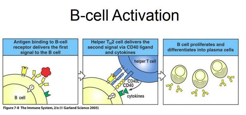
Describe the steps of B-cell activation.
B cells need 2 main signals to become fully activated and produce antibodies
Antigen binds to the B cell
Antigen binds to surface Ig (B-cell receptor aka BCR)
Multiple BCRs bind the antigen and become cross-linked, activating the B cell
The B cell then takes up the antigen by endocytosis, processes it, and displays it on MHC class II
Helper T cell provides the second signal
A CD4 T-helper 2 (TH2) cell recognizes the antigen presented on MHC II
The T cell then provides additional activation signals through:
CD40 ligand (T cell) → CD40 (B cell)
Cytokines
B cell multiplies and becomes an antibody-producing cell
These signals cause clonal expansion (B cell makes many copies of itself)
B cells then differentiate into plasma cells that produce and secrete antibodies

Affinity maturation of activated B-cell:
Once a B-cell is activated, somatic hypermutation of the immunoglobulin ——- regions occurs in the rapidly proliferating germinal center B cells.
This mutation process generates germinal center B cells with either low-affinity or high-affinity surface immunoglobulins.
Low-Affinity Outcome: If a B cell has a low-affinity surface immunoglobulin, its B-cell receptor is not cross-linked, preventing it from presenting the antigen to a T cell. Consequently, the B cell dies by apoptosis.
High-Affinity Selection: If a B cell has a high-affinity surface immunoglobulin, it successfully binds the antigen, leading to BCR cross-linking and interaction with a Helper T cell via CD40 and CD40L.
The combination of T-cell help and B-cell receptor cross-linking sustains the B cell's proliferation and differentiation → these selected B cells differentiate into either ——- B cells (which express IgG) or ——- cells
V, memory, plasma

——- B cells are long-lived, antigen-specific B cells that remain in the body after the first immune response. When the same antigen is encountered again, they are rapidly reactivated and differentiate into ——- cells, producing a faster and stronger antibody response known as the secondary immune response.
memory, plasma
What is the effector function of IgM, IgG, and monomeric IgA?
protect internal tissues of the body
What is the first Aby produced in an immune response? What and where is this Aby secreted?
IgM - secreted by plasma cells located in the bone marrow, spleen, and lymph nodes
As the immune response proceeds, ——- —— and ——- —— in the spleen and lymph nodes produce —— and monomeric ——.
isotype switching, affinity maturation, IgG, IgA

To improve delivery of IgG to tissues, it is actively transported from the blood to extracellular tissues space (i.e. Lamina Propria) through a transport receptor called ——- (aka Brambell receptor) → has a similar structure to an MHC class I molecule.
FcRn

Dimeric IgA protects ——— surfaces throughout the body, including the GI tract, eyes, nose and throat (ENT), respiratory tract, urinary and genital tracts, and mammary glands. It is produced by lymphocytes located beneath the mucosal epithelium.
Transport of IgA:
Dimeric IgA binds to the ——- receptor on the basolateral surface of epithelial cells and is transported across the cell by ——- in an endocytic vesicle. At the apical surface, the receptor is cleaved, leaving a secretory component attached to IgA that helps retain it within the ——-. This allows IgA to provide antibody protection in body secretions such as feces, sputum, tears, and breast milk.
mucosal, poly-Ig, transcytosis, mucus

Mothers provide protective antibodies to their infants both before and after birth.
During pregnancy, maternal ——- is transported across the placenta into the fetal bloodstream by the FcRn receptor. As a result, newborns can have IgG levels similar to their mothers and receive protection against many different antigens. However, maternal IgG gradually decreases after birth, reaching its lowest levels around ——- to ——- months of age, making infants more susceptible to infections during this period.
Infants begin producing ——- soon after birth, while production of their own IgG generally develops during the first year of life (first 6 months).
During breastfeeding, dimeric ——- from breast milk is also transferred to the infant’s gut, where it provides protection against pathogens at the mucosal surface.
IgG, 3-12, IgM, IgA

Antibodies (Aby) itself does not destroy pathogen. Instead they:
——- → covers surfaces on the pathogen, preventing them from binding to host cells, growing, or replicating
——- → coats/tags pathogens to make them easier for macrophages and other phagocytes to recognize and engulf through their —— receptors
interact with ——- proteins to destroy target cells
neutralize, opsonize, complements
Hematopoietic cells such as neutrophils, blood monocytes, and tissue macrophages can endocytose opsonized pathogens. These cells express ——- ——- that recognize and bind to the ——- ——-- of antibodies. Different Fc receptors are specific for different antibody isotypes.
Fc receptors, Fc region

What Fc receptor is present on macrophages that triggers uptake, stimulation, and activation of respiratory burst upon ligation with Fc region of IgG?
Fcγ RI
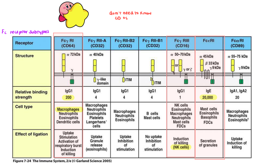
What Fc receptor is present on NK cells that induces killing upon ligation with Fc region of IgG?
Fcγ RIII
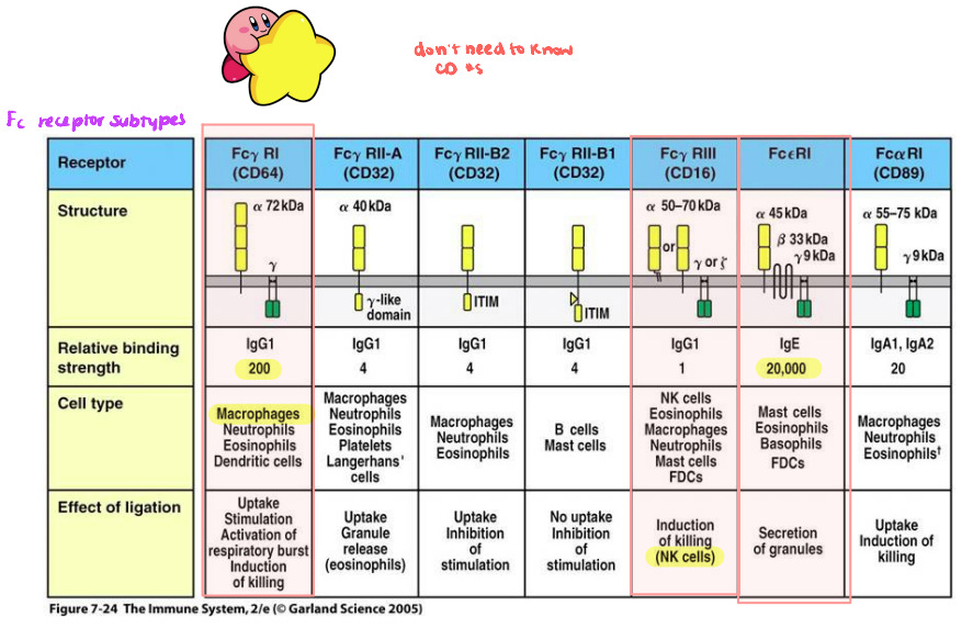
What Fc receptor is present on mast cells that induces secretion of its granules upon ligation with Fc region of IgE?
Fcε RI
IgE antibodies are present in very small amounts in the body, but they bind very tightly to ——- ——- Fc epsilon receptors (FcεRI) on mast cells, basophils, and eosinophils. Because these receptors have very high affinity for IgE, the cells are continuously coated with IgE antibodies (do not dissociate easy due to the high affinity). The IgE antibodies can have many different antigen specificities, allowing these cells to recognize a wide variety of antigens.
high affinity

The granules of mast cells contain —— and other inflammatory mediators → cause increased local blood vessel permeability → molecules, cells, fluid move into tissues → swelling.
They respond very quickly to Ag as the granules are pre-packaged and ready to release.
The natural targets of mast cells are ——- (e.g. helminth worms, protozoa).
histamine, parasites

Natural killer (NK) cells are large, circulating granular lymphocytes that are distinct from B and T cells because they do not express TCRs or surface immunoglobulins. They are primarily part of the ——- immune system and express ——-, which binds IgG1 and IgG3.
This allows NK cells to recognize and kill antibody-coated human cells through ——- ——- ——- ——- (ADCC). NK cells kill target cells by releasing granzymes, which trigger apoptosis.
innate, FcγRIII, antibody-dependent cell-mediated cytotoxicity
The complement (C′) system is a group of serum ——- that are primarily synthesized in the ——-. It is activated during the ——- immune response through several mechanisms and helps destroy foreign organisms through both cell-mediated and antibody-based mechanisms.
glycoproteins, liver, innate
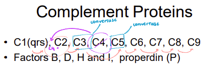
Complement proteins exist in the serum as inactive enzymes called ——-. When activated, ——- cleave the first complement proteins, triggering a cascade of further complement activation and amplification. This leads to the formation of the ——- ——- ——- (MAC), which can destroy target cells, and opsonization, which tags foreign objects and enhances their phagocytosis by macrophages.
zymogens, proteases, Membrane Attack Complex

What are the 3 main pathways of the complement cascade?
classical, alternative, pectin

The complement system can be activated through 3 pathways:
——- pathway → part of the acquired immune response and is activated when complement interacts with antibodies bound to a pathogen
——- pathway → part of the innate immune response and is activated by bacterial cell wall polysaccharides
——- (collectin) pathway → activated when serum proteins (e.g. mannose-binding lectin) bind to carbohydrates on bacterial surfaces
classical, alternative, lectin
The three pathways of complement activation differ in the way they are triggered (and first few reactions). But all pathways converge to the same endpoints, which includes:
——- and/or C4b bind to and tag the pathogen surface → known as complement fixation
formation of the ——- ——- ——- (MAC), which perforates the pathogen’s cell membrane and causes cell destruction
C3b, Membrane Attack Complex

Describe the steps of the classical pathway.
Initiation: The classical pathway begins when antibodies bind to antigens on a bacterial surface. A pentameric IgM molecule can bind to these antigens and adopt a 'staple' form by bending its hinges to grip the antigen, which exposes the C1q binding region. The C1 complex (octopus looking thing) can then bind to a single IgM molecule or multiple IgG molecules at once.
Formation of the C3 Convertase: Once activated, C1s (apart of the C1 complex) cleaves C4 into two subunits, C4a and C4b. This cleavage exposes a thioester bond on C4b → allows it to bind covalently to the pathogen's surface. Activated C1s also cleaves C2 into C2a and C2b. The C2a subunit binds to the surface-bound C4b to form C4b2a, which acts as the classical C3 convertase.
Cleavage of C3 and C5: The C4b2a convertase binds C3 and cleaves it into C3a and C3b. The convertase complex can cleave many C3 molecules, allowing C3b to bind covalently and decorate the pathogen's surface. The association of C3b with the C4b2a complex forms the classical C5 convertase. C5 binds to this complex and is cleaved into C5a and C5b.
Formation of the Membrane Attack Complex (MAC): The soluble C5b fragment initiates the assembly of the MAC. C6 binds to and stabilizes C5b, forming a binding site for C7. C7 binds to the complex and uses its length to grip onto the pathogen membrane by exposing a hydrophobic region. C8 then binds to the C5b,6,7 complex, exposing another hydrophobic region that inserts directly into the cell membrane. Finally, C9 polymerizes onto the C5b,6,7,8 complex to form a ring-like, membrane-spanning channel. This pore disrupts the cell's integrity, causing the pathogen to swell and undergo an osmotic burst that results in cell death.

What is the function of anaphylatoxins (C3a, C4a, and C5a)?
increase vascular permeability → increase flow of fluid, complement components, and antibodies (IgG, IgM) into the infection site

Describe the steps of the alternative pathway.
Initiation via Hydrolysis: In the plasma near a microbial surface, C3 undergoes spontaneous hydrolysis to form iC3 aka C3(H2O).
Formation of Soluble C3 Convertase: The newly formed iC3 molecule binds Factor B. Factor D then cleaves Factor B, releasing the Ba fragment and leaving the Bb fragment attached to form the soluble C3 convertase, iC3Bb. This soluble convertase cleaves C3 into C3a and C3b, allowing C3b to bind directly to the microbial surface.
Formation of the Surface C3 Convertase: The surface-bound C3b binds a new Factor B molecule. Factor D cleaves this bound Factor B, creating the active alternative pathway C3 convertase, C3bBb, directly on the microbial surface.
Amplification and Opsonization: The C3bBb convertase cleaves additional C3 molecules, rapidly decorating the microbial surface with C3b. This coating facilitates the binding of the pathogen to CR1 receptors on macrophages.
Stabilization by Properdin: Properdin (Factor P) binds to the C3bBb complex on the microbial surface, stabilizing the convertase and protecting it from deactivation by host Factor H.
Host Cell Protection: On human cell surfaces, the C3bBb convertase is actively disrupted by regulatory proteins to prevent self-damage. Decay Accelerating Factor (DAF) disrupts the complex by displacing the Bb subunit from C3b. Membrane Co-factor Protein (MCP) also displaces Bb and recruits Factor I, which cleaves C3b into the inactive form iC3b.
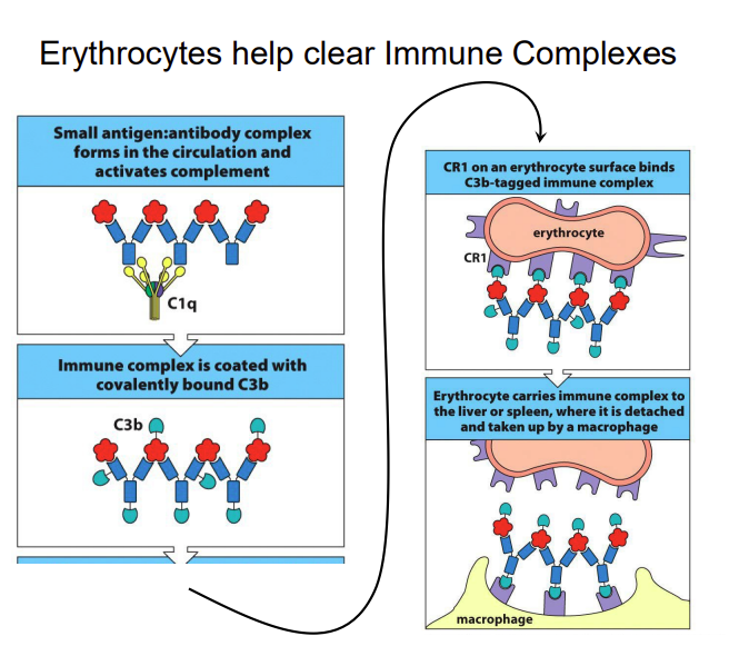
What component of the blood helps to clear immune complexes?
erythrocytes

Match the line to the scenario:
a) lacking innate immunity
b) lacking adaptive immunity
c) both innate and adaptive immunity intact
a) red
b) green
c) yellow
What are the 4 main types of pathogenic organisms?
viruses, bacteria, fungi, parasites

What are direct pathogen mechanisms of tissue damage?
exotoxin release - acts at cell surfaces
endotoxin (LPS) release, after phagocytic digestion of organism - results in cytokine secretion (pyrogenic, leaky vessels)
direct cell killing - results in cell loss
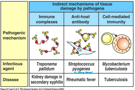
What are indirect pathogen mechanisms of tissue damage?
Ag:Aby immune complexes - results in kidney, blood vessel, joint damage
cross-specificity of pathogen Ag with self-Ag (when pathogen Ag looks like self-Ag) - autoimmune conditions
tissue damage - inflammatory cells release damaging mediators and debris

If you encounter an infection with a virus/bacteria/protozoa/fungi/worms in the interstitial spaces, blood or lymph, which part of the immune system would be activated?
antibodies (IgG), complement, phagocytosis

If you encounter an infection with worms at the epithelial surfaces, which part of the immune system would be activated?
IgA antibodies, antimicrobial peptides
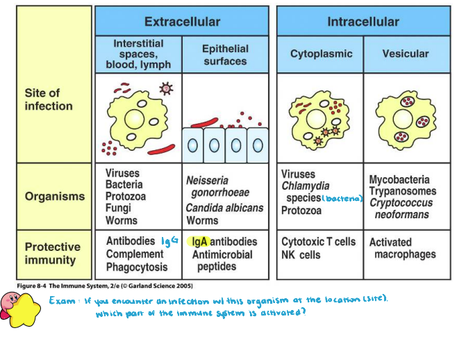
If you encounter an infection of viruses, Chlamydia bacteria, or protozoa in the cytoplasmic (intracellular), what part of the immune system would be activated?
cytotoxic cells, NK cells

If you encounter an infection of mycobacteria or trypanosomes in the vesicular (intracellular), which part of the immune system would be activated?
activated macrophages
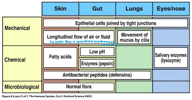
What are the barriers of the immune system in categories: mechanical, chemical, and microbiological?
mechanical - scleroproteins, tight junctions, movement (flow of air/fluid/mucus by cilia - avoidance of stasis)
chemical - enzymes, pH modulation
microbiological - bacterial commensalism (good vs. bad bacteria compete for nutrients and attachments sites)

Phagocytic cells (e.g. macrophages) are decorated with receptors that can induce them to release inflammatory molecules upon receptor binding.
Macrophages has:
receptors for many bacterial components (e.g. mannose, glucan)
—— ——- receptors (TLR) and ——- ——- (LPS or “endotoxin”) receptor activation pathways → triggers cytokine production and further leukocyte recruitment
——- receptors which binds to opsonin-tagged pathogens
toll-like, lipo-polysaccharide, complement

What happens when LPS receptors are activated?
triggers secondary messenger cascade that induces inflammatory cytokines production

On sensing microbial products, macrophages secrete a variety of pro-inflammatory cytokines including:
——- → triggers fever + production of IL-6
——- → triggers fever, mobilization of metabolites, shock
——- → triggers fever, induces acute-phase protein production by hepatocytes (which triggers the lectin pathway of the C’ cascade)
IL-1b, TNF-a, IL-6
CC chemokines are made of 2 cysteine residues side-by-side → they mainly attract ——- and ——-
CXC chemokines has an amino acid residue in between the 2 cysteines → they attract mainly attract ——
monocytes, NK cells, neutrophils
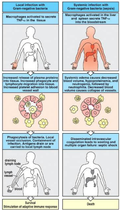
A systemic blood infection is known as ——- and is a result of bacteremia (bacteria present in the bloodstream).
The LSP triggers resident liver macrophages called ——- cells to secrete pro-inflammatory cytokines into the bloodstream causing widespread coagulation and organ failure → septic shock.
sepsis, Kupffer
The 2 dedicated phagocytes are:
——- → long-lived, tissue resident, secrete cytokine mediators
——- → short-lived, more abundant in number, recruited by cytokine mediators at sites of infection
macrophage, neutrophil

Neutrophils are recruited to the site of infection through interactions between adhesion molecules.
Cytokines induce ——- expression on vascular endothelium.
Selectins hook ——- —— —— (S-Lex) on neutrophil to induce leukocyte ——-.
Immunoglobulin-like ——- on endothelium serves as ligands for cellular ——- (LFA-1) binding → this is known as tethering
Neutrophil migrate to infection under the guidance of ——— chemokine gradient.
selectin, sialyl-Lewisx glycoproteins, rolling, ICAMs, integrin, CXC

Neutrophils express receptors that recognize many microbial components. When a microbe binds to these receptors, the neutrophil engulfs it by phagocytosis, forming a ——-. The phagosome then fuses with preformed neutrophil granules and ———, which contain enzymes and antimicrobial peptides that break down and destroy the microbe.
Neutrophils also use a ——- ——-, where NADPH oxidase reduces O₂ → oxygen free radicals → H₂O₂. Finally, myeloperoxidase (MPO) uses H₂O₂ + Cl⁻ to produce → hypochlorite (HOCl), a highly toxic antimicrobial substance that helps kill the microbe.
phagosomes, lysosomes, respiratory burst
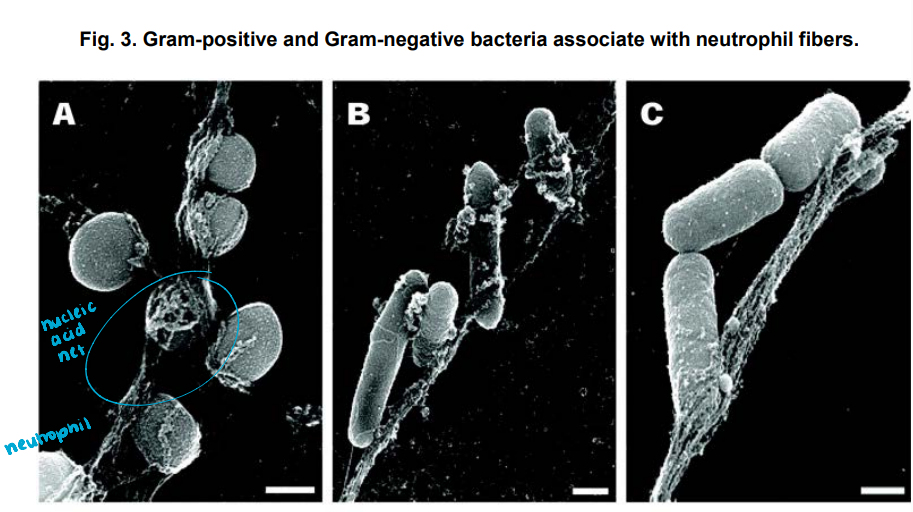
As a final effort to concentrate pathogens and prevent them for further migrating, neutrophils undergo apoptosis or ——- → where they throw out DNA NETs (neutrophil extracellular traps) decorated with cationic proteins that trap and kill pathogens.
The dead neutrophils and debris are then engulfed and cleared by macrophages.
netosis

Which of the following are true regarding the effects of inflammatory cytokines? (multi-select)
a) IL-1 and TNFα acts on hypothalamus to raise body temperature
b) IL-6 acts on hypothalamus to raise body temperature
c) IL-1 and TNFα acts on hepatocytes to make acute phase proteins → notably C-reactive protein (CRP) and mannose-binding lectin(MBL)
d) IL-6 acts on hepatocytes to make acute phase proteins → notably C-reactive protein (CRP) and mannose-binding lectin(MBL)
a, d
What is the purpose of increasing body temperature during a viral infection?
high temperatures interferes with viral replication → makes it inefficient

What are the 2 main acute-phase proteins? What are their functions?
C-reactive protein (CRP) = binds phosphorylcholine (PC) on bacterial surfaces → opsonize it and activates complement
mannose-binding lectin (MBL) = binds residues on bacterial surfaces → opsonize it and activates complement (lectin pathway)

What are the 3 main inflammatory cytokines?
IL-1, IL-6, Tumor Necrosis Factor (TNFα)

What are the effects of IL-1, IL-6, and TNFα on the:
liver
bone-marrow endothelium
hypothalamus
fat, muscle
dendritic cells
liver - acute phase protein production (CRP, MBL) → opsonization activation of complement
bone marrow epithelium - neutrophil mobilization → phagocytosis
hypothalamus - increased body temp
fat, muscle - protein and energy mobilization to generate increased body temperature
dendritic cells → TNFα stimulates migration to lymph nodes and maturation → initiate adaptive immune response
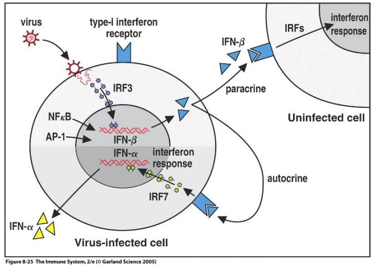
Human cells infected by virus are stimulated to produce certain cytokines called Type ——- interferons → which includes Interferon-α and Interferon-β.
They act interfere with viral replication and signal adjacent cells to prepare defense (all cells have IFN Type I receptors).
I
What is the difference between Type I vs. Type II interferons in the type of cell that secretes them?
Type I = secreted by any cell infected by virus
Type II = secreted by effector lymphocytes
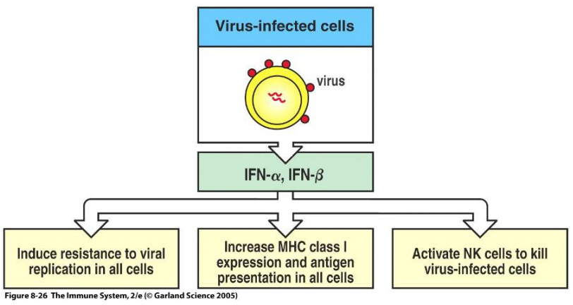
Which of the following are downstream effects on IFN-α and IFN-β? (multi-select)
a) induce resistance to viral replication in all cells
b) increase MHC class I expression and antigen presentation in all cells
c) increase MHC class II expression and antigen presentation in all cells
d) activate NK cells to kill virus-infected cells
a, b, d

B-1 B cells and γ:δ T cells are minority subpopulations of B and T cells that have nonconventional surface phenotypes. Unlike conventional B and T cells, they can respond ——- to a primary infection because they do not require lengthy gene switching and receptor development.
B-1 cells have a general affinity for bacterial ——— antigens, allowing them to provide an early antibody response.
γ:δ T cells respond by secreting cytokines and developing cytotoxic functions, helping to quickly control infected or abnormal cells.
rapidly, polysaccharide
When are symptoms peaks for an influenza infection?
a) barrier phase (hrs)
b) innate phase (Day 1-4)
c) adaptive phase (Day 5-12)
d) resolution phase (Day 10+)
b

What enables for a faster and stronger Secondary Immune Response?
clones of long-lived B- and T-cells + circulating Aby that are produced from the Primary Immune Response

Highly mutable viruses, such as influenza, can change their antigens and evade existing adaptive immunity. During the first infection, the immune system produces memory B cells against the viral antigens it encounters. When the person is reinfected with a slightly different version of the virus, these memory cells can “constrain” the immune response by being preferentially activated to produce antibodies against the original antigen, while the development of new responses to the changed antigens is suppressed. This phenomenon is called ——- ——- ——-.
original antigenic sin

Primary exposure to an antigen: When the body encounters an antigen for the first time, antigen processing and presentation to CD4 T-helper cells takes time, creating a lag phase. The initial antibody response is mainly ——-. Over time, ——- is produced through affinity maturation, resulting in antibodies that are more antigen-specific. Once the infection is controlled, the primary immune response is rapidly downregulated, and most effector cells are cleared, while memory cells remain.
Secondary exposure to the same antigen: On re-exposure, memory cells respond rapidly, resulting in a faster and stronger immune response. —— predominates, and the antibodies have higher affinity and stronger antigen binding because of previous affinity maturation. The secondary immune response is also ——--lasting than the primary response.
IgM, IgG, IgG, longer
——- is the deliberate introduction of an antigen to induce protective immunity against a disease.
vaccination
——— immunity involves stimulation of an immune response using an antigen → creates immunological “memory”
——— immunity involves introduction of Aby (or cells) produced by another individual (human or animal) to provide temporary immunity → does not create memory
active, passive
Distinguish each of the following as either active or passive immunity.
a) Exposure to an infection through natural means
b) Vaccination (traditional)
c) Antibodies transferred from mother to fetus
d) Antibodies transferred through maternal breast milk
e) Antivenin (antivenom) given after a snake bite
f) Bamlanivimab monoclonal antibody treatment
g) Bone marrow transplant, where immune cells are transferred from another individual
a, b - active
c, d, e, f, g - passive
Explain the main difference between these vaccine approaches:
a) killed/inactivated non-replicating vaccines
b) live-attenuated virus
c) non-traditional mRNA vaccine
killed/inactivated non-replicating vaccine → use chemicals, heat, or irradiation to microorganisms
Influenza, rabies, “salk” (killed) Polio vaccine
subunit vaccines w purified pieces of pathogen (proteins, sugars, capsid shell) - Shringrix (shingles), Novavax (COVID-19), Gardasil (HPV), Prevnar (Pneumococcal), DTaP (Pertussis)
live-attenuated → pathogen made “weaker” or less virulent/harmful with chemicat, heat, irradiation or use non-pathogenic “mutated” viruses
MMR, MMRV (Measles, Mumps, Rubella ± Varicella “chicken pox”)
“Sabin” (live attenuated) Polio vaccine
nasal spray Flu vaccine (Flumist)
non-traditional mRNA vaccine → modified viral mRNA used to induce viral protein synthesis and immune response by recipient’s immune system
Pfizer/BioNTech, Moderna

What are the advantages vs. disadvantages of live-attenuated vaccine?
pros:
specific and relevant immune response
small vaccine dose required
lifelong memory
induction of CD8+ cytotoxic T-lymphocyte (CTL) against virus
cons:
possibility of reversion to pathogenic form
contra-indicated for immunocompromised (chemotherapy, advanced HIV) and pregnant women (to protect fetus)

Describe the process of genetically engineering live-attenuated pathogens.
mutate (many, many changes) or completely excise the virulence gene but keep immunogenicity → use in vaccine
Conventional (non-self-amplifying) mRNA vaccines contain mRNA that encodes a specific antigen, allowing cells to produce large amounts of the antigen intracellularly.
——- cells (DCs) take up the mRNA and produce the viral antigen, which is then processed and presented on both MHC class I and MHC class II molecules.
——- —— cells, particularly follicular helper T cells in lymph nodes, recognize the antigen and coordinate the germinal centre B-cell response, stimulating antibody production.
Meanwhile, ——- ——- cells recognize antigen presented on MHC class I and can destroy cells expressing the viral antigen, contributing to cellular immunity.
CD4 T-helper, CD8 cytotoxic T
What is the advantages vs. disadvantages of mRNA vaccines?
pros:
manufacturing process of mRNA does not require cell cultures that could be contaminated with adventitious viruses
short manufacturing time for mRNA
no theoretical risks of infection or integration of the vector into host cell DNA
cons:
instability of mRNA (must be maintained in frozen state)
inefficient in vivo delivery
——- vaccines are made from bacterial toxins, such as those produced by diphtheria and tetanus bacteria. The toxins are treated with formaldehyde, which makes them non-toxic while preserving their antigenicity. This allows the immune system to recognize the toxoid and develop immunity without causing the harmful effects of the toxin. However, toxoids alone may produce an inadequate immune response, so they are given with an ——- to boost immunogenicity.
toxoid, adjuvant
What is the main type of vaccine for bacterial vs. viral diseases?
bacterial → toxoid - bacteria can’t enter our cells, thus use MHC class II for exogenous antigens
viral → attenuated virus - virus infect our cells by getting side, thus use endogenous pathway when developing vaccine
——- are insoluble agents that enhance the antigenicity of co-administered antigen.
adjuvant
Which of the following is the most commonly used adjuvant?
a) Freund’s Complete Adjuvant
b) Aluminum compounds
c) Muramyl dipeptide (MDP) extracts
d) C. parvum extract
b
——- ——- Adjuvant contains dead mycobacteria in oil and detergent and strongly stimulates the immune response, especially macrophages. The oil also creates a depot effect, allowing slow antigen release.
——- and ——- ——- extracts are bacterial extracts that stimulate the immune system but are less potent than Freund’s Complete Adjuvant. Excessive doses can cause macrophage overstimulation.
——- compounds are the main vaccine adjuvants used clinically. They enhance the immune response and provide slow antigen release, but may cause pain at the injection site. Examples include aluminum phosphate and aluminum hydroxide.
Freund’s Complete, Muramyl, C. parvum, aluminum
Which of the following is possible route of administration for vaccines? (multi-select)
a) intra-dermal
b) sub-cutaneous
c) intra-muscular
e) oral
a, b, c, e
——- vaccines are produced by growing them in broth culture. They are then inactivated using chemicals such as formaldehyde or phenol, and for typhoid, heat and phenol. The bacteria are separated by centrifugation, purified, and resuspended in water or 0.9% saline for injection.
——- vaccines are different because they cannot grow on their own in inanimate media. They must be grown using embryonic eggs or living cell cultures. After the virus has grown, it is separated from the host cells and purified to remove unwanted material, which helps reduce hypersensitivity reactions, particularly reactions to embryonic egg components.
bacterial, viral
——- disease refers to an illness or adverse effect that occurs as a result of medical treatment, relating to the principle primum non nocere (“first, do no harm”).
Vaccination can cause temporary febrile symptoms such as fever, aches, and malaise due to immune system activation. In rare cases, live-attenuated vaccines can potentially cause disease if the weakened virus regains pathogenicity through mutation.
iatrogenic
The immune system can over-react, often to harmless environmental antigens → this is known as an ——- or ——- reaction.
allergic, hypersensitivity
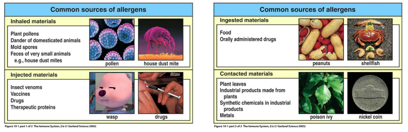
The 4 common sources of allergens include:
——- materials → plant pollen, pet dander, mold spores, feces of house dust mites
——- materials → insect venom, vaccines, drugs, therapeutic proteins
——- materials → food, drugs
——- materials → plant leaves, metals
inhaled, injected, ingested, contacted
What are the 4 types of hypersensitivity reactions and how are they grouped?
Type I, II, III → mediated by antibody effector molecules
Type IV → caused by cytotoxic products of effector T-cells