HDM TBL 9: Glycoconjugates: structure, function, & disease
 Call Kai
 Learn
 Practice Test
 Spaced Repetition
 Match
 Flashcards
 Knowt Play
1/44

There's no tags or description
Looks like no tags are added yet.
45 Terms
glycoconjugates are essential to
connective tissue
2 types of glycan on proteins
proteoglycan
glycoprotein
proteoglycan
core protein, only O-linkage btwn glycan & protein
located only in membranes & ECM
mostly carbohydrate & are structural
Glycoaminoglycan chains [GAG]
glycoprotein
comprised of larger core protein & smaller, discrete glycans
linkages can be N- &/or O-linkage btwn protein & glycan
N-linked is distinct structure to Asn
O-linked can be distinct or variable in size to Ser/Thr
tend to have functions like antibody binding
mostly protein like, have functions [ex. IgG]
*I-cell disease is clinical connect
proteoglycan structure
core protein strand [peptide] has conserved S-G-X-G where GAG is attched
linkage most commonly thru trihexoside directly linked to O-Ser/Thr
many unique GAG chains can be dynamically attched to core protein
glycans are connected to membrane by protein or other anchors or just extracellular
“bottle brush appearance” of proteoglycans
linear structure of GAG chains allows negative charges of sugars to be furthest apart
[carbs]

GAG & its structure
GlycosAminoGlycan
the polysaccharide attched
linear chain of repeated disaccharides
usually repeat unit is an amino sugar + acid sugar
further modified post-attchment w/ epimers + sulfates
sugar acid makes neg charge → high water binding + unfolded structure
sulfate & alcohols provide “stickiness”
epimers change saccharide type
leads to lubricant or mucosal properties
in ECM, each proteoglycan is an…
aggregate
GAG: hyaluronic acid
non-sulfated GAG w/ glucoronic acid + N-acetylglucosamine repeating disaccharide units
largest GAG & retains water
major component of synovial fluid [joint lubricant or shock absorber], vitreous humor of eye, & loose connective tissue

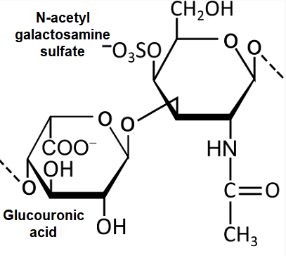
GAG: chondroitin sulfate
glucoronic acid & sulfate-N-Ac-galactosamine
most abundant GAG in body
found in cartilage, bone, heart valves, tendons, ligaments
holds collagen fibers strongly
in joint supplments
lost in osteoarthritis b/c of digestion of proteoglycans by metalloproteinases [in TGF-beta]
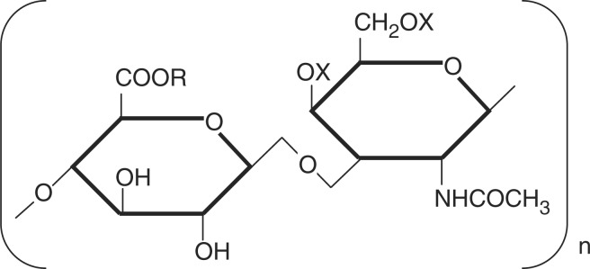
GAG: dermatan sulfate
disaccharide of L-iduronic acid w/ N-Ac-galactosamine w/ sulfate group
in skin, BVs, & heart valves
Hunter’s & Hurler’s syndrome has lack of dermatan degradation in lysosome
mitral valve prolaps has an increase in dermatan sulfate in heart valves
I-cell disease charactered by absence of phospho-mannose being added
GAG: heparin sulfate
disaccharide of iduronic acid + glucosamine
2 sulfate groups attached
Hunter’s & Hurler’s syndrome has lack of degradation in lysosome
an anti-coagulant [enhances antithrombin III activity]
Heparin is an intracellular component of mast cells that line arteries, esp in liver, lungs, skin

GAG: keratan sulfate [KS]
disaccharide of galactose + N-Ac-glucosamine w/ sulfate
KS-1 found in corneas & KS-II found in loose connective tissue proteoglycan aggregates w/ chondroitin sulfate
also found in cartilage, bone, & IV disc

UDP-glycosyltransferases
synthesizes linker region of GAGS [anabolism]
GAG chain then elongated
GAG is degraded in…
lysosome after phagocytosis
*GAGs are unique so have special enzymes in lysozome to catabolize them
complex GAGs
sulfo-iduronic acid
sulfo-glucoronic acid
glucosamine [N-Ac too]
glucocuronic acid
iduronate 2-sulfatase
lysosomal enzyme that takes sulfate off of iduronic acid
alpha-L-Iduronidase
lysosomal enzyme that cleaves carb off & makes GAG 1 shorter
MPS Mucopolysaccharidoses
several hereditary disorders caused by deficiency in GAG specific lysosomal hydrolases
lack of heparan sulfate &/or dermatan sulfate degradation → lysosomal accumulation of GAGs in skin fibroblast or cells in blood
also semi-degraded GAGS detected in urine
abnormalities in bones, joints, ligaments, & intellectual disability, height
Multiple Diseases
MPS 1 = Hurler’s
MPS 2 = Hunter’s
MPS 3 = Sanfilippo’s
MPS 4, 6, 7, based on other GAGs
loss of catabolic enzyme function [enzyme associated w/ each]
Hurler’s Syndrome [MPS I]
alpha-L-Iduronidase deficiency
corneal clouding, dvlpmnt disability, dwarfing, coarse facial features, upper airway obstruction, enlarged liver/spleen, progressive hearing loss
degradation of dermatan sulfate & heparan sulfate affected found in urine
deposition in coronary artery → ischemia & early death
enzyme replacement & HSC transplant therapies
Hunter’s Syndrome [MPS II]
iduronate sulfatase deficiency
X-linked deficiency [ONLY MALES]
NO corneal clouding
mild-severe physical deformity [short neck, broad nose w/ flat bridge], hepatosplenomegaly, dvlpmnt disability, below avg height, aggressive behavior
degradation of dermatan sulfate & heparan sulfate in urine
enzyme treatment to slow progression
“X marks Spot for hunter”
S = sulfatase
Sanfilippo Syndrome [MPS III]
4 subtypes w/ diff GAG degradationn enzyme disrupted
Type A: Heparan sulfatase
Type B: N-Acetylglucosaminidase
Type C: Glucosamine N-acetyltransferase
Type D: N-acetylglucosamine sulfatase deficiency
wide range of severity
symptoms often appear after age 1 w/ slowing learning abilitis ~2-6
coarse facial features
sleep difficulty, behavioral issues, diarrhea, walking problems, stiff joints, below avg height
enzyme test determines subtype
Dolichol phosphate
membrane anchor and transfers sugar molecules during assembly of glycoproteins in N-link pathway
interacts w/ UDP-GlcNac to elongate GAG
which are more diverse: N-glycans or O-glycans?
O-glycans
improper glycan transport [Golgi barcodes]
lysosomal hydrolase enzyme being sorted for transport from golgi → lysosome
substrate: N-acetylglucosamine uridine diphosphate
defect → I-cell disease
key enzyme: N-acetylglucosamine phosphotransferase
mannose phosphate
denotes protein destined for lysosome
needed to form vesicle
I-cell disease
inherited disorder of protein targeting
defective enzyme: N-acetylglucosamine uridine diphosphate
lysosomal hydrolases end up in blood instead of lysosome
diagnostic for I-cell [NO GAG]
no problems arise from extra enzymes in blood as lysosome as low pH
b/c lysosomes lack proper enzyme, they fill up w/ substrates, esp. proteoglycans & glycoproteins
increased need for lysosome autophagy & decrease other organelles in autophagy
→ poorly functioning mitochondria that increase reactive oxygen → cell death
no treatment, death in early childhood
problems catabolizing heparin, dermatan, & keratan sulfate
I-cell disease symptoms
dvlpmnt disability & psychomotor impairments
loss of CNS cells
skeletal issues & joint movement loss
loss of bone & muscle cells
hepatosplenomegaly
from processing molecules for excretion & immune system degradation
I-cell imaging
v similar to Hurler’s syndrome but more mild
“I” = inclusion bodies
key differences
increased lysosomal hydrolase in blood
inclusion bodies in Fbs & B cells [image]
low activity of UDP-N-acetylglucosamine-1-phosphotransferase

Glycan in IgG
maintains Fc domain in an open conformation allowing immune cell surface receptors to bind
stabilizes IgG in blood
in Fc but alters Fab binding strength
imp. in making antibody-based therapies
Glycan Roles
MHC-1 are glycoproteins w/ conserved N-linked glycosylation sites near peptide-binding groove
glycosylation essential for
proper folding & quality control
stability of MHC-I heavy chains
peptide loading & antigen presentation
TCR is a glycoprotein
proper glycosylation ensures
stable TCR-MHC binding
signal transduction fidelity
efficienct immunological synapse formation
immunological synapse
CD8-TCR-MHC-I cluster btwn T cell and DC
*imp for creating a cytotoxic T cell
signaling & expansion of cytotoxic T cell formation
CD28 on T cell bind DC to activate its protein
inside CD8+ cell, Lck kinase activated
it phosphorylates ITAMs on CD3 [part of TCR complex]
this recruits ZAP-70 kinase, which initiates downstream signaling
differentiation to cytotoxic T cell & clonal expansion occur, mediated mostly by IL-2
Fas is also known as
CD95
Bare Lymphocyte Syndome Type I
v. rare condition w/ autosomal recessive inheritance
diagnosed in childhood
mutations occur in TAP1, TAP2, or TAPBP [tapasin] genes
unloaded MHC-1 complexes degraded and don’t reach cell membrane
incomplete T cell dvlpment
recurrent internal bacterial infections
labs show low CD8+ T cells, normal CD4+ helper T cells, & absent MHC-I
ZAP-70 deficiency
defect in ZAP70 gene is a rare SCID like disorder w/ impaired adaptive immunity
autosomal recessive & often diagnosed in 7th yr of life from recurrent infections
mutations impair TCR signaling in CD8+ T cells, so they fail to develop
failure to thrive, large lymph nodes, possible hepatosplenomegaly
CD8+ cell lows, CD4+ cells normal but impaired
further testing will show poor T cell function upon stim.
CD28 overexpression in rheumatoid arthritis [RA]
RA is a chronic autoimmune disease affecting about 1% of global pop.
dysregulated overexpression of CD28 enhances T cell activation, promotes survival of autoreactive T cells, & amplifies inflamm cytokine prod, including IL-2
→ joint inflamm & destruction
Lab findings
positive rhematoid factor
increased ESR [erythrocyte sedimentation rate] or CRP
apoptosis
programmed cell death
phosphatidylserine on inner membrane displayed on apoptotic bodies → macrophages & phagocytic cells to recognize it and engulf them
2 general types
intrinsic
due to internal damage or stress
extrinsic
due to membrane death receptors being acitvated
General events of apoptosis
initiation
death signal received w/ pro-apoptotic signaling
cytochrome c released
execution
cell shrinks
chromatin condenses [pyknosis]
nucleus fragments [karyorrhexis]
caspase cascade active
phagocytosis
cell membrane blebs & apoptotic bodies
done by macrophages w/o inflamm
Intrinsic Apoptotic pathway
ex. radiation induced DNA damage
due to DNA damage, p53 activated
shifts balance toward apoptosis by increasing BAX & decreasing Bcl-2
mitochondrial outer membrane becomes permeable to BAX
cytochrome c binds to Apaf-1 apoptosome & cuts procaspase-9 into caspase-9
caspase-9 cuts execution caspase-3 & others
macrophage engulfs apoptotic bodies
Fas-mediated Extrinsic apoptotic pathway
when CD8+ T cell recognizes a virus-infected cell via TCR it initiates apoptosis via Fas & FasL [& perforin/granzyme]
when bound to infected cell, T cell expresses surface FasL [Fas ligand], which binds to FAS death receptor on infected cell
→ adaptor protein FADD [Fas-associated death domain] & procaspase-8
called death-inducing signaling complex [DISC] → active caspase 8
active caspase 8 connects extrinsic pathway to intrinsic pathway since it cleaves Bid, a Bcl-2 protein
truncated Bid [tBid] translocates to mitochondria, where it activates Bax, opening pore & promoting mitochondrial outer membrane permeabilization
BAX
pro-apoptotic protein
Bcl-2
anti-apoptotic protein
ALPS
Autoimmune Lymphoproliferative Syndrome
rare genetic disorder characterized by defective lymphocyte apoptosis
manifests in early childhood, diagnosis ~3 yrs
caused by mutations in FAS gene
impairs abilitu of lymphocytes to undergo apoptosis → elevated lymphocyte count
large lymph nodes, liver, spleen
autoimmune attack of blood cellls
increased risk of lymphoma