Complex Health of Adult Populations: Exam 3
 Call Kai
 Learn
 Practice Test
 Spaced Repetition
 Match
 Flashcards
 Knowt Play
1/278

There's no tags or description
Looks like no tags are added yet.
279 Terms
upper airway structures
Nose, sinuses and nasal passages, pharynx, tonsils and adenoids, and larynx, which warm, humidify, and filter atmospheric air
Larynx connects the pharynx and the trachea, Larynx has the epiglottis that helps with aspiration
lower respiratory tract
Includes the trachea, mainstem & secondary bronchi, bronchioles, and lungs
blood vessels, thoracic cage, diaphragm, brain (medulla oblongata and pons), central chemoreceptors
What are other parts of the respiratory system?
respiration
requires gas exchange both in the lungs, termed external or pulmonary respiration, and, ultimately, in the tissues, termed internal or cellular respiration
ventilation
inspiration and expiration
diffusion
O2 and CO2 travel across the alveolar–capillary membrane
perfusion
blood flow through the pulmonary circulation
adjusts ventilation based on PCO2 and pH levels
What job does the medulla oblongata and pons do for the repsiratory system?
lungs
_____ exchange CO2 for O2 while kidneys secrete bicarbonate to maintain a pH between 7.35 and 7.45.
fluid balance water
What else is removed during expiration?
alveolar-capillary membrane, pulmonary capillary blood
Oxygen diffuses across the ______________ from the alveoli, where the oxygen partial pressure is higher, into the _________________, where the oxygen partial pressure is lower
98, 2
Oxygen is transported in the blood in two ways:
About __% binds to hemoglobin in red blood cells.
About ___% is dissolved directly in the plasma.
pulmonary capillary blood, alveoli
Carbon dioxide is transported by the blood from the tissues back to the lungs. In the lungs, CO₂ diffuses from the _______________, where its partial pressure is higher, into the _____, where its partial pressure is lower. It is then removed from the body when we exhale.
from the alveoli into the blood, from the blood into the alveoli
Oxygen moves ____________________, while carbon dioxide moves ______________________
80-100 mmHg
What is normal arterial oxygen pressure?
95-100%
What is the normal oxygen saturation?
v/q imbalance
occurs as a result of inadequate ventilation, inadequate perfusion, or both
shunting
ventilation is absent or severely impaired in an area of the lung, while perfusion remains adequate. As a result, blood passes through the pulmonary capillaries without being adequately oxygenated (pneumonia, tumor)
dead space
occurs when air reaches the alveoli, but there is little or no blood flow to pick up the oxygen or remove carbon dioxide ( PE)
hypoxia/hypoxemia
What can shunting and dead space lead to?
hypoxia
Low levels of cellular oxygen
is when the body's tissues do not receive or cannot use enough oxygen to function properly
Presentation: change in mental status, anxiety, restlessness, and/or confusion, dyspnea, tachycardia, and tachypneic, shortness of breath
Causes: PE, anemia, trauma, shock
change in mental status, anxiety, restlessness, and/or confusion, dyspnea, tachycardia, and tachypneic, shortness of breath
How does hypoxia present?
PE, anemia, trauma, shock
Name causes to hypoxia?
hypoxemia
Low levels of oxygen in the arterial blood
PaO2 < 60 mmHg or SaO2 < 90% on RA
May persist even with supplemental O2
Hypoxemia is often a primary cause of hypoxia
PaO2 < 60 mmHg and SaO2 < 90%
What are parameters that indicate hypoxemia?
hypoxemia
______________ is often a primary cause of hypoxia.
hypoxia
Hypoxemia is often a primary cause of _________.
disruption in oxygenation (either with ventilation and/or perfusion)
Hypoxia and hypoxemia are caused due to a ____________________________________________.
chief complaint, factors that improve or worsen symptoms, PMH, social factors, family recent travel smoking status, occupation, ADLs, weight, medications, allergies
What is the initial assessment or patients with respiratory alterations?
do you take meds, do you wear oxygen?
What are important questions for initial assessment of a respiratory patient?
look at speech, use of accessory muscle, orientation, and anxiety levels
How do you inspect respiratory alteration?
dyspnea (SOB), cough, sputum production, chest pain, wheezing, clubbing, and hemoptysis
What are clinical signs of respiratory alteration?
chronic indicators
What are chronic indicators of respiratory alteration?
pulse ox and respiratory rate
What vital signs do you assess in respiratory alterations?
limited palpation for crepitus; percussion is generally not performed
Do you palpate and percuss for respiratory alteration?
evaluation of normal lung sounds
What do you auscultate for in respiratory alteration?
neck and trachea
Where do you auscultate the bronchial breath sounds?
first to second intercostal spaces
Where do you auscultate the bronchovesicular breath sounds?
below the second intercostal space and peripheral lung fields
Where do you auscultate the vesicular breath sounds?
pulse ox, look at waveform, assess immediate need for oxygen, ABGs, d-dimer, consider end tidal CO2, chest x-ray, sputum
What will you do first for respiratory alteration?
muscle twitching, hand tremors, muscle cramps, nausea
What indicates a high CO2 level?
rapid or heavy breathing, confusion, fatigue, weakness, general malaise
What indicates a low CO2 level?
capnometry
used for basic, quick, or routine spot-checks where continuous pattern tracking is not strictly required:
Endotracheal Tube Confirmation: Immediately verifying proper placement of an endotracheal or supraglottic airway tube right after intubation.
Routine Anesthesia & Sedation: Getting baseline or periodic ETCO₂ checks during low-risk or short-duration procedures.
Basic Ventilation Checks: Quickly assessing whether a patient is clearing carbon dioxide adequately during standard transport or basic mechanical ventilation.
capnography
used for continuous, real-time clinical monitoring whenever detailed analysis of ventilation mechanics, gas exchange, or circulatory status is needed:
General Anesthesia & Procedural Sedation: Mandated or recommended as a standard of care by medical societies (such as the American Society of Anesthesiologists) for general anesthesia and moderate-to-deep sedation to catch early signs of hypoventilation or apnea.
Intensive Care Units (ICUs) & Emergency Medicine: Continuous monitoring of critically ill patients, mechanical ventilation, and artificial airways.
Cardiopulmonary Resuscitation (CPR): Evaluating the effectiveness of chest compressions (target ETCO₂ generally ≥ 10-20 mmHg) and detecting an immediate spike when a patient achieves a Return of Spontaneous Circulation (ROSC).
Differentiating Respiratory Distress: Analyzing waveform shapes to spot clinical problems like bronchospasm ("shark-fin" pattern), partial airway obstruction, pulmonary embolism, esophageal intubation, or rebreathing.
Transport Monitoring: Continually tracking patients on transport ventilators or those receiving high-risk sedatives/opioids.
chest x-ray, CT/CTA Scan, MRI/PET, VQ Scan, pulmonary function test, bronchoscopy, thoracentesis, lung biopsy
What are the later diagnostic tools for respiratory alterations?
loss of lung elasticity, enlargement and rigidity of the chest wall, decrease in muscle strength, and decrease in alveolar surface area
What happens to our older patients?
less depth of cough, decreased lung expansion, look at their history
What does loss of lung elasticity lead to?
prevent or correct tissue hypoxia. cardiac output, hemoglobin concentration, and metabolic requirements. inspired oxygen (FiO2), percentage of oxygen, or liters per minute (L/min)
Indications for Oxygen
The goal of using oxygen therapy is to ______________________, which is decreased oxygen supply to the tissues
Oxygen transport to the tissues is dependent not only on the arterial oxygen content but also on __________________ or ______________.
Each system and oxygen flow rate is designed to provide a specific fraction of ___________________________. The appropriate form of oxygen therapy is best determined by ABG levels, which indicate the patient’s oxygenation status (POX and/or PaO2)
physical assessment, what do they look like, pulse oximetry, ABGs, acute vs chronic need for oxygen, 93% pulse ox or below
What are the aspects of your patient’s assessment when giving them oxygen?
nasal cannula, simple face mask, partial rebreather, non rebreather
Name types of low flow oxygen managements.
Venturi mask, aerosol mask. Tracheostomy collar, T-piece adaptor, face tent, Vapotherm blended with compressed air
Name types of high flow oxygen managements.
1-6
How much L does the nasal cannula deliver?
5-10
How much L does the simple face mask deliver?
8-10
How much L does the partial rebreather deliver?
12-15
How much L does the non rebreather deliver?
oxygen toxicity, mucous membrane dryness, infection
What are complications of oxygen therapy?
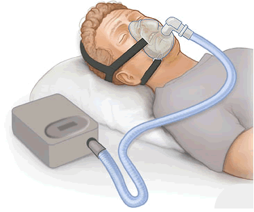
continuous positive airway pressure (CPAP)
a medical device that treats sleep-related breathing disorders, most commonly obstructive sleep apnea (OSA).
bilevel positive airway pressure (BiPAP)
It is a non-invasive ventilation machine that delivers pressurized air through a mask (nasal or full-face) at two distinct alternating levels:
IPAP (Inspiratory Positive Airway Pressure): Higher pressure when you inhale to help open the airway and deliver oxygen.
EPAP (Expiratory Positive Airway Pressure): Lower pressure when you exhale so it is easier to breathe out against the flow
alveoli
What do CPAPs and BIPAPs keep open?
endotracheal (ETT) tube, tracheotomy
What are forms of invasive airway management?
endotracheal tube (ETT)
A flexible plastic tube placed through the mouth or nose into the windpipe (trachea) to maintain an open airway and support breathing.
Mechanical Ventilation: Connects to a ventilator to breathe for a patient during critical illness, surgery, or severe trauma.
Airway Protection: Prevents saliva, blood, or vomit from entering the lungs (aspiration) in unconscious, heavily sedated, or neurologically impaired patients.
Oxygen & Medication Delivery: Delivers high concentrations of oxygen or inhaled anesthetics directly to the lungs
tracheotomy
a surgical procedure that creates a direct opening into the windpipe (trachea) through the front of the neck. A breathing tube (known as a tracheostomy or "trach" tube) is inserted into the opening so air can bypass the mouth and nose.
Airway blockage: Treats severe obstructions caused by tumors, trauma, laryngeal swelling, or foreign objects.
Prolonged ventilation: Helps patients who need mechanical breathing support for weeks or months.
Mucus clearance: Makes it easier to suction thick fluids and secretions out of the lower lungs.
Nerve or muscle conditions: Assists people with paralysis, vocal cord issues, or progressive neuromuscular diseases.
temporary or long-term
Airway management solutions are selected based on whether the need is _________________.
continuous monitoring of patient (cardiac monitor, vital signs), assist the HCP (set up, bagging, suctioning, sedation, capnography), look at the rise of the chest, end-tidal Co2, assessment (breath sounds, chest movement), chest x ray (tube placement), documentation (where is it? moving), communication
What is your role as a nurse during intubation?
full head to toe assessment (complex respiratory assessment, neuro, skin), continuous monitoring ( vital signs, cardiac monitor ), sedation, suctioning, repositioning, tube feedings, move ET tube per protocol
What do you do with a patient now since they are on a vent?
hand hygiene, head of bed elevation, oral care protocol, endotracheal tube, endotracheal cuff pressure 20-30 cm H2O, minimize duration
How can you prevent an infection with a vent?
analgesics (fentanyl, morphine), sedatives (propofol, dexmedetomidine), bronchodilators (albuterol), corticosteroids (dexamethasone), antibiotics, mucolytics (acetylcysteine)
What medications would you maybe give a patient with a vent?
unplanned extubation, aspiration, ventilator acquired pneumonia, hypotension, barotrauma
What are complications of mechanical ventilation?
tracheostomy may be either temporary or permanent, replaces the ET tube, cuffed or uncuffed
If I need to be on a vent, long term…
outer cannula, inner cannula
_____________ is the tube that holds the tracheostomy open; the neck plate holds the straps. ________________ locks in the outer cannula
suction based on policy (based on assessment), suctioning (< 15 seconds, waiting at least 1 minute between suctioning events), good respiratory assessment, look at vital signs, good positioning (semi-Fowlers), provide trach care (cuffed, make sure there is air), communication
What is my nursing care for a trach patient?
decannulation
What would you do if the tracheostomy tube fell out?
decannulation
is the planned removal of a tracheostomy tube once a patient no longer needs it to breathe.
you need a trach set and intubation set up
obstruction, infection, subcutaneous emphysema
What are the complications of a trach tube?
therapeutic modalities for the respiratory system

atelectasis
Atelectasis is the collapse of alveoli, leading to the loss of lung volume
Classificated by its cause
open, ventilation, shunt-like
Atelectasis
Alveoli must remain _____ for gas exchange. Collapse happens when _____ is lost, preventing fresh air from replenishing oxygen and allowing carbon dioxide to accumulate
The collapsed alveoli cannot ventilate properly, so oxygen cannot enter the blood effectively in that area. If blood continues flowing past them, a _____ effect can occur
airway secretions, mucus plugs, foreign bodies, bronchial tumors, or oxygen toxicity
What are mechanical obstructions of atelectasis?
ARDS, pleural effusions, and lung tumors
What are clinical conditions for atelectasis?
post-op patients, chronic lung disease, morbid obesity, CVA
What are risk factors of atelectasis?
chest X-ray (initial), followed by CT scan and maybe ultrasound, later bronchoscopy, ABG
What are the diagnostics of atelectasis?
dyspnea, cough, sputum production, and leukocytosis (elevated WBC), crackles, decreased/diminished breath sounds; wheezing/stridor (if obstruction is the cause), if large enough can cause develop significant respiratory distress (can be a problem)
What is the s/s of atelectasis?
significant respiratory distress
If large enough, atelectasis can develop _______________________.
O2, bronchodilators, CPT, mucolytic agents (acetylcysteine Mucomyst)
What is the treatment of atelectasis?
incentive spirometry, education of patients
What is the prevention of atelectasis?
pulmonary edema
Abnormal accumulation of fluid within the lung tissue, the alveolar space, or both
Can be life threatening (if it is acute “flash”)
ARDS
What is the most common cause of pulmonary edema?
cardiogenic and noncardiogenic
What are the types of pulmonary edema?
capillary fluid leaks or is forced by high hydrostatic pressure into alveolar space (they don’t work, causing intrapulmonary shunting of blood and hypoxemia)
What is the pathophysiology of pulmonary edema?
respiratory distress, anxiety + agitation, foam/frothy sputum
What are the findings of pulmonary edema?
distinct crackles or wheezing
What is in the assessment of pulmonary edema?
oxygen therapy and treat underlying cause
What is the treatment of pulmonary edema?
pleurisy
inflammation of both layers of the pleurae (parietal and visceral)
It is with something else
sharp knife-like pain, stabbing, worsens with deep breath, coughing, or sneezing or movement. On one side
How is the pain in pleurisy?
pleural friction rub
What do you find upon auscultation with pleurisy?
found with pneumonia, infection, trauma, PE, or cancer
What is the etiology with pleurisy?
lie on painful side, NSAIDs, lidocaine patches
What is the treatment of pleurisy?
pleural effusion
A collection of fluid in the pleural space
Too much fluid and it can be infectious
complication of heart failure, TB, pneumonia, PE, or tumors
What are the causes of pleural effusion?