Post Seg Exam 3 - Pictures & Cases
 Call Kai
 Learn
 Practice Test
 Spaced Repetition
 Match
 Flashcards
 Knowt Play
1/266

There's no tags or description
Looks like no tags are added yet.
267 Terms

41 YO Male
Systolic BP 260
(-) DM
Diagnosis?
Hypertensive Retinopathy

finding?
Elschnig’s Spots

Finding?
Elschnig’s Spots

Finding?
Siegrist’s Streaks

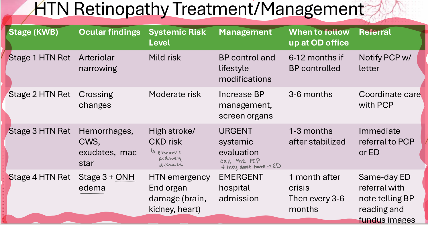
Grade this HTN retinopathy based on Keith-Wagener Barker classification
Grade 1


(-) DM
BP 132/ 81
management?
BP control,
lifestyle modification

(-) DM
BP 132/ 81
how do you proceed with referral?
letter to PCP


(-) DM
BP 132/ 81
currently taking lisinopril for hypertension
when do you want to see them back?
6-12 months


You find this vessel change on retinal examination. They also show generalized arteriolar narrowing.
No exudates or hemes or other relevant findings.
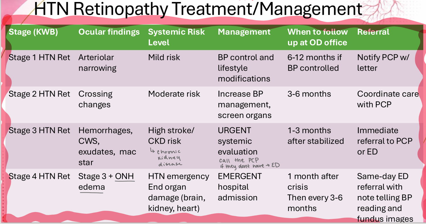
Grade this HTN retinopathy based on Keith-Wagener Barker classification
Grade 2


You find this vessel change on retinal examination. They also show generalized arteriolar narrowing.
No exudates or hemes or other relevant findings.
(-) DM
BP 132/ 81
management?
Increase BP management, screen organs (ie kidney)

You find this vessel change on retinal examination. They also show generalized arteriolar narrowing.
No exudates or hemes or other relevant findings.
(-) DM
BP 132/ 81
how do you proceed with referral?
coordinate care with PCP


You find this vessel change on retinal examination. They also show generalized arteriolar narrowing.
No exudates or hemes or other relevant findings.
(-) DM
BP 132/ 81
when do you want to see them back?
3-6 months

Finding?
Salus Sign


Finding?
Guns Sign


Finding?
Bonnets Sign


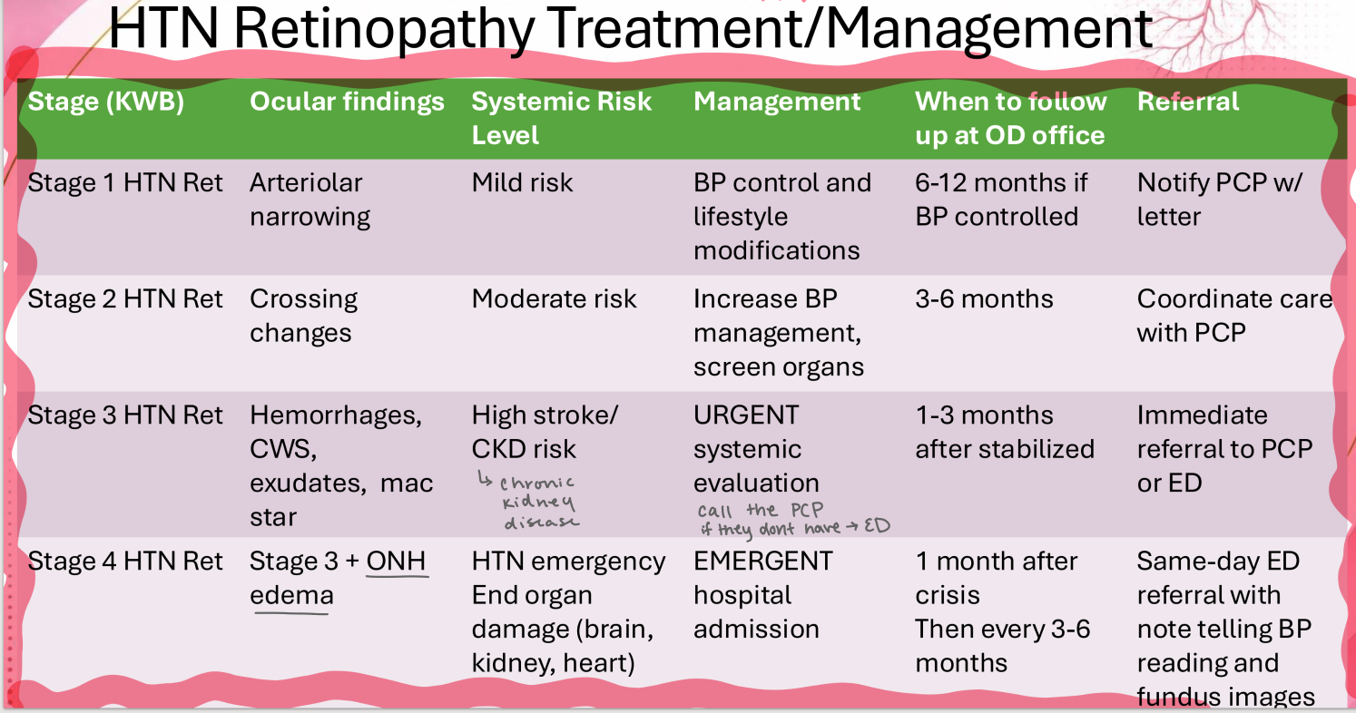
Grade this HTN retinopathy based on Keith-Wagener Barker classification
Grade 3


(-) DM
BP 162/ 87
management?
urgent systemic evaluation
(call PCP, or send to ER/urgent care if they don’t have PCP)


(-) DM
BP 162/ 87
how do you go about referal?
immediate referral to PCP or ED

(-) DM
BP 162/ 87
when do you want to see them back?
1-3 months after stabilized


Grade this HTN retinopathy based on Keith-Wagener Barker classification
Grade 3
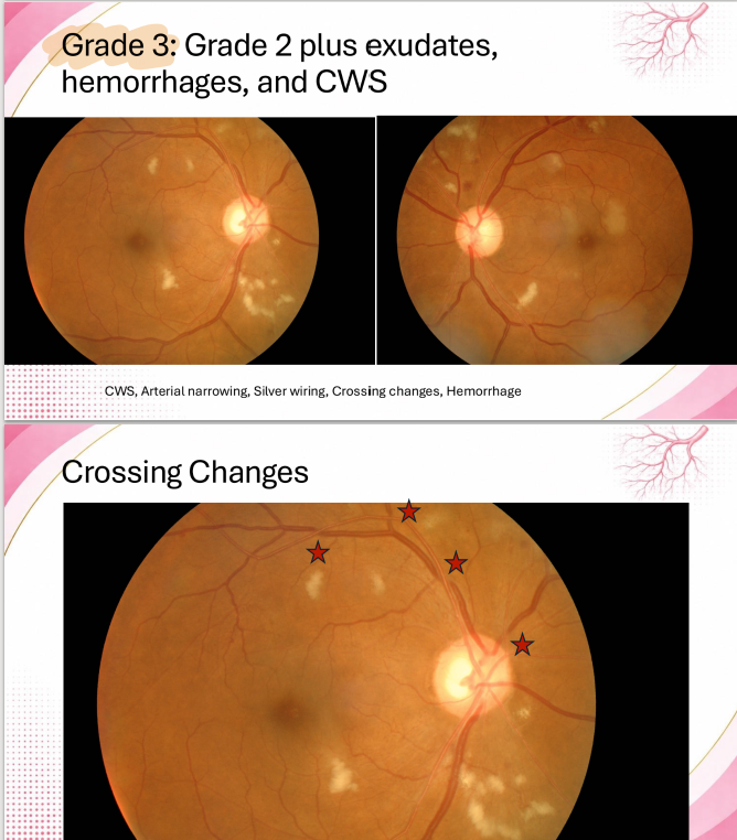

(-) DM
BP 142/ 92
management?
urgent systemic evaluation
(call PCP, or send to ER/urgent care if they don’t have PCP)


(-) DM
BP 142/ 92
how do you go about referral?
immediate referral to PCP or ED


(-) DM
BP 142/ 92
when do you want to see them back?
1-3 months after stabilized
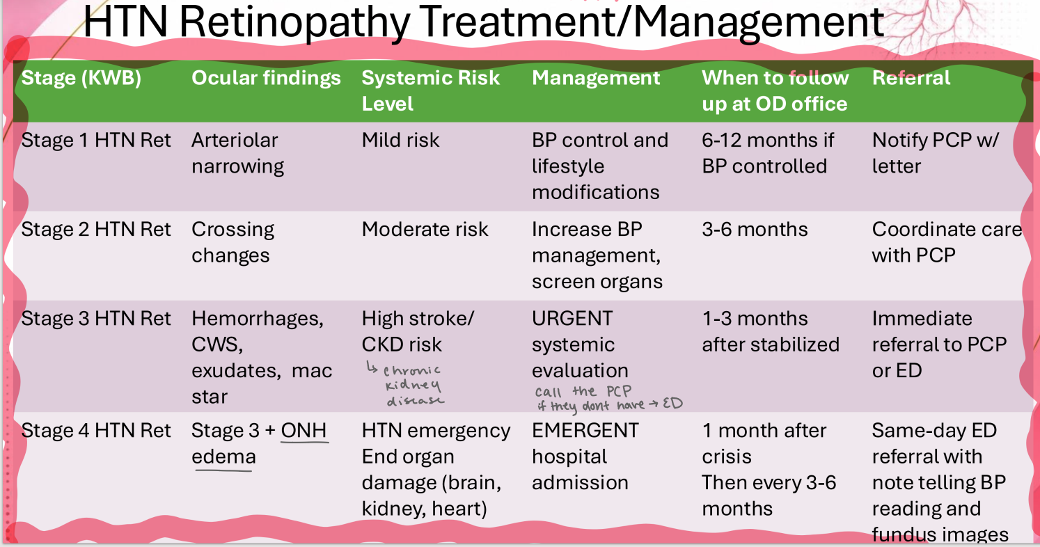

finding?
Gunn’s Sign


Grade this HTN retinopathy based on Keith-Wagener Barker classification
Grade 3

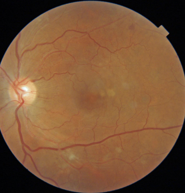
(-) DM
BP 140/ 82
management?
urgent systemic evaluation
(call PCP, or send to ER/urgent care if they don’t have PCP)


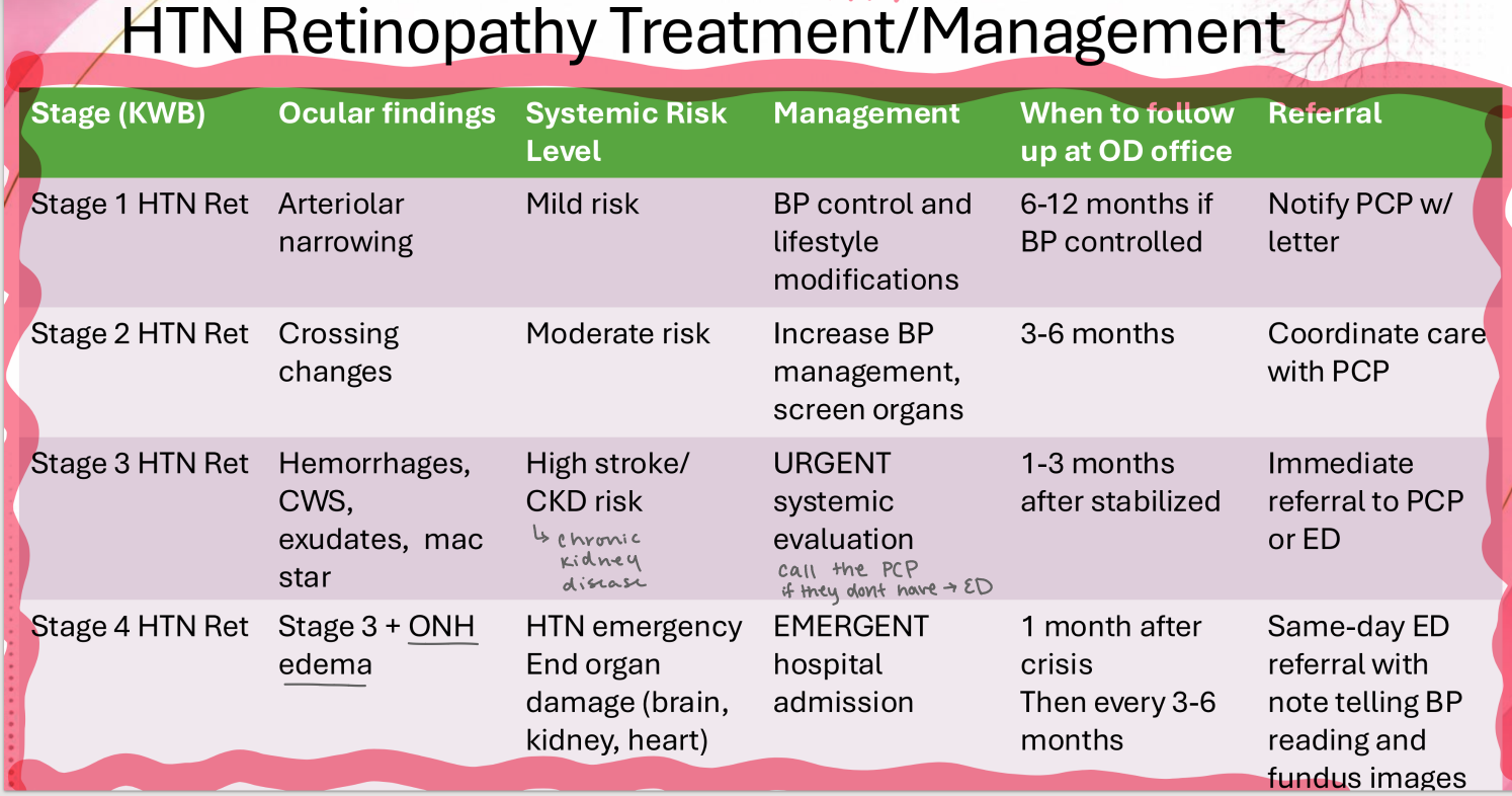
Grade this HTN retinopathy based on Keith-Wagener Barker classification
Grade 4

(-) DM
BP 196/ 97
Management?
Referral to ER


(-) DM
BP 196/ 97
When do you want to see them back?
1 month, then every 3-6 months


(-) DM
BP 196/ 97
How do you go about referral?
same day ED referral + note telling BP reading & fundus images


Grade this HTN retinopathy based on Keith-Wagener Barker classification
Grade 4
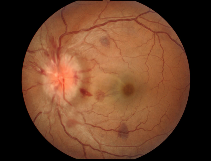
Management?
Referral to ER


Vessel Finding?
Silver Wiring


Vessel Finding?
Silver Wiring


Vessel finding?
Copper Wiring

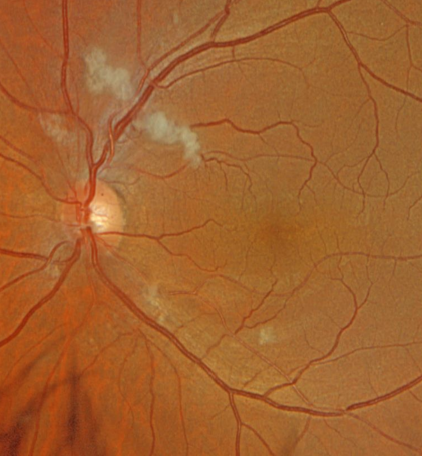
Vessel finding?
Copper Wiring


Diagnosis? (2)
Severe HTN retinopathy;
Serous macular detachment

(-) DM,
Uncontrolled blood pressure - 160/102 today
Diagnosis? (2)
Severe HTN retinopathy;
Serous macular detachment

Diagnosis + give 2 findings
severe HTN retinopathy;
ONH edema, mac star

(-) DM,
Uncontrolled blood pressure - 160/110 today
Diagnosis?
HTN retinopathy
(-) DM,
Uncontrolled blood pressure - 160/110 today


Diagnosis + finding?
HTN retinopathy;
macular edema

diagnosis?
retinal arterial microaneurysm (RAM)

diagnosis?
retinal arterial microaneurysm (RAM)

diagnosis?
retinal arterial microaneurysm (RAM)

diagnosis?
retinal arterial microaneurysm (RAM)

finding?
resolved RAM;
scar + exudates


finding?
resolved RAM;
ring of exudates (surrounding old RAM location)

finding?
platelet-fibrin emboli


finding?
Hollenhorst plaque


finding?
calcific clot (on superior ONH)


Finding?
calcific emboli (moved distal)

Patient reports to your clinic with a CC of intermittent vision loss of the left eye, that had sudden onset yesterday. Patient denies any pain. Patient has VAs of 20/400. You find a (+) APD.

Patient comes into the clinic with right eye vision loss that comes and goes, when it comes it last a few minutes with gradual recovery. It happens several times a day. Denies pain.
How should you manage this patient?
immediate stroke work-up,
see them back in 1-2 weeks then 1-3 months

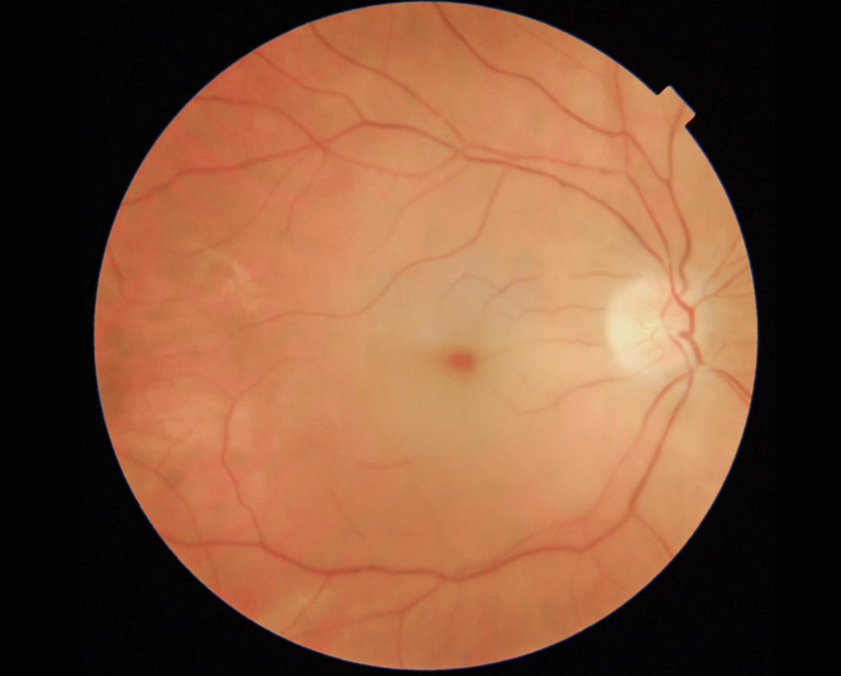
Likely Dx?
Central Retinal Artery Occlusion
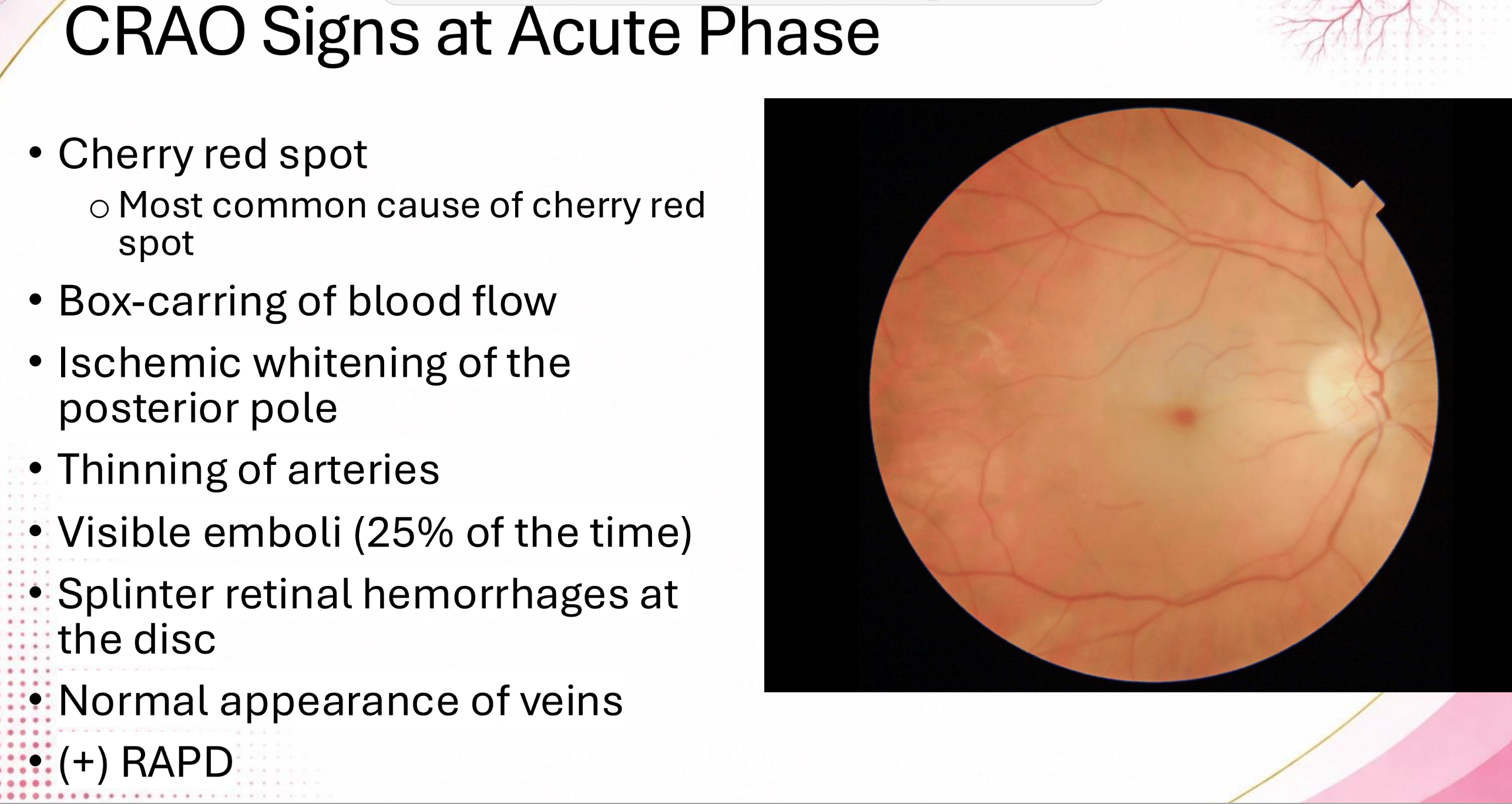

Finding?
Choroid; NFL
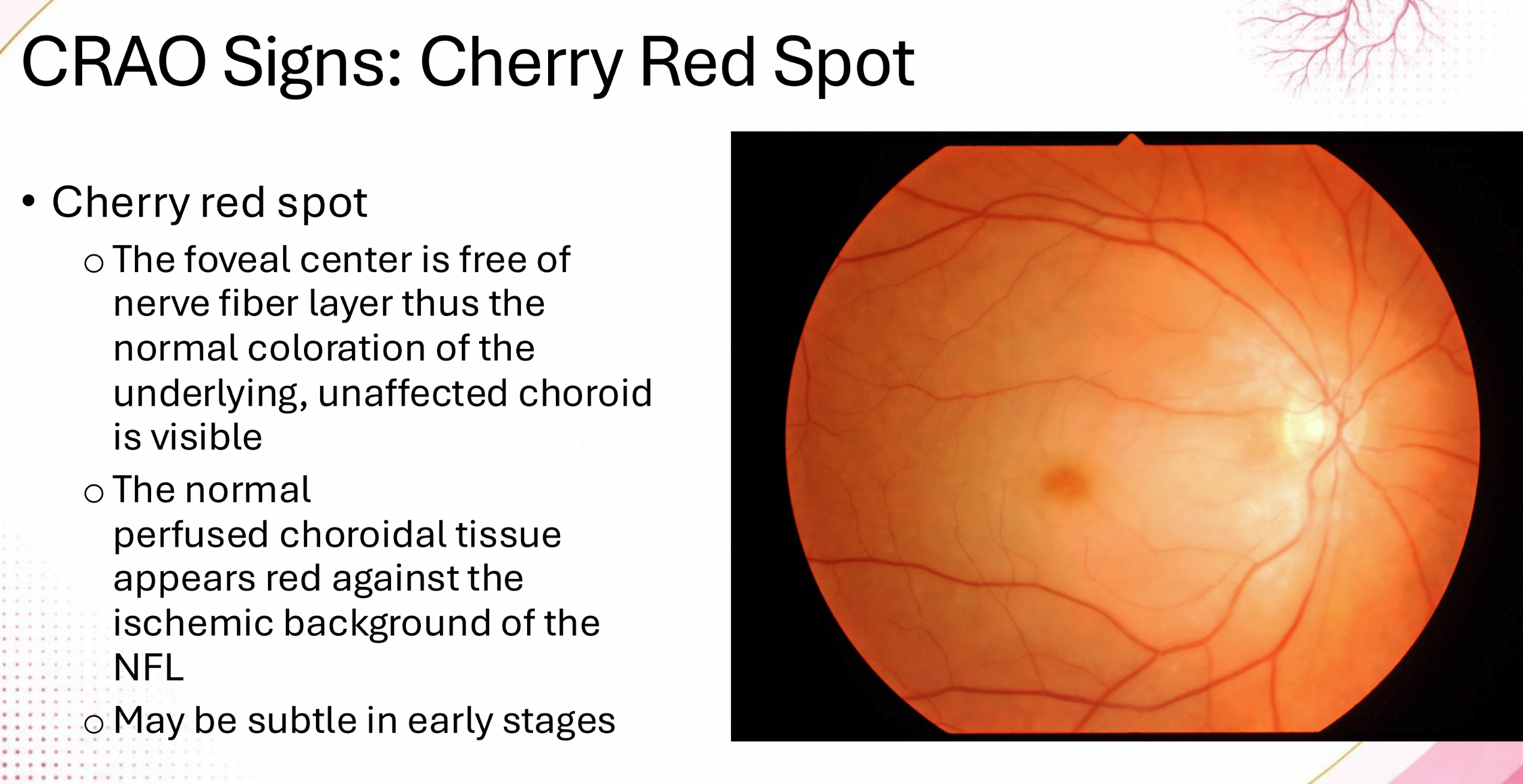

Finding?
Box-carring
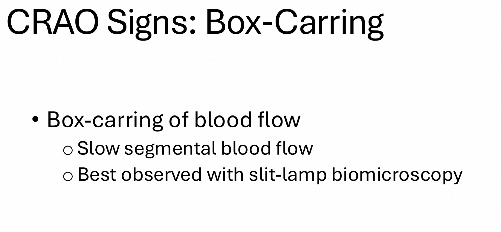

Finding?
Collateral


Finding?
Collateral


Patient presents with amaurosis fugax. You find these findings after OCT and on FA there is a delay in arterial filling. OCT angio shows arterial thinning.
Findings (3) & Likely Dx?
i. thickening of inner retina,
ii. paracentral acute middle maculopathy (PAMM),
iii. inner nuclear layer hyperreflectivity;
Central Retinal Artery Occlusion (acute phase)


What needs to be ruled out in this patient?
Giant Cell Arteritis (GCA)


Finding?
Cilioretinal Artery,
Spared Central Vision in a CRAO

Patient experienced abrupt inferior vision loss. Likely dx?
branched retinal artery occlusion (BRAO) - acute
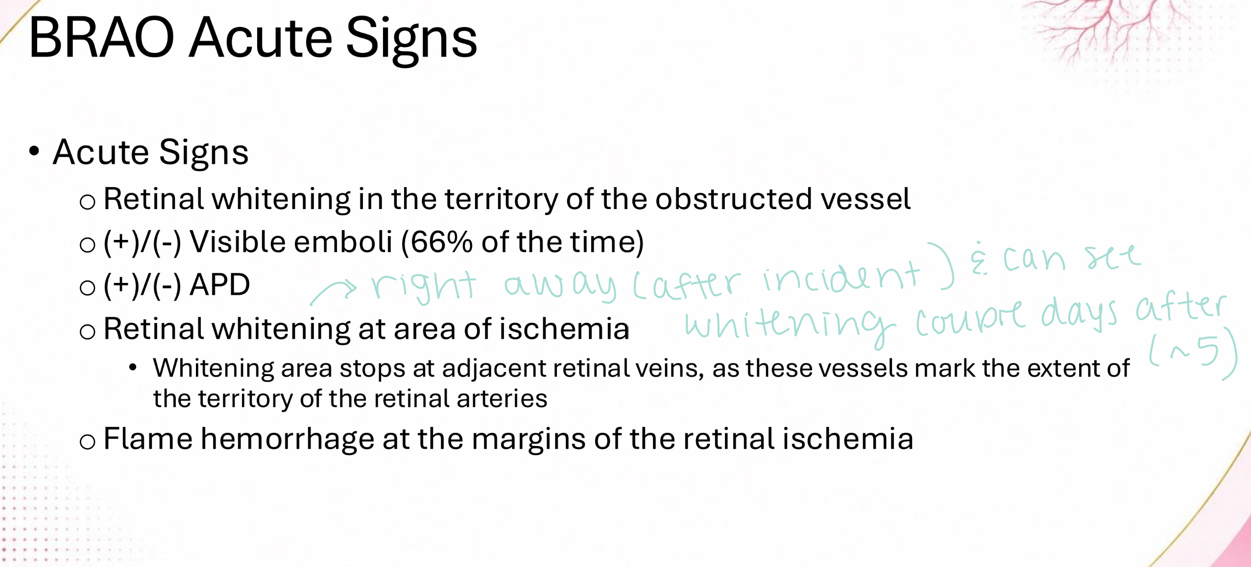

branched retinal artery occlusion (BRAO) - chronic


Finding?
delay in FA (at sight of BRAO)


Finding?
thickening of inner retina (during acute phase of BRAO)


Finding?
Collateral


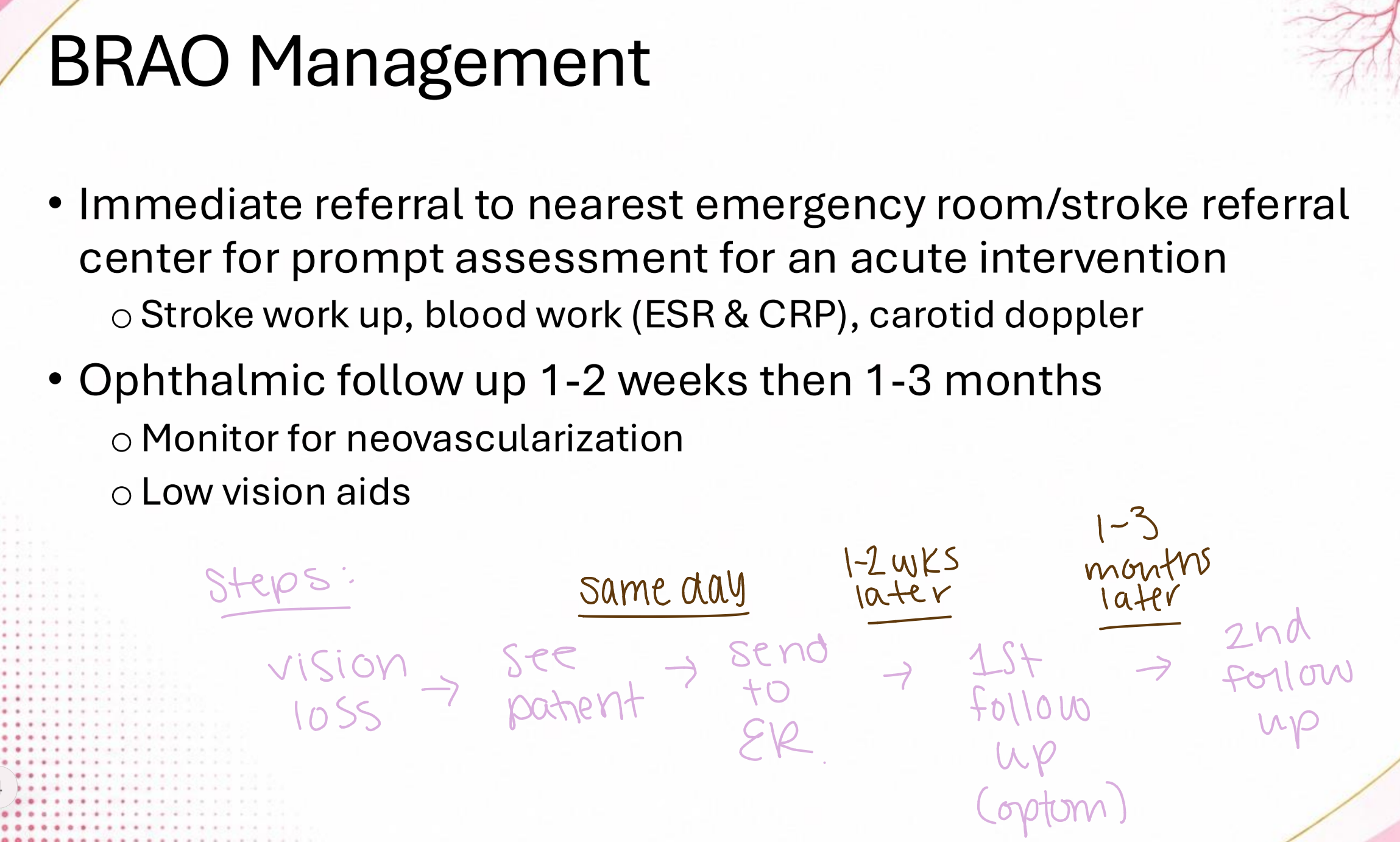
Patient experienced abrupt inferior vision loss. How should you manage this patient??
immediate referral to ED/ stroke work-up;
follow up 1 - 2 weeks then 1 - 3 months
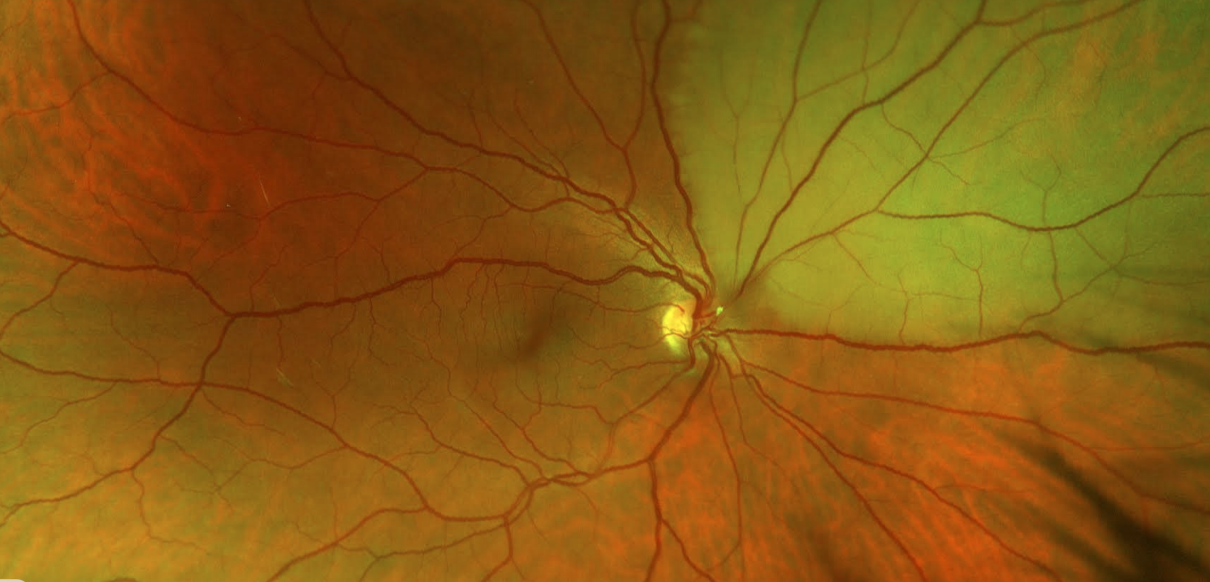
finding?
Hollenhorst plaque

Patient comes in with acute vision loss and is seeing hand motion. You find a (+) APD. Likely dx?
Ophthalmic Artery Occlusion


Patient comes in with acute vision loss and is seeing hand motion. You find a (+) APD. Management?
Immediate stroke evaluation,
GCA work-up

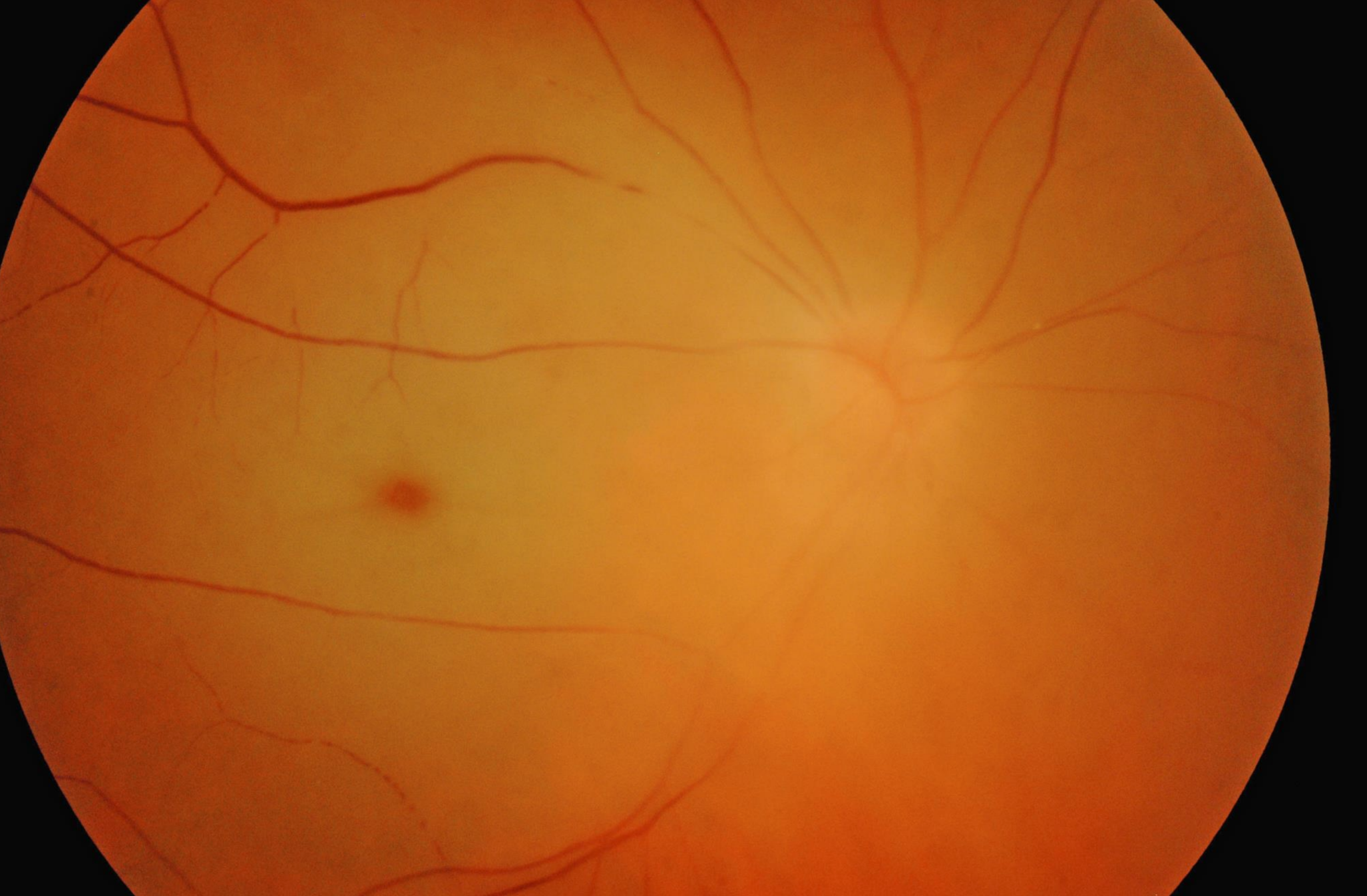
Patient comes in with acute vision loss and is seeing light perception. You find a (+) APD. Likely Dx?
ophthalmic artery occlusion

Likely dx?
Cilioretinal artery occlusion
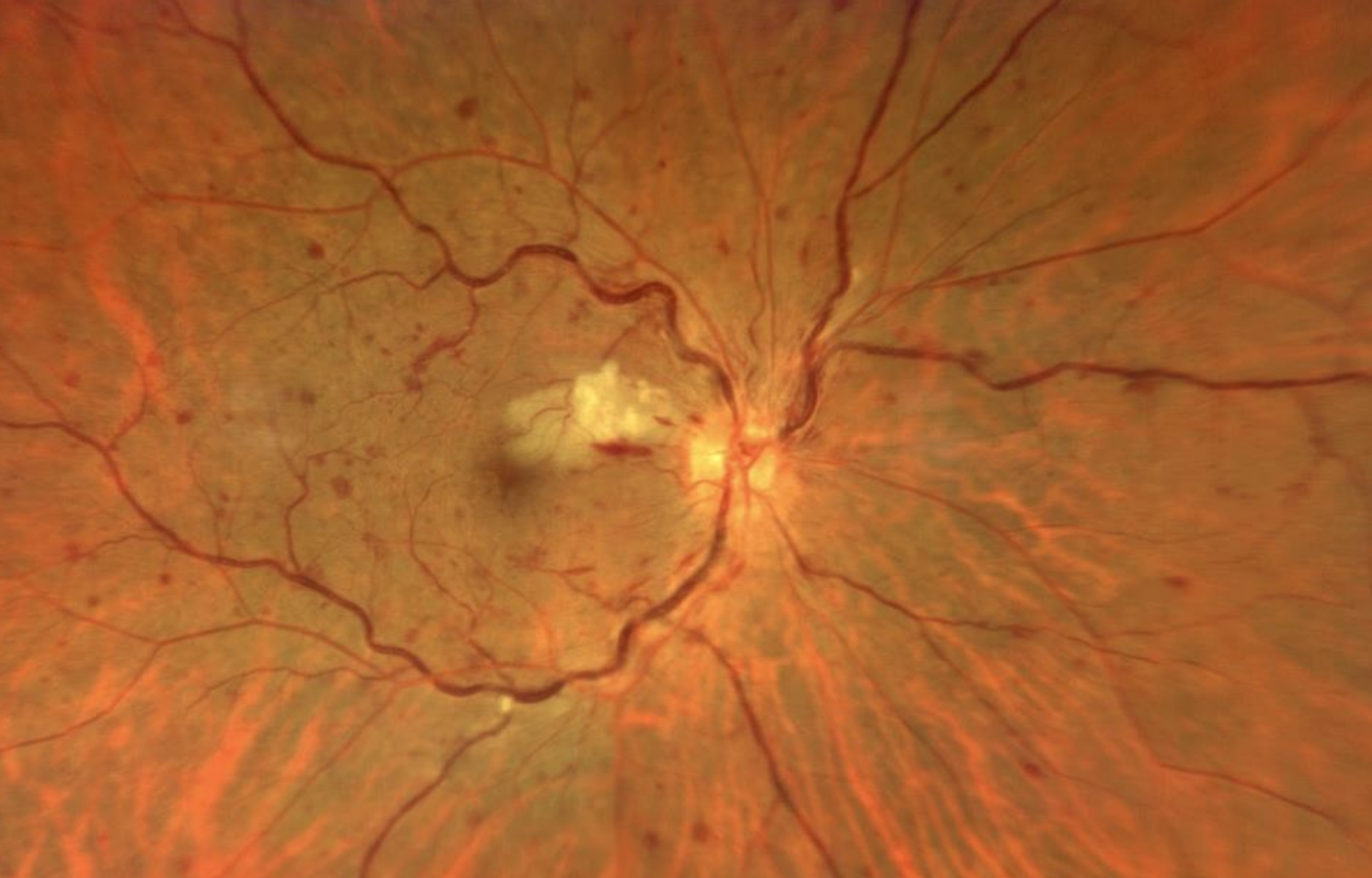


Likely dx? (2)
CRVO,
Cilioretinal Artery Occlusion


VA <20/400, sudden in onset
likely dx?
combined CRVO + CRAO


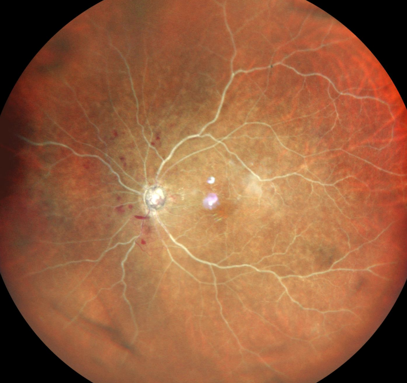
VA = LP, sudden in onset
likely dx?
combined CRVO + CRAO
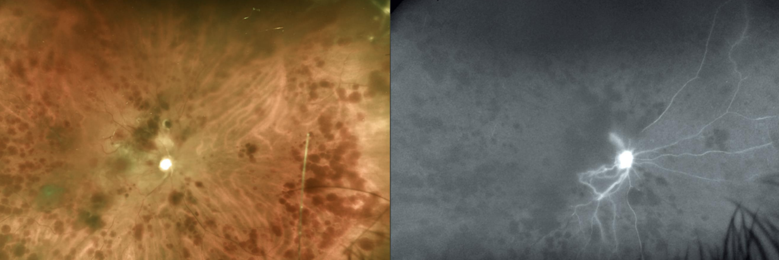
VA <20/400, sudden in onset
likely dx?
combined CRVO + CRAO

Likely dx?
BRVO
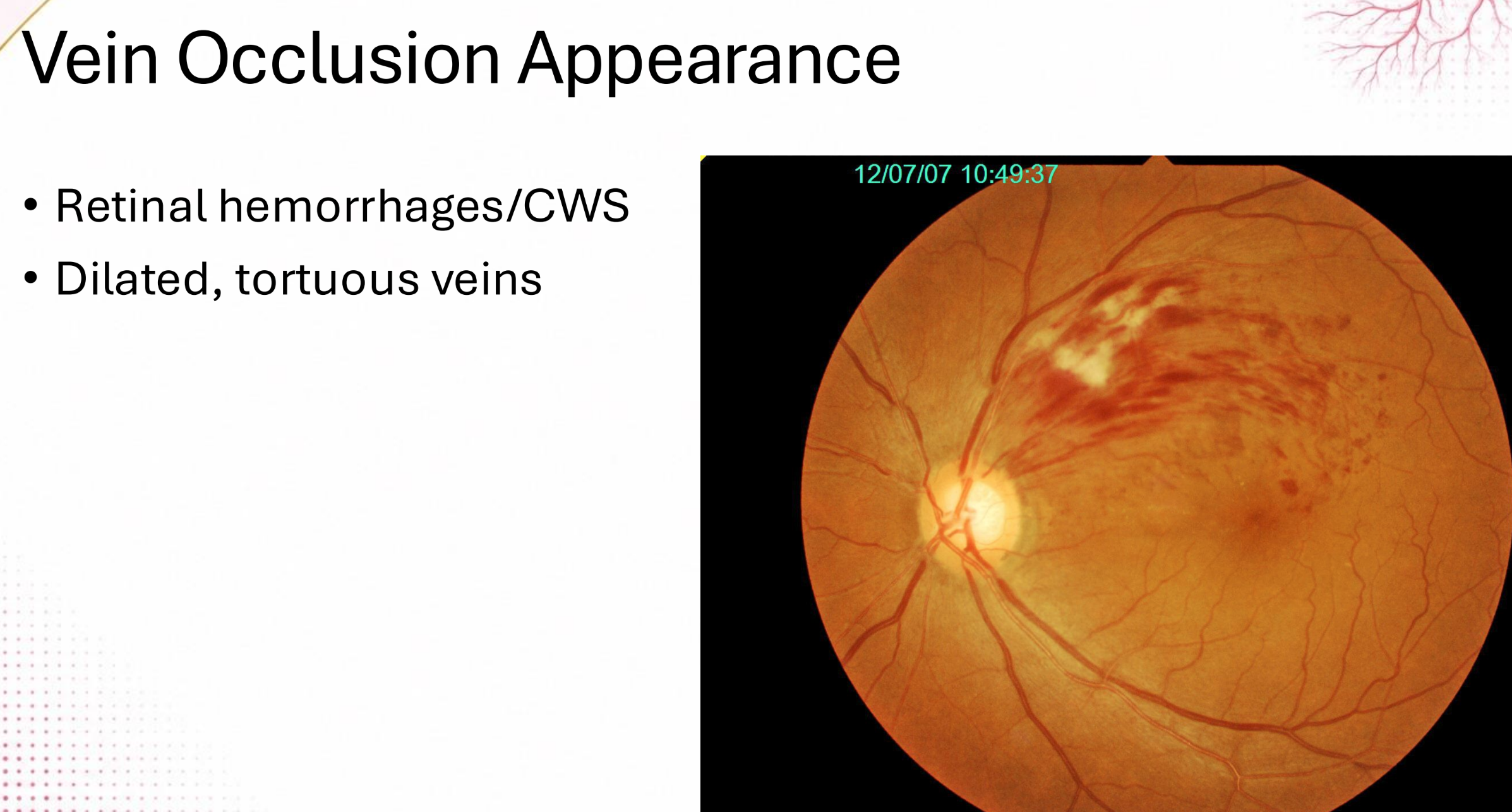
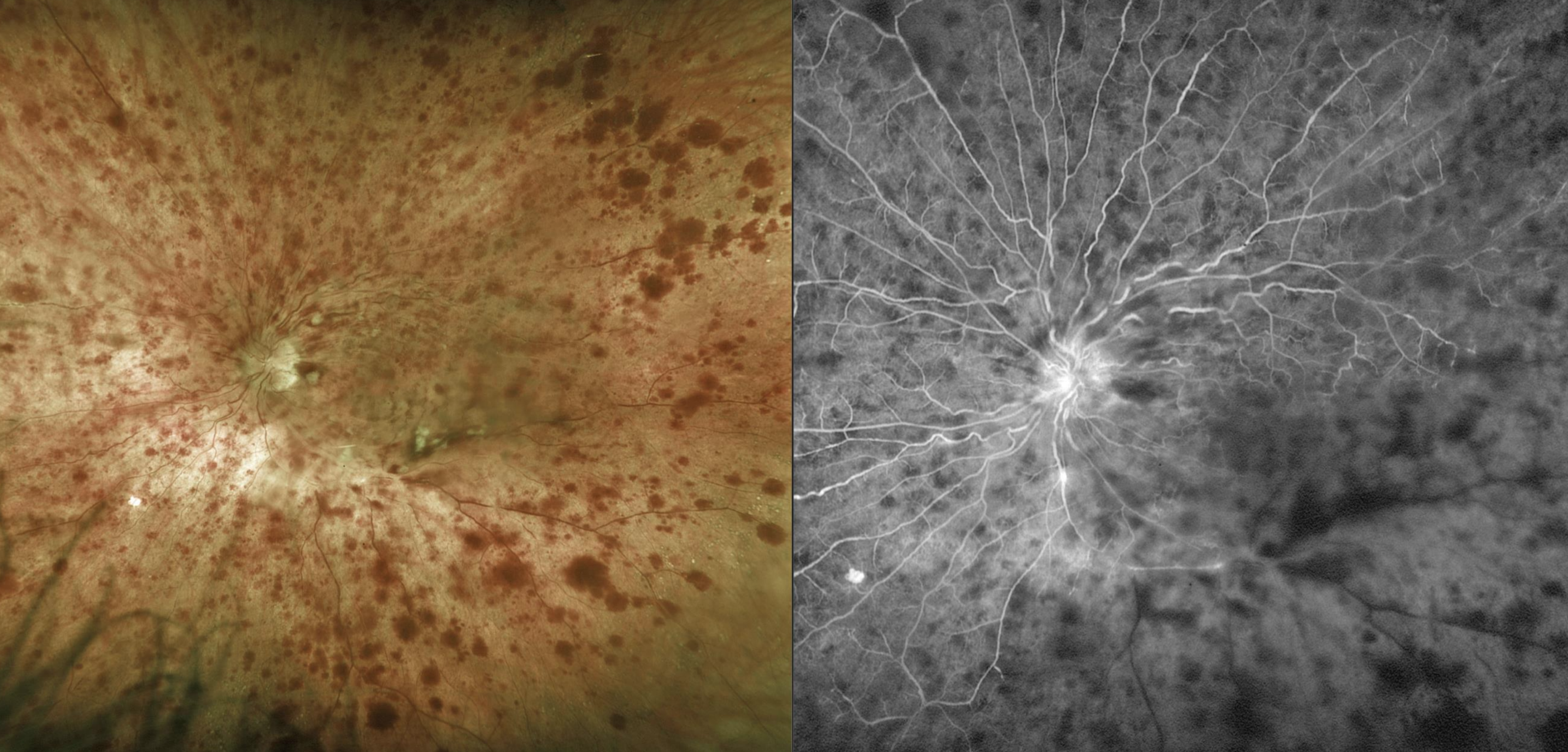
Likely dx?
CRVO
A patient comes in with a CC of acute onset blurry vision. they deny any pain. VAs are 20/100. (+) APD. On dilated exam you see flame hemes in all 4 quadrants, 10 disc areas of capillary nonperfusion, and signs of disc edema. Likely dx?
Ischemic CRVO

VAs are 20/50. (-) APD. On dilated exam you see 5 disc areas of capillary nonperfusion as well as a flame hemes in 4 quadrants. Likely dx?
Non-ischemic CRVO

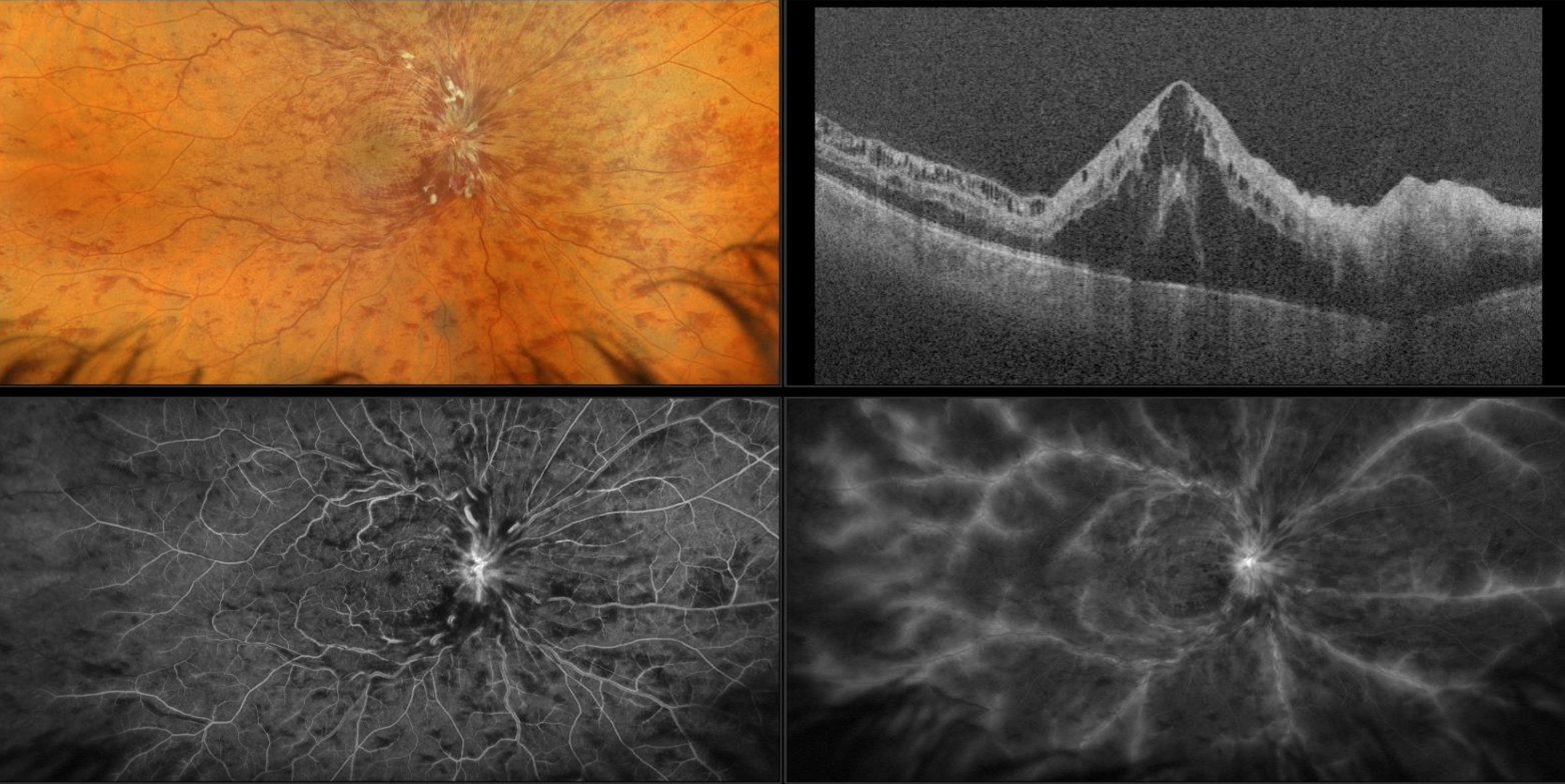
Likely dx?
Ischemic CRVO


Likely Dx?
Non-ischemic CRVO

A patient comes in with a CC of acute onset blurry vision. they deny any pain. VAs are 20/100. (+) APD. On dilated exam you see flame hemes in all 4 quadrants, 10 disc areas of capillary nonperfusion, and signs of disc edema. Likely management?
anti-vegF


likely dx?
CRVO

likely dx?
CRVO

likely dx?
non-ischemic CRVO

finding?
NVI

likely dx?
(HRVO)


likely dx?
hemi-retinal vein occlusion (HRVO)

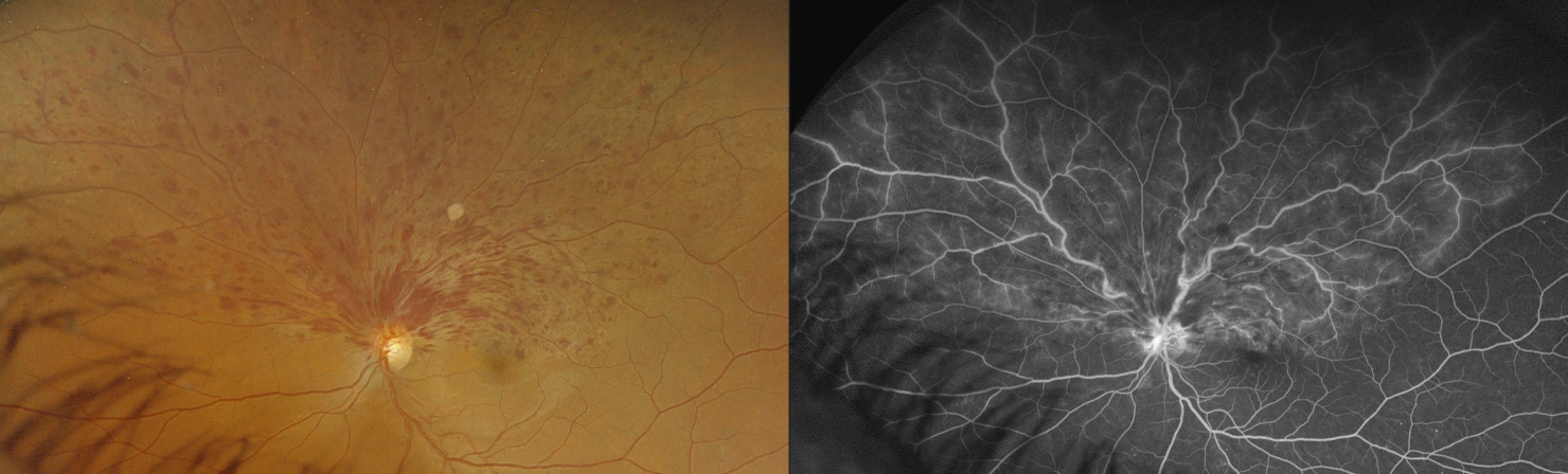
likely dx?
hemi-retinal vein occlusion (HRVO)

A patient comes into your clinic for pain and vision loss of his left eye. In his ocular hx, you note he had a CRVO 9 months ago. His IOP is 30.
What is the likely dx?
Where should you examine to confirm the diagnosis?
How should you monitor them in the future?
Neovascular glaucoma,
pupillary border,
routine gonioscopy

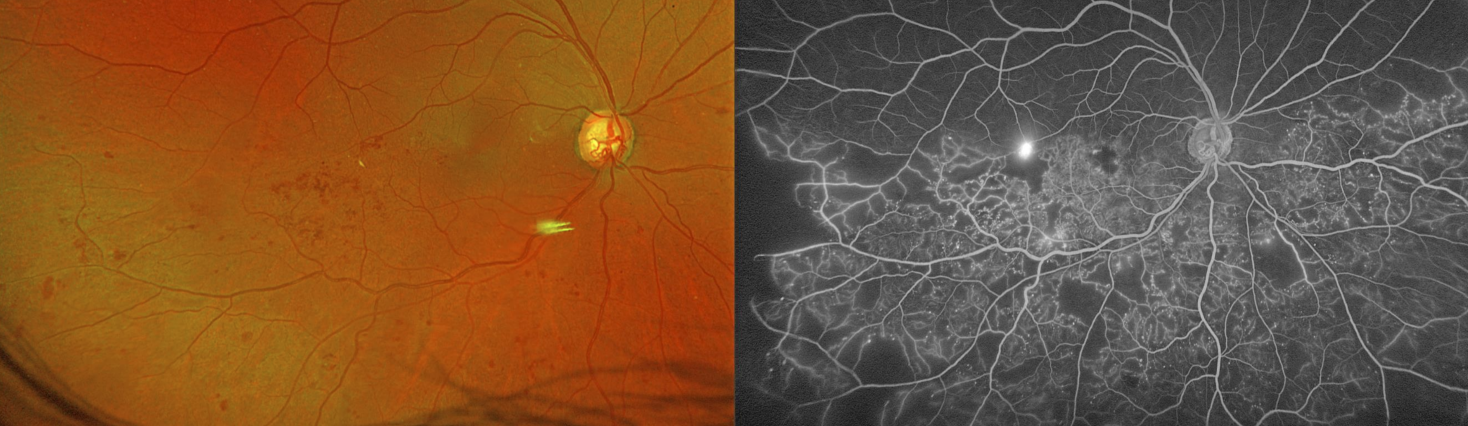
likely dx?
hemi-retinal vein occlusion (HRVO)


a 62 yo man comes in for sudden onset blurred vision of OS. VAs are 20/50-. Pt has slight superior VF defect. likely dx?
Branch Retinal Vein Occlusion (BRVO)


a 62 yo man comes in for sudden onset blurred vision of OD. VAs are 20/25-. Pt has slight superior VF defect. likely dx?
Branch Retinal Vein Occlusion (BRVO)

a 62 yo man comes in for sudden onset blurred vision of OS. VAs are 20/60-. Pt has slight inferior VF defect. likely dx?
Branched Retinal Vein Occlusion (BRVO)
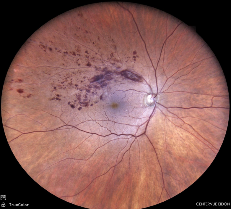
a 62 yo man comes in for sudden onset blurred vision of OD. VAs are 20/25+. Pt has slight inferior VF defect. likely dx?
Branched Retinal Vein Occlusion (BRVO)

a 62 yo man comes in for sudden onset blurred vision of OS. VAs are 20/20-. Pt has superior VF defect. likely dx?
Branched Retinal Vein Occlusion (BRVO)
a patient has macular edema secondary to a BRVO. How should it be treated?
Anti-VEGF (ie: ranibizumab)
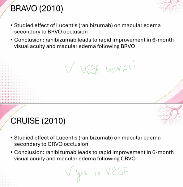
a patient has macular edema secondary to a CRVO. How should it be treated?
Anti-VEGF (or laser or corticosteroids)

A 67 yo male patient presents with a CC or transient vision loss in the right eye with associated symptoms or a dull ache over the brow. He’s noticed his vision gradually getting worse the last few weeks. VAs are 20/70. Upon slit lamp exam you note some neo of the iris and dot/blot hemes confined to the mid periphery. Likely dx?
Ocular ischemic syndrome (OIS)
