The Brain and Cranial Nerves
 Call Kai
 Learn
 Practice Test
 Spaced Repetition
 Match
 Flashcards
 Knowt Play
1/118

There's no tags or description
Looks like no tags are added yet.
119 Terms
regions of the brain
Cerebrum, Cerebellum, Diencephalon, Brainstem
the cerebrum is responsible for-
high-level functions like thought, memory, emotion, speech, and voluntary movement.
the cerebrum has _ hemispheres
2
the cerebrum has _ lobes
4
the Cerebellum is responsible for-
Coordination of body movements, controls posture, balance and fine motor movement, and Involved in motor learning
the Diencephalon is made up of-
the thalamus and hypothamalus

identify
Diencephalon

identify
thalamus
function of the thalamus
transmits sensory information to the appropriate areas in the cerebral cortex except for olfactory and subconscious proprioceptive information
function of the hypothalamus
major autonomic center that controls body temperature, appetite, thirst, fight or flight response, production of hormones that regulate anterior pituitary hormones, and produces ADH and oxytocin

identify
hypothalamus
the brainstem includes-
Midbrain, Pons, and Medulla oblongata
the brainstem is responsible for-
Processes and relays information between the spinal cord and the cerebrum or cerebellum
what is the ventricular system?
A connected network of four fluid-filled cavities that produce and circulate cerebrospinal fluid (CSF) to cushion and protect the central nervous system.
the Ventricular System has _ ventricles and the ____________ ___________
4; interventricular foramen

identify A
First & second Ventricles

identify
third ventricle

this facilitates Communication between lateral ventricles and 3rd Ventricle
interventricular foramen

the ventricle located between the cerebellum and the dorsal pons, in the center of the metencephalon
fourth ventricle
Dura Mater
The tough, thick outer fibrous layer attached to the skull that provides structural stability and protection.

arachnoid mater
The web-like middle layer forms the subarachnoid space and contains CSF

Circulating through the ventricles and subarachnoid space that allows the brain to float, reducing its effective weight by about 95% and cushioning against sudden movements.
Cerebrospinal Fluid (CSF)
Blood-Brain Barrier
A highly selective cellular barrier formed by tight junctions that shields the brain tissue from harmful pathogens and toxins circulating in the blood.
Pia Mater
The delicate, tender inner layer that tightly hugs the surface of the brain and supports blood vessels.

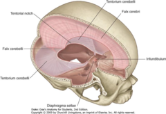
extensions of the meningeal cranial dura into the cranial cavity
- Compartmentalization: Support and separate major regions of the brain to reduce physical pressure.
- Stabilization: Limit excessive movement and rotational displacement
of the brain during sudden head movements.
- Venous Drainage: Enclose the dural venous sinuses between their layers to collect blood and cerebrospinal fluid from the brain.
Dural folds
Dural Sinus
Large venous channel that sits between the layers of the dura mater, and drains blood and CSF fluid from the brain into the internal jugular veins.
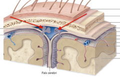
function of Arachnoid Villi
Act as natural one-way valves allowing CSF flow out of the subarachnoid space and into the bloodstream inside the dural sinuses.
function of arachnoid aranulations
Formed by aggregation of arachnoid villi most commonly found along the superior sagittal sinus and transverse sinuses.
function of choroid plexus (CP)
Produces CSF via Ependymal cells that surround the capillaries of choroid plexus and secretes CSF
location of choroid plexus
present in all four ventricles of the brain
function of ependymal cells
surround the capillaries of the choroid plexus and secretes CSF. And also removes wastes from the CSF and adjusts its composition
Blood CSF barrier
Specialized ependymal cells connected by tight
junctions. Fenestrated capillaries within the choroid plexus
the abnormal accumulation of fluid (CSF) in the brain is known as
Hydrocephalus
describe the Circulation of CSF
1. the lateral ventricles, 3rd ventricles, and 4th ventricles of the choroid plexus form the CSF
from superior to inferior-
2. lateral ventricle though the interventricular foramina to
3. the third ventricle through the cerebral aqueduct to
4. the 4th ventricle and goes through the lateral and median apertures
5. to the subarchnoid space to
6. the arachnoid villi od the dural venous sinuses to the
7. venous blood which goes back to the lungs and heart to be reoxygenated

How would decreased diffusion across the arachnoid granulations affect the volume of cerebrospinal fluid in the ventricles?
a) Volume would increase.
b) Volume would decrease.
c) Volume would remain the same.
d) Volume would fluctuate erratically.
a) Volume would increase.
the blood brain barriers formed by:
tight junctions between capillary endothelial cells, Astrocytes, and Pericytes

what can diffuse into interstitial fluid of CNS?
only O2, CO2, and, lipid-soluble compounds like Steroids, prostaglandins, alcohols and general anesthetics can diffuse
an alzheimers brain is a disorder in the__?
Blood brain barrier
what arteries supply blood flow to the brain? what veins remove the blood? where from?
Internal carotid arteries and vertebral arteries. Most blood is removed from dural venous sinuses by internal jugular veins
a ________ is considered a Cerebrovascular accident (C V A) which Stops blood flow to a portion of the brain and Affected neurons begin to die within minutes
stroke
ischemic stroke is what type of stroke?
a type of stroke that occurs when the flow of blood to the brain is blocked
hemorrhagic stroke is what type of stroke?
occurs when a blood vessel in the brain leaks or ruptures; also known as a bleed
what are the parts of the brainstem?
midbrain, pons, and medulla oblongata
functions of the Midbrain
responsible for visual and auditory reflexes (tracking moving objects or reacting to sounds) and contain The red nucleus and substantia nigra that coordinate fine motor movements, posture, and muscle tone. also maintain consciousness

the red nucleus and substantia nigra are found where?
midbrain
function of superior colliculi
part of the midbrain that process visual signals and coordinate reflexes like tracking moving objects, head turns and eye movement and controlling pupil dilation and lens shape.
function of inferior colliculi
part of the midbrain that process auditory signals, help localize the source of sounds, and relay hearing information to the thalamus
function of substantia nigra and red nucleus
part of the midbrain that coordinates fine motor movements, posture, and muscle tone. The red nucleus is part of the rubrospinal tracts.
the midbrain contain nuclei of which cranial nerves?
nuclei of Cranial nerves III & IV

reticular formation
network of nuclei that spans much of the brainstem and is crucial for maintaining consciousness, arousal, and sleep cycles.
the headquarters of the reticular activating system is located where?
midbrain
function of Ascending Reticular Activating System (ARAS)
Sends signals upward to the thalamus and cerebral cortex to control wakefulness, sleep-wake cycles, and general alertness.
function of Descending Reticular System
Sends signals downward via the reticulospinal tracts to the spinal cord to manage posture, balance, and coarse limb movements.
pneumotaxic center
a neural network in the upper pons of the brainstem that acts as an inspiratory off-switch to regulate breathing rate and depth
micturition center
located in the pons. It acts as the primary switchboard coordinating the micturition reflex, triggering the contraction of the bladder muscle while relaxing the urethral sphincters to allow urination
function of pons
white matter carries information to and from the cerebellum. Gray matter is the location of the pneumotaxic center and the micturition center

The ________ ______ in Pons act as a smooth override system. Makes breathing controlled and adaptable to metabolic needs
pnemotaxic/pontine centers
the pons house nuclei of which cranial nerves?
Houses the nuclei for cranial nerves V through VIII

how does the feedback loops of the pontine/pneumotaxic centers operate?
These centers receive signals from lung stretch receptors and blood chemoreceptors to adjust how speed and depth of breathing
function of medulla oblongata
gray matter is location of of respiratory, cardiac, and vasomotor centers. control reflexive actions such as vomiting and swallowing

what does the Cardiovascular center of the medulla oblongata do?
adjusts heart rate and the force of heart contraction.
what does the vasomotor center of the medulla oblongata do?
changes blood vessel diameter to regulate blood pressure.
what does the respiratory center of the medulla oblongata do?
generate the respiratory rhythm and control the rate and depth of breathing.
the medulla contains nuclei of which cranial nerves?
Contains nuclei of cranial nerves IX through XII
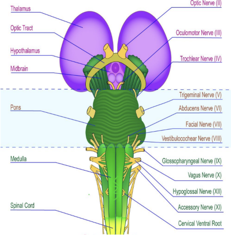
Parinaud's syndrome is a ocular motility issue where a person will have the Inability to look upward while downward gaze is typically preserved. where would the brainstemlesion be located to cause this?
superior colliculi of the midbrain
Oculocephalic Reflex (Doll's Eyes)
turn the head quickly, first to one side then to the other (in the non- neck-injured patient). Absence of doll's eye movements suggests a lesion of the midbrain or pons.
how does the swallowing reflex work?
Breathing stops momentarily, closes the airway (via the larynx/epiglottis), and triggers rhythmic peristaltic waves down the esophagus while relaxing esophageal sphincter
Each cerebral hemisphere controls the voluntary movements of muscles on _______ side of the body.
opposite
describe the decussation of sensory and motor signals, and where it takes place
sensory and motor fiber will cross at either the spinal cord or the medulla to innervate or control the opposite side of the body
functions of the cerebellum
A portion of the hindbrain that maintains posture and balance and coordinates body movements; involved in storage of procedural memories
Cerebellar peduncles
massive white matter tracts that link the cerebellum to the brainstem, acting as the primary pathways for all incoming and outgoing neural signals.

Dysmetria, a type of ataxia characterized by the inability to judge distance, speed, or range of motion is a disorder of what part of the brain?
cerebellum
Ataxia is uncoordinated, shaky, or clumsy movements caused by nervous system damage to which part?
cerebellum
function of the thalamus
transmits sensory information to the appropriate areas in the cerebral cortex except for olfactory and subconscious proprioceptive information
function of the Hypothalamus
Major autonomic center that controls body temperature, appetite, thirst, fight or flight response, production of hormones that regulate anterior pituitary hormones, and produces ADH and oxytocin
what is the limbic system?
A complex network of structures located deep within the brain, sitting underneath the cerebral cortex and above the brainstem, that acts as the brain's primary center for emotions, memory, motivation, and survival behaviors.

the limbic system contains
the hippocampus, amygdala, thalamus, and hypothalamus
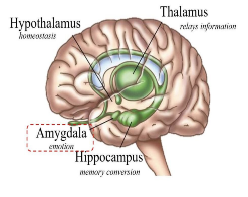
Dysfunctional interaction between the amygdala, hippocampus, and prefrontal cortex can cause the excessive fear responses and emotional flashbacks characteristic.
PTSD
function of Hippocampus
structure that converts short term memory into long term memory
what is the pathway of stimulus to the hypothalamus?
1. sensory stimuli
2. sensory cortex
3. limbic system (attaches emotion
4. hypothalamus
5. autonomic, somatic, immune, endocrine systems

Amygdala
A limbic system structure involved in memory and emotion, particularly fear and aggression.
_______________ Translates Emotional Signals From The Limbic System Into Physiological Responses.
Hypothalamus
Cerebrum
Largest part of brain. Controls all conscious thoughts and intellectual functions. Processes SOMATIC sensory and motor information
function of Corpus Callosum
contain commissural fibers that allow communication between the 2 cerebral hemispheres
the ______ hemisphere is responsible for spatial relationships (understanding a map or locating objects).
right
______ hemisphere is responsible for logical thought and language
Left
Hemispheric lateralization
Functional differences between left and right hemispheres Each cerebral hemisphere performs certain functions that are not ordinarily performed by the opposite hemisphere
what happens with Split-Brain Syndrome?
the corpus callosum is severed. The two cerebral hemispheres cannot communicate with each other
a patient with their corpus callosum severed is shown the word ‘face’ in their right visual field and is asked what they see. What would they be able to do in this situation? why?
the patient would be able to verbalize what they see when asked because the left hemisphere is processing the information of the right visual field and the hemisphere is dominant for verbal processing.
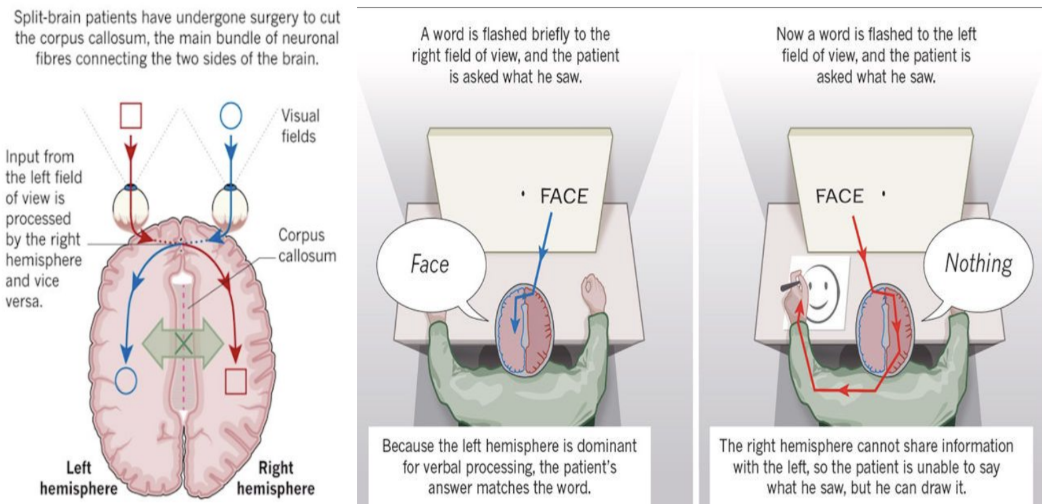
a patient with their corpus callosum severed is shown the word ‘face’ in their left visual field and is asked what they see. What would they be able to do in this situation? why?
the patient would not be able to verbalize what they see but they can draw a picture of it because the visual information goes to the right hemisphere which doesnt have wernickes or brocas areas however it is dominant in spatial visualization
where is grey matter in the brain?
the periphery of the brain and the basal nuclei (center

basal nuclei
Islands of grey matter embedded in white matter of cerebrum important in motor coordination
Functions of basal nuclei
Directs subconscious activities such as regulating rhythmic movement related to skeletal muscle, controlling the timing and sequencing of motor actions in synchronization with external stimuli, and Coordination of movement patterns (swinging arms during walking)
In Parkinson's disease, what area of the basal ganglia has a decreased amount of dopamine?
substantia nigra
what type of fibers are in the white matter?
association, commissural, and projection fibers

association fibers
horizontal running fibers that connect different parts of same hemisphere

3 area of cerebral cortex
Motor areas, Sensory areas, and Association areas.
Motor Areas Of The Cerebral Cortex
prefrontal cortex, premotor cortex, and primary motor cortex
prefrontal cortex main roles
high-level cognitive control of behavior such as Decision making, Goal setting, Planning, Working memory (mental scratch pad), Evaluating consequences of actions, Attention/focus on a goal and direct behavior toward achieving that goal.
which cortex decides what action should be taken and why it should be taken?
prefrontal cortex