cardiovascular - blood vessels and circulation
 Call Kai
 Learn
 Practice Test
 Spaced Repetition
 Match
 Flashcards
 Knowt Play
1/40

There's no tags or description
Looks like no tags are added yet.
41 Terms
does smooth muscle have sarcomeres?
no, actin & myosin are not organized into sarcomeres in smooth muscle = non-striated appearance
approx actin:myosin in smooth muscle vs straiated muscle cells
smooth = ~15:1, far greater than striated = ~2:1
dense bodies
function like Z-lines of striated muscle cells
where actin is anchored

contraction speed of smooth muscle
slow
portal systems (parts of circulatory system)
large vessels otw to heart become
smaller vessels that supply tissues become
larger vessels that return to the heart
*breaks normal hierarchy of vessels becoming continuous smaller or larger

major example of portal systems in mammals - hepatic portal system
venous blood returning to the heart from the digestive tract via the hepatic portal vein first pass through a capillary network in the liver before entering the vena cava of the heart via the hepatic vein

lymphatic system - subsystem of the circulatory system
network of lymphatic vessels, lymph nodes, and associated organs/tissues (e.g. bone marrow, spleen, and thymus)
function of lymphatic system
drain interstitial fluids and returns leaked fluids (collected by lymph capillaries) and proteins back to the cardiovascular system via the subclavian vein
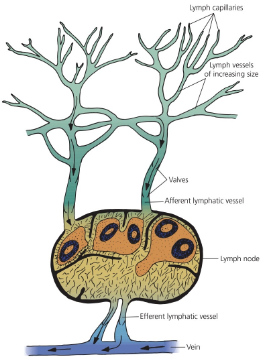
lymph
the fluid traveling in lymphatic vessels
*very similar in composition to interstitial fluid
endothelium
the inner lining of the entire circulatory system composed of a specialized type of simple squamous epithelium called endothelial cells
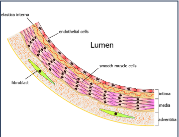
function of endothelium
to form the barrier between vessels and tissue to control the movement of substances, fluid, and white blood cells

vascular endothelial cells
line the interior of all blood vessels and the interior of the heart (endocardium)
lymphatic endothelial cells
line the interior of all lymphatic vessels
endothelial cells are a specialized type of
simple squamous epithelium
arteries carry
oxygenated blood away from the heart
*exceptions: pulmonary and umbilical arteries that carry deoxy blood to the organs that oxygenate it (the lungs and placenta, respectively)

artery sizes
elastic artery: largest, ex: aorta →
muscular arteries: can change in diameters via contracting & dilating due to smooth muscle →
arterioles →
capillaries →

function of capillaries
site of exchange of substances between vasculature and surrounding interstitial fluid
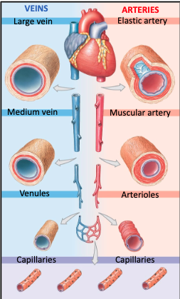
vein sizes
capillaries
venules →
medium veins →
large veins: return blood to the heart, ex: vena cava

veins carry
deoxygenated blood towards the heart
*exceptions: pulmonary and fetal veins that carry oxygenated blood

tunica intima
the innermost layer of an artery or vein consisting mainly of endothelial cells and a supporting internal elastic lamina
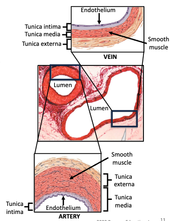
tunica media
the middlemost layer of an artery of vein made up of smooth muscle cells and elastic connective tissue

tunica externa
the outermost layer of an artery or vein made up of connective tissue comprised of collagen and elastin fibers
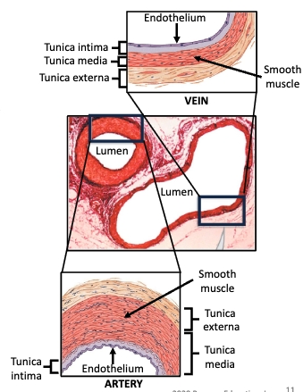
traits of artery walls vs veins
artery walls contain more smooth muscle, are more elastic, and often have a smaller diameter than veins
which part of circulatory system generally has higher blood pressure?
arteries
*highest during ventricular systole
what is special about veins?
they have one-way valves to prevent backflow of blood while the blood is moving back up toward the heart
skeletal muscle pump
skeletal muscle contraction that squeeze on intramuscular veins, pushing venous blood back toward the heart
microcirculation
blood circulation in the smallest blood vessels (pre-capillary aterioles, metarterioles, capillaries, and post capillary venules) present in most tissues of body

function of microcirculation
delivers O2 & nutrients to tissue cells
removes CO2 & waste products formed by cells
regulates local blood flow and tissue perfusion → affecting BP, edema, and thermoregulation

pre-capillary sphincters is composed of
smooth muscle cells

pre-capillary sphincters determines
amount of blood that enters capillary beds
pre-capillary sphincters react to
level of O2/CO2 in capillary bed
low O2/high CO2 = opens sphincters (dilates vessels coming in)
high O2/low CO2 = closes sphincters (constricts vessels coming in)
where is BP highest? lowest?
highest: in ascending aorta as blood leaves the left ventricle
lowest: in vena cava before returning blood into right atrium
systolic arterial blood pressure
highest blood pressure measured when ventricles contract (ventricular systole)
diastolic arterial blood pressure
lowest BP measured when ventricles relax (ventricular diastole)
pulse pressure =
difference between systolic and diastolic BP
autoregulation
process by which lack of tissue oxygen or increase in carbon dioxide creates local vasodilation of vascular smooth muscle thereby increasing local blood flow
when cardiac output is increased via sympathic neural stimulation of SA node, what happens to blood vessels?
skeletal muscle & heart: vasodilation
other organs (like intestines): vasocontriction
baroreflex - high BP
increased activation of cardioinhibitory center = decreased cardiac output
decreased activation of cardiostimulatory center = decreased cardiac output
decreased activation of vasomotor center = vasodilation
capillary hydrostatic pressure
pressure at capillary bed determined by ratio between pressure in artery and vein
high arterial BP = high capillary bed pressure
blockage in veins = high capillary bed pressure
can lead to edema
blood osmotic (colloid oncotic) pressure
osmotic pressure exerted by molecules too large to leave capillaries, such as proteins
large molecules are what draw fluid in