HAP- exam 2 study guide
 Call Kai
 Learn
 Practice Test
 Spaced Repetition
 Match
 Flashcards
 Knowt Play
1/94

There's no tags or description
Looks like no tags are added yet.
95 Terms
Epithelial Tissue (Epithelium)
2 types (by location)
Covering/lining
external/internal surfaces
Glandular
secretory tissue in gland
Functions of Epithelial Tissue
physical protection
selective permeability
secretions
sensations
Epithelial Tissue- Physical Portection
Protects from dehydration, abrasion, destruction
Epithelial Tissue -Selective permeability
Allows passage of some subtsnaces while preventing passage of others
Epithelial Tissue - secretions
Some cells specialized to secrete
Epithelial Tissue - Sensations
Supply information to nervous system
Characteristics of epithelial tissue
Cells have polarity—apical (upper, free) and basal (lower, attached) surfaces
Apical surfaces may bear microvilli (e.g., brush border of intestinal lining) or cilia (e.g., lining of trachea)
Noncellular basal lamina of glycoprotein and collagen lies adjacent to basal surface
Types of cells
Squamous
Cuboidal
Columnar

Layers
Simple
Stratified

Simple columnar epithelium Function
Absorption
secretion of mucus
enzymes , and other substances
ciliated types propels mucus (or reproductive cells ) by ciliary action
Simple columnar epithelium location
along the lines of most digestive tract ( stomach to anal canal )
gallbladder
excretory ducts of some glands
Stratified squamous epithelium function
protects underlying tissues in areas subjected to abrasion
Stratified squamous epithelium location
moist linings of esophagus , mouth, and vagina
Loose connective tissue
fewer fibers, more ground substance
areolar
adipose
reticular
Dense connective tissue
more fibers , less ground substance
dense regular
dense irregular
elastic
Support connective tissue - cartilage
hyaline
fibrocartilage
elastic
Supporting connective tissue Bone
compact
spongy
Fluid connective tissue
Blood
Lymph
Major functions of connective tissue
binding/ support
ligaments/tendons
Protection
bones of cranium, fat for kidneys
Storage
fat for energy, bone for calcium/ phosph.
Transportation (blood)
Structural elements of connective tissue
cells
fibers
ground substance
Collagen fibers ( white )
strongest/ most abundant type
25% of body’s protein
provides high tensile strength
Elastic
Networks of long, thin, elastin fibers that allow for stretch (skin, arteries)
Reticular fibers
Short, fine, highly branched collagenous fibers
abundant in the stroma of lymph nodes/ livers
Ground Substance
Nonliving material
medium through which solutes diffuse b/t blood capillaries and cells
solid/semisolid
viscous
Loose connective tissue
Wraps/ cushions organs
plays important role in inflammation
holds/conveys tissue fluid

Dense Irregular connective tissue
able to withstand tension exerted in many directions; provides structural strength
major cell in fibroblast

Supportive connective tissue - Resist Compression
No role in fat storage or disease Matrix 80% water ( why it can resist compression
Hyaline
Supports/ reinforces
has resilient cushioning properties
resist compressive stress
located in costal cartilages

What is the bodies Largest Organ ?
Skin
covers 1.5→ 2.0 m2
Composes 15% of body weight
Another name for skin is the ______ membrane
Cutaneous
Functions of skin
Resistance to trauma/ infection (protection)
immune defense
vitamin D(3) synthesis (metabolic reg)
sensation
thermoregulation
nonverbal communication
Thick Skin
Covers front of hands, bottoms of feet
epidermis 0.5 mm thick
Thin Skin
covers rest of the body
epidermis about 1.0mm thick
Epidermis what layer
Outer layer of skin
Dermis what layer
inner layer beneath epidermis
Epidermis
05-.1mm thick
Made up of stratified squamous epithelial cells.
Provides a protective barrier against environmental
damage (like pathogens, UV radiation, and water loss).Constantly renews itself through cell division at the base and shedding at the surface.
How often do we shed ours epidermis?
You lose about 30,000 to 40,000 skin cells every minute, which adds up to millions per day
Keratinocytes
Most abundant cells in the epidermis.
Produce keratin, a tough protein that provides protection and waterproofing.
Form multiple layers as they move from the base to the surface
Dendritic Cells (Langerhans cells)
Immune cells that detect and fight pathogens.
Found in the stratum spinosum.
Act like sentinels, alerting the immune system to invaders.
Tactile Cells (merkel Cells)
Located in basal layer
Connected to nerve endings / help detect touch/pressure
important for sensory perception
Melanocytes
also found in basal layer
Produce melanin, pigment gives skin its color
protect against UV radiation by absorbing harmful rays
Stem cells (basal cells )
Found in the stratum basale (deepest layer).
Continuously divide to produce new keratinocytes.
Essential for skin regeneration and repair
The dermis
4mm thick
Composed of connective tissue, rich in collagen and elastin fibers.
Contains blood vessels, nerve endings, hair follicles, sweat glands, and sebaceous (oil) glands.
Provides strength, elasticity, and nutrients to the skin.
Supports thermoregulation and sensory perception
Hypodermis
Subcutaneous tissue
has more areolar/adipose than dermis has
Subcutaneous fat
energy reservoir
thermal insulation
Integumentary system
Largest organ in body
nails
hair
sebaceous oil glands
sweat glands
Hair
Hair found almost everywhere on the body
Keratinized cells form slender filament
Grows from hair follicles
Hair 3 types
lanugo
vellus
terminal hair
Lanugo
fine, unpigmented, downy hair. Appears in last trimester
Vellus
fine hair
Primary human hair
Found on upper and lower limbs
Terminal Hair
coarser, pigmented, longer
On scalp, eyebrows, eyelashes, beards
During puberty, replaces vellus hair in axillary and pubic regions
3 zones of a hair follicle
hair bulb
root
shaft
Hair matrix
keratinization
new cells divide
pushed up
Hair follicle
oblique tube around root
extends down to dermis
Hair functions
protection
heat retention
sensory
Burns
Major cause of accidental death
Caused by heat, radiation, chemicals, sunlight, electrical shock
Threat to life from fluid loss, infection, effects of burned tissue
1st degree burns
involve only epidermis
slight redness/pain
immerse burned area in cool water
2nd degree burns
involve epidermis/ part of dermis
skin blistered /painful
slight scarring
3rd degree burns
involve epidermis, dermis, and subcutaneous layer
Require hospitalization
Treatment for dehydration and infection
Require additional caloric intake
Severe scarring
May need debridement and skin graft
Skeletal System
composed of bones, cartilages, and ligaments
bone
cartilage
ligaments
tendons- attach muscle to bone
Bone is ________ and contains mainly ________ nerves.
highly vascularized
sensory nerve
Types of bones
compact
spongy
ligaments
Connect bone to bone
Tendons
connect muscle to bone
The bones of the fingers and toes (phalanges)
are categorized as _____ bones.
A) long
B) irregular
C) short
D) flat
E) sesamoid
A) long
Long bone
humerus
Short bone
talus
Flat bone
Sternum
Irregular bone
Vertebra
General Features of bones
Epiphysis Diaphysis, Metaphysis
Compact and spongy bone
Marrow cavity
Articular cartilage
Periosteum
Epiphyseal plate/line

Bone Marrow
bone marrow
red marrow (myeloid tissue)
yellow marrow
Bone cells: Osteoblast
under endosteum/periosteum
synthesize organic matter of bone matrix
nonmitotic
Osteocytes
former osteoblast in matrix
reside in lacunae
cytoplasmic process that reach into canaliculi
connect to osteoblasts
importance of canals
Oseteoclasts
ruffled border
increase cell surface area
reside in pits: resorption bays
Bone matrix: Ground substance/ fibers
Bone matrix has Organic and inorganic components
organic compounds
Osteoid produced by osteoblasts
Histology of Osseous Tissue
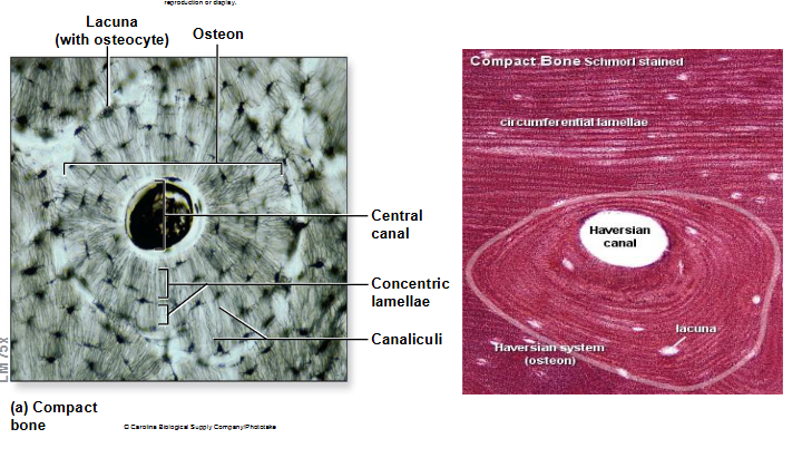
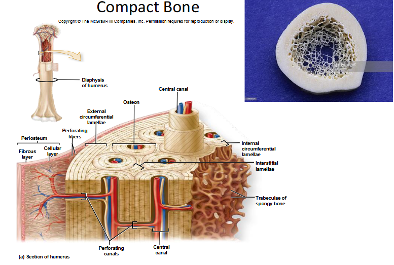
Compact bone
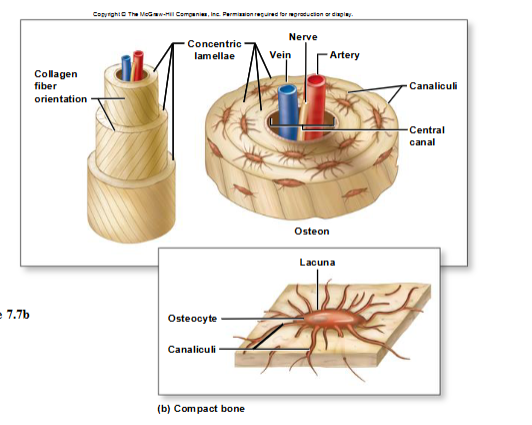
Osteon Structural Unit
Growth of Cartilage
Interstitial Growth
occurs within internal regions of cartilage
Appositional Growth
occurs on cartilage’s outside edge

Bone Development
Ossification or Osteogenesis- the formation of bone
in human fetus/ infant, bone develops by 2 methods
intramembranous
endochondral
Intramembranous Ossification
bone growth within a membrane
produces:
flat bone of skull
some of facial bones
mandible
central part of clavicle

Endochondral Ossification
Within cartilage model
most bones of skeleton
long bones
Step 1 of long bone development in limb
fetal hyaline cartilage model develops
chondroblasts secrete cartilage matrix during 8th→12th week of development
Step 2 of long bone development in limb
Cartilage calcifies. a periosteal bone collar forms
chondrocytes in cartilage model produce holes in matrix
Matrix calcifies, chondrocytes die
produces calcified cartilage shaft with large holes
Step 3 of long bone development in limb
Periosteal; bud extends from periosteum into cartilage shaft , growth of capillaries /osteoblasts
Osteoblasts produce osteoid on calcified cartilage template
primary ossification center
Bone development extends in both directions toward epiphyses
Step 4 of long bone development in limb
Secondary ossification centers form in epiphyses
hyaline cartilage calcifies / degenerates
blood vessels / osteoprogenitor cells enter
secondary ossification centers form
Step 5 of long bone development in limb
Bone replaces cartilage, except articular cartilage/ epiphyseal plates
Step 6 of long bone development in limb
Lengthwise growth continues until epiphyseal plates ossify/ form epiphyseal lines
Lengthwise bone growth continues into puberty
growth continues until epiphyseal plate is converted to epiphyseal line indicates bone has reached adult length
Occurs between ages of 10 and 25
Bone remodeling
Continues throughout adulthood
Occurs at different rates
20% of skeleton replaced yearly
Dependent upon the coordinated activities of osteoblasts, osteocytes, and osteoclasts
Influenced by hormones and mechanical stress
Bone growth/ remodeling
Ossification continues throughout life with the growth and remodeling of bones
Bones grow in two directions
length
width
Interstitial in growth plate
Appositional in perichondrium
Epiphyseal Plate zone cartilages

Cartilage Growth Plate

Appositional Bone Growth
Osteoblasts build bone at the circumferential lamellae
Osteoclasts widen the medullary cavity
Osteopenia
a condition where bone mineral density is lower than normal, sitting in the warning stage between healthy bone and osteoporosis. [1, 2]
