All David Cattle
 Call Kai
 Learn
 Practice Test
 Spaced Repetition
 Match
 Flashcards
 Knowt Play
1/201

There's no tags or description
Looks like no tags are added yet.
202 Terms
Topic 1: Conditions of the Bovie Head
Topic 1: Conditions of the Bovie Head
Describe The Bovine Head in terms of:
1. Oral cavity Anatomy
2. Oral Cavity Disease Clinical Signs
3. Examination of the Mouth
4. Saliva
1. Teeth, Buccal mucosa, Tongue, Jaw, Oesophagus
2. Drooling saliva, tongue protrusion, prehension issues, mastication issues, regurgitation following difficult swallowing, foul smell, swelling of cheeks
3. Examine using torch and mouth gag, the tongue (if not painful) can be caught by the hand and slightly pulled to the side to view parts of inside the mouth
4. Responsible for rumen pH Buffer + rumen hydration. Excessive salivation reduces these functions

Describe Excess Salivation Without Oral Vesicles/Erosions in terms of:
1. Many Animals Affected
2. Single Animals Affected
1. Heat stress, botulism, Ephemeral Fever
2. Woody tongue, foreign body, hypomagnesemia, mal-eruption, choke, botulism,

Describe Excess Salivation With Oral Vesicles/Erosions in terms of:
1. Many Animals Affected with Vesicular Lesions
2. Many Animals Affected with Erosions
3. Single/Few Animals Affected with Erosions
1. FMD, Vesicular stomatitis
2. Bluetongue, Chemical irritants, toxic plants
3. BVD, Mucosal disease, MCF
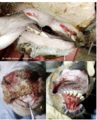
Describe Aging Cattle Using Permanent Incisor Teeth Eruption in terms of:
1. 2 Teeth
2. 4 Teeth
3. 6 Teeth
4. Full Mouth
1. 2y
2. 2.5-3y
3. 3.5-4y
4. 4-5y

Describe Teeth Abnormalities as a Oral Disease in terms of:
1. Common Abnormalities
2. Fluorosis Definition
3. Causes of Fluorosis
4. Diagnosis of Fluorosis
5. Pain From Erupting Incisors
1. Malocclusions of Incisors, Loose Incisors, Tooth Root Abscess
2. Chronic ingestions of excess fluoride (pollution, ash, contaminated feeds). Teeth become brittle and wear fast. Causes Low BCS + worn teeth
3. Cattle exposed from 6m-30m are more resistant (ameloblasts still active). Causes Osteoporosis, seen as bone weakness throughout life (lameness)
4. Bone fluoride content evaluation (commonly tail amputation)
5. Metabolic disorders due to low food intake. Common in heifers.

Describe Cleft Palate as a Oral Cavity Disease in terms of:
1. Definition
2. CS
1. Failure of midline fusion during foetal development, Heritable. Sporadically found in sheep + cattle
2. Milk Reflux through nostrils w/ consequential aspiration pneumonia, Difficulty feeding + breathing, poor growth/ill thrift

Describe Actinobacillosis (Wood Tongue) as a Oral Cavity Disease in terms of:
1. Causative Bacteria/Pathogenesis
2. Clinical Signs
3. DDx
4. Other organs affected
5. Atypical Cutaneous Actinobacillosis
6. Treatment
1. Actinobacillus lignieresii. Environmental bacteria, normal inhabitant of mouth, infection follows abrasion/traumatic injury of oral mucosa (Following teeth eruption, grazing apprasive feeds)
2. Salivation, tongue swollen/hard, lymphadenitis, snoring
3. Abscess, trauma, causes of salivation
4. Soft tissue of head, regional LNs, hair loss, oesophageal groove involvement
5. No salivation, cutaneous lesions
6. Sodium Iodide (IV injection with sterile water - Watch for iodine toxicity (skin scaling, lacrimation, coughing) followed by antibiotics (oxytetracyclines). Potentially surgical curette.

Describe Actinomycosis (Lumpy Jaw) as a Oral Cavity Disease in terms of:
1. Causative Bacteria/Pathogenesis
2. Clinical Signs
3. Treatment
4. Prognosis
1. Actinomyces Bovis. Normal inhabitant of mouth, gains access to bony structures through lymphatic drainage from oral traumatic buccal injury - also through tooth root injury or molar tooth eruption.
2. Pyogranulomatous osteitis / osteomyelitis of mandible and maxilla. Sporadic but common.
3. As per actinobacillosis (Sodium Iodide + oxytetracyclines) Potentially surgical curette.
4. Ok if just enlarged, poor if animal is having difficulty eating due to mal-alignment of cheek teeth. Grave if jaw is fractured
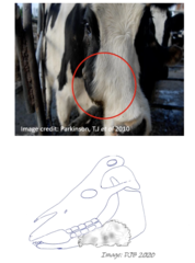
Describe Trauma, FBs, and Swellings as a Oral Cavity Diseases in terms of:
1. Oral Injuries
2. Grass Seed Abscess
3. Phlegmonous Cellulitis
4. Choke
1. Under tongue/pharynx, often caused by drenching gun. Treat with Oxytetracycline
2. Seed penetrates buccal mucosa → abscess Formation. Surgically drain and iodine lavage
3. Caused by Fusobacterium Necrophorum. Causes submandibular swelling (can extend to briskit), high T + HR due to septicaemia, death. Palpation reveals fluid rather than oedema. Establish drainage and treat with high doses of procaine penicillin
4. Associated with Salivation, Poor Nutrition, and secondary (free-gas) bloat

Describe Enzootic Nasal Granuloma (+ Nasal Foreign Bodies) as a Head Disease in terms of:
1. Pathogenesis
2. Clinical Signs
1. Acute onset bilateral nasal and ocular discharge → nasal mucosa swelling causes noisy breathing → irritation causing cow to rub nose and shake head → gets foreign bodies stuck in nostril
2. Unilateral nasal discharge, Palpation of nostril shows small firm polyps/nodules. Not fatal

Describe Diseases of the Head in terms of:
1. Laryngeal Necrobacillosis (Calf Diphtheria)
2. Sinusitis
1. Caused by Fusobacterium Necrophrum Causes necrotic laryngitis, common in bucket fed calves.
2. Trueperella pyogenes mostly isolated. Usually associated with dehorning of older cattle.
CS: Anorexia, lethargy, frontal bone distortion, nasal/horn stump purulent discharge, exophthalmos, dull sound on frontal sinus percussion
Tx: Flush sinuses + systemic ABs
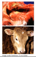
Describe Infectious Bovine Keratoconjunctivitis (IBK/Pinkeye) as a Eye Condition in terms of:
1. Causative Agents
2. Disease Characteristics
3. Predispositions
1. Haemolytic Moraxella bovis is major causative organism. (Mycoplasma, Rickettsia, and Chlamydia may be involved)
2. Highly contagious disease, spreads rapidly in summer month's. CS exacerbated by BoHV-1 infection
3. Damage to Cornea Predisposed by UV light, Dust, Long grass (irritation), Flies. Host factors include bos taurus cattle, pigmentation around the eye, and increasing active immunity with age

Describe Infectious Bovine Keratoconjunctivitis (IBK/Pinkeye) as a Eye Condition in terms of:
1. Bacteriology
2. Transmission
1. Different serotypes. M. bovis use pili to adhere to epithelium of cornea, Infected animals carriers for up to a year, outbreaks often occur due to purchased cattle.
2. Direct contact, Face flies (mechanical vector), fomites (grass, seeds)
Describe the Pathogenesis of Infectious Bovine Keratoconjunctivitis (IBK/Pinkeye)
Pilli attach to cornea → Ulceration → Neutrophils migrate to infection site and release enzymes → corneal ulceration worsens → blood vessels grow into eye producting redpink colour of eye → granulation tissue fills defects → neutrophil accumulation results in creamy purulent material in centre of lesion . Then 2 Outcomes
a. Ulcer may rupture causing eye collapse (permanent blindness)
b. often heals spontaneously in 60 days → mild or sever scarring can remain → can cause vision problems

Describe Infectious Bovine Keratoconjunctivitis (IBK/Pinkeye) as a Eye Condition in terms of:
1. Clinical Signs
2. Treatment
3. Preventative Management
1. Corneal oedema, Conjunctivitis, Photophobia, with blepharospasm. Lacrimation plus discharge from eyes, tear staining of eyes.
2. Pink Eye eye ointment (cloxacillin). Subconjunctival injection with procaine penicillin. Parenteral antibiotics (injectable) - oxytetracycline. Severe Cases: Cover eye with eyepatch, third eyelid flap, closure of eyelid temporarily via suture. Metaphylactic Injection (long-acting tetracycline off all at-risk cattle may be conside red in severe outbreaks)
3. Fly control, isolate infected animals, avoid overcrowding, manage new stock separately from rest of herd, UV protection (shade), Vaccinate. Treat both eyes if infected

Describe Bovine Iritis (Silage Eye) as a Eye Condition in terms of:
1. Definition
2. Clinical Signs
3. Treatment
4. Prevention
5. Ddx
1. Common cause of uveitis in cattle being fed silage. Associated with conjunctival infection with listeria monocytogenes ehen animal puts its head in feed
2. Uveitis, fibrin in anterior chamber causing white material beneath cornea (no conjunctivitis or corneal ulceration)
3. Regresses without treatment (but painful), responds well to subconjunctival injection of oxytet + dexamethasone
4. Rolling/spreading big-bale silage
5. IBK, MCF, FB, uveitis from bacteraemia

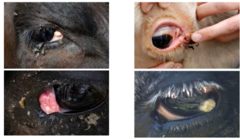
Describe Cancer Eye (Squamous Cell Carcinoma) as a Eye Condition in terms of:
1. Definition
2. Prevalence
3. CS
4. Treatment
1. Occurs in poorly pigmented areas at mucocutaneous junctions (Mainly eye, but can be vulva/anus). Typically involves third eyelid. Follows prolonged periods of UV radiation
2. Most common in cows >5y, Hosteins + herefords. Animals with non-pigmented eyelids and protruding eyes most susceptible
3. Unilateral blepharospasm and ocular discharge, swelling lesion, local invasion of ocular tissue common but metastatic spread to regional LN rare
4. Surgical excision, 3rd eyelid excision, eye removal, cryosurgery plaque removal from cornea
Describe Orbital Exenteration (Enucleation) in terms of:
1. Definition
2. Considerations
3. Sedition
4. Skin Prep
5. Anaesthesia
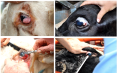
1. Removal of the eye and the contents of the orbit
2. Required when lesions of eyeball, 3rd eyelid, or eyelid are >3cm, always assess for bony involvement/metastases
3. Xylazine/acepromazine
4. Clip/shave around infected eye, wash/clean, surgically prepare with chlorhexidine or povidone iodine
5. Auriculopalpebral block, then Paterson eye block,

Describe Orbital Exenteration (Enucleation) in terms of:
1. Surgical Procedure
2. Aftercare
3. Complications
1. Cut around eyelid → cut deeper until eye is removed → close skin with sutures
2. Do not ligate vessels, do not use gauze to pack eye, orbit will fill with blood to control significant haemorrhage, provide systemic antibiotics, remove sutures after 2-3 weeks or when cattle are mustered (in case of nylon sutures)
3. Cattle with poor temperament more difficult to handle in yards, re-growth of cancerous tissue, infection/abscessation

Describe Ablation of 3rd Eyelid in terms of:
1. Definition
2. Surgical Procedure
1. Surgical removal of the third eyelid. For cancerous growth/injury isolated to 3rd eyelid
2. Sedate cow → restrain head → apply topical eye anaesthetic → prolapsed 3rd eyelid and infiltrate lignocaine into 3rd eyelid well beneath tumour using thin needle → grasp 3rd eyelid with hemostats and complete lignocaine → excise 3rd eyelid deep to cartilage using scalpel blade

Describe Cryotherapy of Eyeball Lesions in terms of:
1. Definition
2. Surgical Procedure
1. For treatment of superficial eye lesions where the eye can be spared
2. Sedate cow and restrain head → anaesthetise eye (petersons/auriculopalpebral) → proptosed eye (apply pressure to rim of eye until eyeball protrudes/emerges) → shave lesion back to healthy corneal tissue → Freeze spot on cornea for 10-20 seconds
Describe Conditions of the Ear in terms of:
1. Otitis Externa
2. Otitis Media/Interna
1. Affected ear droops with foul smelling, purulent discharge from ear. Treat with antibiotics. Caused by nematodes, mites, or foreign bodies
2. Head is rotated → animal ataxic and may drift/fall to affected side. Purulent discharge from ear. Facial nerve paralysis. Treat with broad spectrum antibiotics for several weeks.
Ddx: Listeriosis, Thrombotic meningoencephalitis (H. somni)

Describe Dehorning in Cattle
- Removal of horns
- Desensitise via nerve blocks
- Once adult horns are removed a large sinus opening remains, be caution of infection

Topic 2: Conditions of the Bovine CVS
Topic 2: Conditions of the Bovine CVS
Describe Examination of the Bovine Heart in terms of:
1. Auscultation
2. Normal Newborn HR
3. Normal Calve/Adult HR
4. Jugular Pulse
5. Jugular Vein Engorgement
1. Deep in the axilla region. Rate, intensity, rhythm, abnormal murmurs
2. Up to 120
3. 60-80. Handling+stress can raise HR by 50%
4. Extending 1/3 of neck when head In normal position (Negative jugular pulse). Positive Pathological jugular pulse extends further up the neck
5. Suggestive of Right heart failure (pericarditis, endocarditis, myocarditis, Pimelea spp. toxicity) or Space occupying lesions in thoracic inlet (abscess, neoplasia)

Describe Examination of the Bovine Heart in terms of:
1. Pericardiocentesis
2. Left Side
3. Right Side
1. Collection of fluid from pericardial sac (on L + R side between 5-6th rib)
2. Mitral, aortic, and pulmonary valve
3. Tricuspid valve (Most affected in cattle)

Describe Examination of the Bovine MM in terms of:
1. Pink
2. Pale
3. Petechial
4. Jaundice
5. Cyanotic
6. Brown
7. Bright Red
1. Healthy/Normal
2. Anaemia/shock
3. Brachen fern
4. Liver damage, sever haemolysis
5. Heart failure, severe resp. distress
6. Nitrate/nitrite toxicity
7. Cyanide toxicity

List the Common Clinical Signs Suggestive of Cardiovascular Disease
Age of Animal, Onset, Duration, progression, and No. animals affected Important for Disease Diagnosis
- Depression and weight loss
- Reluctant to move
- Oedema of brisket, submandibular space, ventral abdomen, or udder
- Distended/engorged jugular veins
- Dyspnoea, tachypnoea, cough
- Weakness, unwilling to walk, excersise intolerance
- Heart murmur
- Elbow abduction while standing (thoracic pain) and arched back

Describe Traumatic Reticulopericarditis (Hardware Disease) in terms of:
1. Definition/ General Pathogenesis
2. Heart Failure Pathogenesis
1. Occurs as a result of foreign body penetration of reticular wall then migrating through the diaphragm into the pericardial sac → results in pericardial sac filling with exudate → constricts heart action → bacterial flora variable (Staphylococcus, Streptococcus, TP, E.Coli). Can also follow septicaemia, or severe pneumonia
2. Exudate and fibrin results in friction sounds and epicardium and pericardium rub together → pericardial sac fills with exudate (heart sounds muffled) → If bacteria produce gas, the pressure of gas causes "washing machine noise" → adhesions restrict heart even further → Congestive heart failure

Describe Traumatic Reticulopericarditis (Hardware Disease) in terms of:
1. Reticular Foreign Body Origin
2. Clinical Signs
3. Diagnosis
1. Recent fencing (cut of wires), feeder wagon bails, old wood piles, bale wire, old tires (have wires)
2. Early Stages (Traumatic reticulopericarditis) show anorexia, severe drop in milk production, rumen tympanyreluctant to move, abducted elbows, arched back → Once pericardial effusion is present, heart sounds will be muffled and fluid splashing sounds may be heard near heart base → eventually CHF develops (Distended jugular veins, ventral oedema, enlarged liver, ascites, diarrhoea)
3. Grunt Test for Abdominal pain (pole tests, withers pinch)
Cardiac auscultation (washing machine sounds - pericardial gas + fluid)
Pericardiocentesis
Lab Results (Leukocytosis with left shift, high fibrinogen, increased globulin and decreased albumin)

Describe Traumatic Reticulopericarditis (Hardware Disease) in terms of:
1. Post Mortem
2. Treatment + Management
1. Distension of pericardial sac with foul smelling contents, pericardial sac very thickened due to fibrin and fibrosis, possible ventral oedema
2. Not effective in chronic cases (CHF), can consider draining and flushing pericardial sac and administering antibiotics, Left side ex-laparotomy and rumenotomy (removal of foreign body), rumen control methods related to TRP (Magnets per os when herd problems arises)

Describe Endocarditis (Vegetative Endocarditis) in terms of:
1. Aetiology and Pathogenesis
2. Clinical Signs
1. Follows chronic infection or bacteraemia resulting from remote sepsis (infected joint, umbilical, abscess ect.). Results in infective lesions on heart valves and endocardium, causes vegetative lesions and subsequent valvular regurgitation commonly on the tricuspid valve
2. Oedema, Painful, weight loss, murmur, Venous distention, Oedema, other organs infected due to septicaemia, fluctuating temperature, shifting lameness (Due to Joint Effusion)

Describe Congestive Heart Failure in terms of:
1. Common Causes
2. Pathogenesis
3. RS CHF CS
4. LS CHF CS
1. Pericarditis, Vegetative endocarditis, myocarditis, myocardial degeneration/contractility (Monensin; Pimelea), congenital heart defects
2. Reduced myocardial contractility → reduced CO and pulmonary oedema with left sided CHF. Pitting oedema and impaired venous return with RSHF
3. Congestion and oedema of submandibular area, ventral neck and body, distention/engorgement of veins, ascites, enlarged liver (nutmeg liver)
4. Lung congestion, HR increase, dyspnea

Describe Myocardial Disease in terms of:
1. Myocarditis Definition
2. Bacterial Myocarditis
3. Viral Myocarditis
4. Dilated Cardiomyopathies:
1. Inflammation of the myocardium
2. Histophilus somni / S. aureus infection, or following septicaemia, pericarditis, or endocarditis
3. FMD (Acute deaths in Calves)
4. Ventricular dilation → decreased CO, CHF. Inherited in 2-3y Holstein Frisians + Australian polled Hereford calves
Describe Myocardial Disease in terms of:
1. Selenium/Vitamin E Deficiency
2. Copper Deficiency (Falling Disease)
3. Poisoning
1. White muscle disease.. Lack of glutathione peroxidase leads to accumulation of peroxidases in muscle tissue causing myonecrosis and mineralization
2. Primary or secondary (excessive molybdenum/sulphur. Cattle collapse and die and have "flabby hearts" on PM
3. Fluoroacetate: Gastrolobium, Gossypol Cardiac Glycosides: Oleander, rubber vine
Describe Myocardial Disease in terms of:
1. Clinical Signs
2. Post Mortem Examination
1. Cardiac failure (Peripheral oedema, jugular distention), murmurs/arrhythmias, HR increased, Sudden Death
2. Pallor of the heart muscle, focal myocardial necrosis, ascites, oedema of the brisket
Describe Udder Oedema in terms of:
1. Definition
2. Possible Causes
3. Importance
4. Treatment
5. Prevention
1. Affects periparturient high producing dairy cows, in particular heifers. Exact cause unknown but has a multifactorial etiology
2. Pregnancy (Sodium retention → Increased ECF), High energy / concentrate feeding, dietary sodium or potassium excess, increase in hydrostatic pressure as a result of increased blood flow to udder, compression of abdominal veins + Lymph vessels by large foetus
3. Causes discomfort, can cause rupture of suspensory ligament and udder attachments
4. Not usually needed. Can use Diuretics (Frusemide), induction of parturition in severe cases
5. Restrict Salt in diet, avoid high energy feeding during late gestation

Describe Congenital Cardiac Defects in terms of:
1. Definition
2. Clinical Signs
3. Common Conditions
1. Sporadic, calves usually present with cardiac failure at birth
2. Murmurs, If calves survive and is not born dead they are weak, stunted, poor growth, cyanosis
3. Interventricular septal defect, tetralogy of fallot
Topic 3: Conditions in Cattle Associated with the Hematopoietic System and Blood Cells
Topic 3: Conditions in Cattle Associated with the Hematopoietic System and Blood Cells
Describe Bovine Leukosis (Lymphosarcoma, Bovine Leukaemia) in terms of:
1. Definition
2. 2 Types/Forms
1. Systemic Neoplastic disease of Cattle
2. a. Enzootic Bovine Leukosis (EBL)
b. Sporadic Bovine leukosis (Juvenile/thymic/cutaneous forms):
Describe Enzootic Bovine Leukosis (EBL) as a form of Bovine Leukosis in terms of:
1. Definition
2. Transmission
3. Aetiology
4. Diagnosis
1. Caused by bovine leukaemia virus (BLV). Disease of blood forming organs. Long incubation period
2. Horizontal + vertical
3. Infected animals become seropositive, but only some of these progress to lymphocyte proliferation → develop a persistent lymphocytosis/leukocytosis → even smaller numbers of animals continue to develop tumours (Lymphosarcoma involving abomasum, intestine, heart, lung ect.)
3. Milk/blood serology, Bulk milk screening
Describe Sporadic Bovine Leukosis (SBL) as a form of Bovine Leukosis in terms of:
1. Definition
2. Juvenile Form
3. Thymic Form
4. Cutaneous Form
1. Not virus related, non-contagious. Classified into syndromes based on age of animal and distribution
2. Calves
Describe Anaemia in Cattle in terms of:
1. Definition
2. CS/Consequences
3. Causes
1. Deficiency in RBCs or Hb resulting in reduced capacity of blood to transport O2. Can be regenerative or non-regenerative. Not a diagnosis or disease (it is a clinical finding with many potential causes)
2. Lethargy, exercise intolerance, Increased HR + RR, Pale MM
3. Blood loss, haemolysis, or defective RBC production.
Describe Bracken Fern Toxicity as a Cause of Anemia in terms of:
1. Definition
2. Associated Syndromes
3. Conditions for Toxicity
1. Contains ptaquiloside (cardiogenic), Causes bone marrow suppression (Leukopenia, anaemia, thrombocytopenia)
2. a. Acute Haemorrhagic syndrome associated with leukopenia, thrombocytopenia and anaemia (Acute toxicity)
b. Bovine enzootic haematuria with multiple urinary bladder tumours
3. Only eaten when pasture is sparse, Large amounts need to be eaten before poisoning occurs, newly sprouted young fronds most toxic

Describe Bracken Fern Toxicity as a Cause of Anemia in terms of:
1. Acute Toxicity Clinical Signs
2. Diagnosis
3. Post Mortem
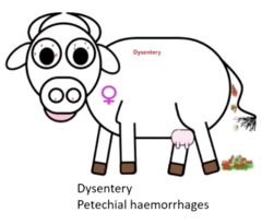
1. Sudden death, high fever (40.5-42℃), haemorrhage, bleeding from orifices, petechiae on MM, bacteraemia from secondary infections due to leukopenia
2. History of access to broken fern, CS, PM findings, prolonged bleeding + clotting times, loss of condition, anorexia
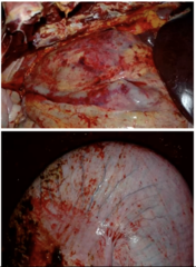
3. Haemorrhage in all tissues, red/yellow bacterial infarcts in liver/heart/kidney, ulceration of intestines with severe inflammation
Describe Bracken Fern Toxicity as a Cause of Anemia in terms of:
1. DDx
2. Treatment
3. Control
1. Babesiosis, anaplasmosis, BEF, Lantana poisoning
2. Most cases die, milder cases survive if supported by administration of broad-spectrum-antibiotics and removed from plant
3. Adequate feeding should ensure cattle do not graze bracken fern
Describe Copper Deficiency as a Cause of Anemia in terms of:
1. Definition
2. Causes
3. Clinical Signs
1. Important in the formation of haemoglobin, anaemia occurs after prolonger and/or severe periods of deficiency
2. Deficiency due to inadequate levels in diet, or are secondary due to excess molybdenum or sulphur
3. Ill-thrift, change in coat colour, chronic diarrhoea, chronic lameness, spontaneous fractures (humerus) anaemia
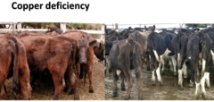
Describe Cobalt Deficiency as a Cause of Anemia in terms of:
1. Definition
2. Causes
3. Clinical Signs
1. Cobalt is a constituent of vitamin B12
2. When dietary cobalt inadequate, rumen microbes are unable to synthesize sufficient B12 → Vitamin B12 deficiency
3. Gradual loss of appetite, poor growth/loss of condition, poor coat, inappetence, muscle wasting, anaemia, death
Describe Anaplasmosis as a Cause of Anemia in terms of:
1. Definition
2. Transmission
3. Natural Immunity
4. Clinical Signs
1. Caused by Anaplasma marginale, A Arthropod transmitted rickettsial disease, Only seen in cattle tick areas (Source of infection is blood)
2. Transmitted by cattle tick or iatrogenic (needles). 3-4w incubation period
3. Young cattle are relatively resistant as they have both maternal and innate resistance. Cattle 2-4 years mostly affected by serious illness if not previously exposed/transported into endemic area
4. Anaemia, jaundice, no hemoglobinuria, fever, pale udder, acute cases die, pregnant animals abort, High RR + HR

Describe Anaplasmosis as a Cause of Anemia in terms of:
1. Diagnosis
2. Treatment
3. Control
1. Location, CS and Blood Smear showing large numbers of reticulocytes (indicative of regenerative anaemia). Small round dot like rickettsial organisms may be seen at edges of the RBCs
2. Tetracyclines, Imidocarb dipropionate.
3. Exposure to ticks carrying Anaplasma organisms in their first year of life. Vaccinate calves between 3-9m - reaction to vaccine may occur from 30-60 days after vaccination

Describe Theileriosis as a Cause of Anemia in terms of:
1. Definition
2. Pathogenesis/Clinical Signs
3. Benign Theileriosis
4. Clinical Theileriosis
5. Treatment
6. Control
1. Tick borne protozoan disease associated with Theileria spp, Spread by bush tick
2. Sporozoites are injected into host via tick saliva → Disease involves fever with varying degrees of anaemia and leukopenia
3. Infection causes slightly reduced PCV due to damage of parasitized RBCs, but not significant enough to cause anaemia
4. Caused by specific strains. Causes anaemia, lethargy, inappetence, excersise intolerance, jaundice, abortion, stillbirth (Not haemoglobinuria
5. Usually poor response. Oxytetracycline, Imidocarb, blood transfusion.
6. Maintain good bush tick control
Describe Babesiosis in terms of:
1. Definition
2. Causative Protozoa
3. Transmission
4. Prepatent Period
1. Protozoan disease, develops in RBCs and transmitted by Rhipicephalus australis (cattle tick). Disease restricted to geographical areas where cattle ticks are endemic
2. Caused by B. bovis + B. bigemina (Bigemina more prevalent in infected herds, however B.bovis more important as causes 80% of outbreaks and higher mortality)
3. Engorged adult tick picks up infection → pass to proginy via eggs → larvae then pass onto animal when feeding
4. Bovis = 6-12d, Bigemina 12-18d
Describe Babesiosis in terms of:
1. Pathogenesis
2. Clinical Signs
3. Diagnosis
1. Overproduction of cytokines causing vascular collapse and coagulation disorders, Infected erythrocytes aggregate in capillary beds causing damage to brain and lungs. Causes massive intravascular haemolysis causing haemoglobinuria
2. Haemoglobinuria, fever, anaemia/jaundice, nervous signs (circling, head pressing, aggresion, paralysis), diarrhoea, rapid weight loss, recumbancy, coma, death
3. Examination of blood smears from capillary blood (tail)
Bovis: Looks like bowtie
Bigemina: seperated pair of structures

Describe Babesiosis in terms of:
1. Post Mortem
2. Treatment
3. Natural Immunity
4. Control
1. Enlarged spleen, swollen liver, congested kidneys, jaundiced/anaemic
2. Imidocarb dipropionate, Vaccination, blood transfusion, Supportive therapy
3. In endemic areas passive immunity from maternal antibodies and innate immunity means castle exposed at a young age develop immunity. Possible to have "endemic stability" with infection presence but no clinical signs in a herd
4. Natural immunity, vaccination, genetic resistance (bos indicus), tick control

Describe Indications for Blood Transfusions in terms of:
1. Cases
2. Clinical Findings
3. PCV Values
4. BLood Loss
1. Severe haemolytic anaemia or Acute haemorrhagic anaemia event (e.g torn mammary vein)
2. Weakness, increased HR + RR, weak pulse, Cold extremities, knowledge of ongoing haemorrhage
3. PCV of
Describe Blood Transfusions in terms of:
1. Cross Matching
2. Donor Selection
3. Quantity of Blood
1. Generally, not required for first time transfusion as chance of acute reaction is low. If additional transfusions are required cross-matching is advised
2. Need to be quiet, healthy, not pregnant, large, same breed, preferible recipients dam
3. 4-6L will raise PCV from 8%-12% in a 600kg cow → of which there will be significant improvement in the animal

Describe the Process of Blood Collection in terms of:
1. Donor Collection
2. Recipient Administration
1. Sedate, secure head, apply tourniquet to raise vein → Make skin incision over jugular → collect (collection vessel must have anticoagulant → collect 5-8L (20% of blood volume can be safely collected) → Close jugular + skin → administer blood within 2h
2. Sedate, secure head, apply tourniquet to raise vein → Skin incision, insert catheter and suture into place → give first 200mL and watch for transfusion reaction (Tachypnoea, tachycardia, hives, oedema → treat any reaction with adrenaline) → if no reaction can administer → 5-20mL/kg/h

Topic 4: Conditions of the Bovine Respiratory System
Topic 4: Conditions of the Bovine Respiratory System
Describe Respiratory Anatomy in terms of:
1. Upper Respiratory Tract Structures
2. Lower Respiratory Tract Structures
1. Nasal passages, Sinus, Pharynx, Larynx, Trachea
2. Bronchial Tree, Lung Parenchyma

Describe Respiratory Disease Examination in terms of:
1. Differentiating Upper from Lower Resp Tract
2. Potential Presenting Clinical Signs
1. If respiratory sounds can be heard without using a stethoscope, they are most likely upper respiratory. can auscultate over the trachea at the thoracic inlet to distinguish
2. Polypnoea, tachypnoea, dyspnoea, coughing, crackles, wheezes, absent LS, nasal discharge, coughing blood, fever, foul breath, depression, lacrimation, loss of BCS, sudden death
Describe Respiratory Disease Examination in terms of:
1. Normal RR
2. Physiological Causes of Increased Resp Rate
3. Clinical Exam
4. Additional Special Examination/Diagnostic Techniques
5. Post Mortem Inspection
1. Normal 10-40, adults 10-30, calves 15-40
2. Environmental temperature, Exercise, pregnancy, young animals
3. Head exam, auscultation of lung field, percussion of chest wall
4. Broncho-alveolar lavage, Nasopharyngeal swabs, transtracheal wash, Bacterial culture, Radiology, U/S, endoscopy, PM
5. Cases should correlate with herd problem, early stages of disease before treatment, collect samples

Describe Bovine Bacterial Pneumonia as a Respiratory Disease in terms of:
1. Predisposing Factors
2. Primary Viral Causes
3. Secondary Bacterial Involvement
4. 3 Classifications of Pneumonia
1. Weather, Feedlot dust, Commingling, weaning, transport, ammonia levels
2. BHV-1, BRSV, BVDV: Cause Immune system alteration + mucosal erosion
3. Normal commensals with virulence factors
4. Bronchopneumonia, Interstitial Pneumonia, Metastatic/Embolic Pneumonia

Describe Bronchopneumonia as a Bacterial Pneumonia in terms of:
1. Definition/Characteristics
2. Suppurative Bronchopneumonia Distribution
3. Fibrinous Bronchopneumonia Distribution
4. CaseoNecrotic Distribution
1. Cranioventral distribution. Organisms gain entry through pulmonary tree
2. P. Multocida. Causes atelectasis. E.g enzootic pneumonia in calves
3. M. haemolytica, H. Somni. Causes marbled appearance. E.g shipping fever
4. M. bovis. Most cases combined with other bacteria. patchy areas of atelectasis, later chronic areas of caseous necrosis with nodular feel

Describe Interstitial Pneumonia as a Bacterial Pneumonia in terms of:
1. Definition/Characteristics
2. Causes
3. Secondary Interstitial Pneumonia
1. Usually non-infectious and atypical, alveolar septa damage through inhalation of causative agent, diffuse distribution
2. - Chemical compounds: Lush green pasture "fogfever" (L-tryptophan converted to 3-methylondole)
- Larvae migration
- Immunological reaction
- Gases
- Allergens
3. Broncho-interstitial combination. Rare

Describe Embolic/Metastatic Pneumonia as a Bacterial Pneumonia in terms of:
1. Definition/Characteristics
2. Causes
1. Caseous necrosis, can cause erosion of pulmonary vessels → pulmonary haemorrhage
2. Distant chronic infection (liver abscess, CVC, amstits, endometritis). Haematogenous spread. F. necrophrum common

Describe Respiratory Distress as a Resp. Disease of Young Cattle in terms of:
1. Asphyxia Secondary to Dystocia
2. Severe Respiratory Distress
3. Aspiration Pneumonia
1. Swollen head + tongue, depressed, may bellow, gasping respiration
2. Born premmy or delivered by C-section
Describe Enzootic Calf Pneumonia as a Resp. Disease of Young Cattle in terms of:
1. Definition/Characteristics
2. Viral Contributions
3. Secondary Bacterial Infections
1. Occurs most in housed dairy calves 2w-5m old. Bronchopneumonia seen, infectious agents are normal flora of the nasopharynx. Multifactorial disease of infectious agents, environmental conditions, and impaired host immunity
2. Parainfluenza type 3, BRSV, BHV-1. BVDV can contribute to suppressed immunity
3. T. Pyogenes Common

Describe Enzootic Calf Pneumonia as a Resp. Disease of Young Cattle in terms of:
1. Risk Factors
2. Clinical Signs
3. PM Findings
1. Poor ventilation, weather change, overcrowding, draughts causing chills, stress, comingling of different age groups, high humidity, Noxious gases (ammonia+methane), viral infections impairing cellular defences
2. Dull, anorexic, temp 40-40.5, coughing, mucopurulent nasal discharge, abnormal lung sounds cranio-ventally
3. Consolidation of cranioventral lung lobes, areas of collapsed, dark red lung tissue instead of healthy pink colour of normal lung tissue

Describe Verminous Pneumonia as a Resp. Disease of Young Cattle in terms of:
1. Causes
2. Cattle Affected
3. Clinical Signs
4. Post Mortem
5. Treatment/Prevention
1. Lungworm (dictyocaulus viviparus), Less common nowadays due to effective anthelmintics
2. Young cattle develop immunity. Disease usually occurs in naive cattle that are exposed to a large challenge of infestation (moved onto heaving infected pasture
3. Acute cases have rapid shallow breathing, fever, frequent coughing progressing to resp. distress. subacute cases more common, causing fever + coughing and may develop bronchopneumonia
4. Adult thread-like worms in airways, consolidation of lung tissue in caudoventral lobes, Bloody froth in bronchi and trachea
5. Broadspectrum anthelmintics, severe cases need ABs + NSAIDs. Prevent by strategic anthelmintics as animals need to acquire immunity, implement rotational grazing
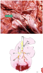
Describe The Histophilus somni Disease Complex as a Resp. Disease of Young Cattle in terms of:
1. H. Somni Characteristics
2. Associated Syndromes
1. Gram -ve rods, commensal inhabitant of nasopharynx and causes a range of disease syndromes associated with septicaemia
2. Thrombotic meningoencephalitis, repro failure, myocardial infection, joint infection, resp. disease
Describe The Histophilus somni Disease Complex as a Resp. Disease of Young Cattle in terms of:
1. Respiratory Form Characteristics
2. Thrombotic Meningoencephalitis (TME) Form Characteristics
1. URTI + LRTI. resp tract common entry point before septicemia. Occurs primarily in feedlot calves 6-12m old Causingfibrinous suppurative bronchopneumonia. Also causes fibrinous pleuritis in feedlot stears and causes acute deaths. can be part of shipping fever
2. Occurs 1-2w following resp. form of H. Somni. Animals found dead or profoundly depressed (sleeper cattle), with fever, neuro signs (ataxia, tremors, convulsions, blindness). Rapid onset and often fatal. PM shows pathognomonic red/brown foci of hemorrhage and necrosis (infacts) in the brain + spinal cord
Describe Fucobacterium necrophrum as a Resp. Disease of Young Cattle in terms of:
1. Characteristics
2. Laryngeal Necrobacillosis (Calf Diptheria)
1. Infection occurs following MM damage, ubiquitous around cattle, have differing clinical presentation
2. Necrotic laryngitis/pharyngitis, common in bucket fed calves. CS include Moist painful cough, dyspnoea, salivation, fould breath, poor response to treatment, and animal is ill-thrifty

Describe Bovine Respiratory Disease Complex (BRD) as a Resp. Disease of Adult/Grower Cattle in terms of:
1. Definition
2. Aetiological Agents
3. Conseuqences of Infection
1. Important Disease in feedlot cattle responsible for most treatments and deaths. Caused by a combination of infectious agents, environmental stressors and impaired host immunity.
2. Infectious bovine rhinotracheitis virus (BoHV1), BVDV, Bovine resp. syncitial virus, Parainfluenze type 3, M. haemolytica, P. Multpocida, H. Somni
3. Reduced weight gains, Pneumonia, dyspnoea, nasal discharge, coughing,
Describe Bovine Respiratory Disease Complex (BRD) as a Resp. Disease of Adult/Grower Cattle in terms of:
1. Primary Viral Causes
2. Secondary Bacterial Involvement
3. PRedisposing Factors
1. BHV-1, BRSV, BVDV, PI3: Cause Immune system alteration + mucosal erosion
2. Normal commensals with virulence factors (M. haemolytica, P. multocida, H. somni, T. pyogenes, M. bovis)
3. Weather, Feedlot dust, Commingling, weaning, transport, ammonia levels

Describe Bovine Respiratory Disease Complex (BRD) as a Resp. Disease of Adult/Grower Cattle in terms of:
1. Prior to Feedlot Stressors
2. Entry into Feedlot Stressors
3. After Arrival on Feedlot Stressors
1. Weaning, transport, time off feed/water, co-minging, no colostrum
2. Handling, co-mingling, pen density, weather extremes, dust, injury, adaptation to feed/water, husbandry
3. pen add-ons, pen movement/changes, mixing cattle, handling, ration changes
Describe Bovine Respiratory Disease Complex (BRD) as a Resp. Disease of Adult/Grower Cattle in terms of:
1. Individual Clinical Signs
2. Herd Observations/CS
3. PM
1. Depression, slow coming to feed bunk (aggresion score), head+neck outstretched, drooling, dyspnoea, nasal+ocular discharge, fever. Increased lung sounds with crackles and wheezes
2. Reduced interest in feeding (Decreased rumen fill), Overal decreased food intake (feed left in bunk), Drooling in water trough, coughing and increased resp. noises
3. Severe cranioventral pneumonia, fibrous adhesions between lung lobes, mav have abscesses, bronchopneumonia, Upper respiratory tract lesions

Describe the Treatment of Bovine Respiratory Disease Complex (BRD)
Specific for pulled animals, early aggressive treatment is essential for high cure rate → pen checkers/riders must be able to detect early cases.
- ABs, Preferably injectables (Tilmicosin, Oxytet, ceftiofur, Trimethoprim)
- NSAIDs (Flunixin)
- Transfer of Affected animals to suitable hospital pen with good shade, water, and palatable feed

Describe Prevention of Bovine Respiratory Disease Complex (BRD) in terms of:
1. Preconditioning Prevention
2. Backgrounding Prevention
3. Management of Feedlot Prevention
4. Vaccination
5. Mass Medication (Metaphylaxis)
1. Husbandry before weaning, vaccinate, yard weaning (train to feed from bunk), treat for parasites
2. Calves from a variety of sources are assembled and processed prior to feedlot entry
3. Minimise transport, control dust, limit mixing of cattle, limit SR, careful management, consistent ration delivery
4. Available for IMR, BVDV, and M. haemolytica. Best to vacciante prior to entry
5. Treatment of whole groups of cattle with ABs showing clinical signs of BRD, as some apparently healthy animals might already be incubating the pathogens of concerns

Describe Infectious Bovine Rhinotracheitis (IBR) as a Resp. Disease of Adult/Grower Cattle in terms of:
1. Definition/Characteristics
2. Transmission
3. Clinical Signs
1. Bovine herpesvirus type 1. Highly contagious oculonasal discharge. Limited to the URT but can predispose to more severe respiratory disease
2. Direct contact with infected cattle/feed/water contaminated with discharges from clinical/subclinical shedders. Spread in genital and respiratory secretions
3. Conjunctivitis (red eye), excessive salivation, laboured breathing/coughing, white pustular lesions on oral mucosa, diphtheritic plaques, tracheitis (white pustular lesions enlarging to necrotic/diphtheritic plaques
Describe Infectious Bovine Rhinotracheitis (IBR) as a Resp. Disease of Adult/Grower Cattle in terms of:
1. Treatment
2. Control
3. Genital Form
1. ABs and NSAIDs, Live intranasal vaccine available (rapid protection within 2-3d)
2. Hygiene + closed herds not very effective due to ease of spread and wide virus distribution
3. Causes Infectious pustular vulvovaginitis in cows and infectious pustular balanoposthitis in bulls. Spread vinearealy. CS are small pustules and mucopurulent discharge from vulva (cows), and severe ulcerative balanoposthitis in bulls. Cuases abortions and associated with mastitis

Describe Shipping Fever as a Resp. Disease of Adult/Grower Cattle in terms of:
1. Definition/Characteristics
2. Causative Organisms
3. Clinical Signs
4. PM
5. Treatment/Control
1. AKA Bovine pneumonic pasteurellosis (mannheimiosis). Sudden/acute onset following transport and characterised by severe acute exudative fibrinous pleuropneumonia. Mostly affects intensively reared cattle. Viral infection and stressors contribute to risk
2. Mannheimia Haemolytica (90% of cases), followed by P. multocida
3. Within 1m of feedlot arrival, Lasts 2-4d, sometimes sudden death. high fever, painful chest, ocular/nasal discharge, increased RR with abdominal effort, reluctant to move, Little coughing
4. Exudative fibrinous pneumonia in cranioventral lobes, fibrinous pleurisy, lungs dark red/purple and firm with extensive consolidation
5. Severe cases may die despite aggressive treatment. prevent with vaccine
Describe Caudal Vena Cava Syndrome as a Resp. Disease of Adult/Grower Cattle in terms of:
1. Aetiology
2. Clinical Picture
3. Pathology
4. Diagnostic tests
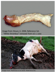
1. Complete/partial obstruction of CVC by white thrombus. Caused by liver abscesses of F. necrophorum/T. pyogenes. but can result from emboli caused by other infections. Resp disease is only 1 part of disease
2. History of no/transient improvement with AB, weight loss, fever, tachypnoea/tachycardia, suppurative nasal discharge, epistaxis, blood from mouth, faecal occult blood
3. Found dead in a pool of blood, metastatic pulmonary abscessation and haemorrhage, ascites and hepatic congestion
4. CBC shows anaemia, leukocytosis, elevated GGt + GLDH. modified transudate on abdominocentesis. Endoscopy, US, Rads
Describe Bovine Interstitial Pneumonia in terms of:
1. Acute Bovine Pulmonary Oedema and Emphysema (Fog Fever)
2. Atypical Interstitial Pneumonia
3. Plant Poisoning
1. Due to toxic amount of L-tryptophan ingestion in lush pasture → converted to 3-methylindole in the rumen → toxic injury to lungs. Causes acute resp distress, extended neck, tongue out, and acute deaths. No treatment. pretreat with ionophores before putting onto known toxic pasture
2. Cause unclear, inhalation of antigens with dust/mould suggested, BRSV possible initiator
3. Forage Brassica, sweet potato infected with Fusarium solani fungus,

Describe Pneumomediastinum in terms of:
1. Definition
2. Clinical signs
1. Leakage of air into the mediastinum. Most common in postpartum cows due to exertion during calving. Air migrates along the aorta → through the lumbar fascial planes → subcutaneous emphysema of the back
2. Dyspnoea, SC emphysema, bilateral muffled heart sounds
Describe Aspiration Pneumonia in terms of:
1. Severity
2. Clinical Signs
3. PM Exam
1. Depend on nature and amount of aspirated material. Rumen contents/irritant medications may result in severe necrotizing pneumonia within days. Smaller amounts of fluids may be resolved without problem
2. Dull, inappetence, coughing, fever, dyspnoea, toxaemia, increased RR
3. Presence of aspirated material in lungs usually obvious (needs to be distinguished from rumen contents aspirated prior to death). Over cause a severe suppurative pneumonia

Describe Lung Abscessation in terms of:
1. Definition
2. Causes
1. Occurs following bronchopneumonia of following spread form other conditions
2. Mastitis, metritis, navel-ill, caudal vena cava thrombosis. May be multiple of single abscesses
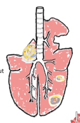
Describe Mycotic Pneumonia in terms of:
1. Definition
2. Clinical Signs
1. Fungal infection caused from eating mouldy silage/hay. Caused by Mortierella wolfii/Aspergillus
2. Pregnant cattle abort as fungus spreads to bloodstream, Pneumonia, usually fatal
Topic 5: Conditions of the Bovine Urinary System
Topic 5: Conditions of the Bovine Urinary System
Describe Examination of the Bovine Urinary System in terms of:
1. Rectal Examination
2. Urine Collection
1. Palpation of the left kidney, bladder (when full or when wall is thick), Ureters (when thickened)
2. - Catheter (Cow, use epidural)
- Manually: Stroke perineal area (don''t hold tail)
- Bulls: Preputial hair stimulated
- Diuretic: urinate in 10-20m, suitable from microbiology but composition changes

Describe Examination of the Bovine Urinary System in terms of:
1. Gross Urine Colour
2. Gross Foreign Material in Urine
1. Normally clear / yellow. red/red-brown "wine" colour = haematuria / haemoglobinuria/ myoglobinuria
2. Crystalluria (usually not significant), Pyuria

Describe Urine Strips as a Examination of the Bovine Urinary System in terms of:
1. Glucose
2. pH
3. Ketones
4. USG
5. Urea (BUN)
6. Creatinine
7. Others
1. High in clostridium diseases, parenteral glucocorticoids)
2. 7-9, often false +ve for protein when urine is alkaline
3. Commonly used to check for presence of acetoacetate (Not B-OHB, which is in plasma)
4. Usually 10.015 - 10.040
5. Produced by liver from ammonia. High levels follow intake of protein in the diet, and muscle catabolism. Not specific
6. Not influenced by diet, better guide to renal function, only rises when 60-75% of nephrons destroyed
7. Hyponatremia / hypochloremia in chronic renal failure + hyperphosphatemia in acute renal failure
Describe Nephritic Lesions in terms of:
1. Interstitial Nephritis
2. Pyelonephritis
3. Septic Embolic Nephritis
4. Granulomatous Nephritis
5. Glomerulonephritis
1. Seen at slaughtered but not usually associated with clinical disease, chronic cases result in renal atrophy and fibrosis. Classical lesion of leptospirosis and MCF
2. Always involves and infectious agent, usually originating from the lower gut or perineal skin
3. Bacteria/fungi arise from the bloodstream and localise at distant sites (kidneys)
4. Inflammatory Masses
5. Clinical Disease very rare in cattle, type 3 hypersensitivity

Describe Leptospirosis as a Kidney Disease in terms of:
1. Definition
2. Associated Syndromes
3. 2 Main Species in Australia
1. Caused by gram -ve spirochetes, many species and serovars. Important zoonosis
2. Abortion/stillbirths.weak calves, Chronic interstitial nephritis, mastitis, haemolytic crisis
3. L. Hardjobovis (Cattle Hots Species) + L. Pomona (Pig Host Species)
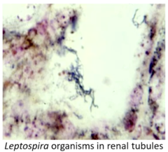
Describe Leptospirosis as a Kidney Disease in terms of:
1. Cattle as Maintenance Hosts for L. Hardjobovis
2. Cattle as Accidental Hosts for L. Pomona
Signs mild with host-adapted serovar vs. Severe with non-adapted serovar
1. Easily infection, mild/no CS, chronic disease, persist in kidney (tubules) and genital tract. Sheds, Low humoral antibody response
2. Low susceptibility, high pathogenicity. Acute, sporadic transmission between pig → cow, short kidney phase, marked antibody response, high efficacy of vaccination. Not very common due to vaccination. Risk associated with feral pigs

Describe Leptospirosis as a Kidney Disease in terms of:
1. Bacterial Characteristics
2. Pathogenesis
1. Survive long periods in moist conditions (soil, water, following flooding), extremely infectious (readily penetrates MM), Urine most important source of infection
2. Infection through MM → localised infection (2-20d incubation phase) → Bacteremia phase (Disseminated throughout the body, lasts 9d, Abs detected at this time) → Organism settles in kidneys, repro tract, CNS, Eyes → Invades immune system (convalescent phase) → sheds via urine (if Hardjobovis)

Describe Leptospirosis as a Kidney Disease in terms of:
1. L. Harjobovis CS in Cows
2. L. Pomona CS in Cows
3. L. Pomona CS in Cows
1. Clinical infection may not be apparent. Can cause transient fever, Dairy cattle (sudden drop in milk, high SCC, soft flabby udder, poor quality blood tinged milk), abortion/stillbirths/weak calves common. Most common scenario is no CS, with carrier animals shedding lepto in urine for 2y
2. Mild fever + inappetence, abortion possible from 4m, RFM, milk drop, mastitis, mild interstitial nephritis
3. High fever, interstitial nephritis and tubular nephrosis, haemolytic anaemia with red urine "red water" Haemoglobinuria, Jaundice, liver failure, death

Describe Diagnosis of Leptospirosis as a Kidney Disease in terms of:
1. Detection/isolation of Organism
2. Serology
3. Biochemistry
1. Difficult. Dark field microscopy, fluorescent Ab test, culture, PCR of foetal material
2. Microscope agglutination test, Abs in serum, rising antibody titres, herd serological surveys
3. Serum creatinine may be elevated
Describe Leptospirosis as a Kidney Disease in terms of:
1. Why Treatment is Used
2. Treatments
3. Control
1. Used for clinically sick animals, to eliminate shedding in chronically infected animals, for export animals, for semen collection centers
2. Oxytetracyclines, Ampicillin, cephalosporins, amoxicillin,
3. Vaccination, cattle vaccines usually contain Hardjobovis + Pomona serovars. Vaccinate calves from early age (from 6w, before acquired passive immunity starts to wane). Separate cattle from pigs, try to reduce watery environments

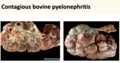
Describe Contagious Bovine Pyelonephritis as a Kidney Disease in terms of:
1. Aetiology/Epidemiology
2. Causes
3. Pathogenesis/Clinical Signs
1. Usually single animals affected, usually affects mature cows in late pregnancy. Causes chronic purulent inflammation of the bladder and kidneys.
2. Corynebacterium renale. Lives on vaginal and penile mucosa of carrier cattle. E.coli uncommon cause
3. Disease develops as an ascending infection from the lower urinary tract, carrier cows are the principle source of infection. Can commence acutely with UT obstruction and may produce colic signs (abdominal pain). More common gradual onset with loss of condition, reduced milk, small amounts of urine passed, slow urine flow, arched back following urination