Clinical Diagnosis Quiz 5 + FISHBONE + Ultrasound
 Call Kai
 Learn
 Practice Test
 Spaced Repetition
 Match
 Flashcards
 Knowt Play
1/367

There's no tags or description
Looks like no tags are added yet.
368 Terms
1. Inspection
2. Auscultation
3. Percussion
4. Palpation
***Must be in this order
What are the four components of the abdominal exam? (ie listening, tapping)
Cullen's sign (bruising around the umbilicus). This indicates that there is bleeding within the abdomen.
What is this called? What does this indicate?

Cullen's sign
_____ sign is bruising around the umbilicus that indicates that there is bleeding in the abdomen.
Grey Turner's Sign (streaky bruising around the flank). This indicates that there is some form of intra-abdominal bleeding.
What is this called? What does this indicate?

Grey Turner's sign
_____ sign is streaky bruising around the flank that signifies the presence of intra-abdominal bleeding.
Sister Major Joseph's Nodule. This is metastatic carcinoma of the umbilicus, which indicates that there is metastatic cancer in the abdomen (stomach, colon, pancreas, ovary)
What is this called? What does this indicate?

Sister Mary Joseph's Nodule
(Cancer usually originates in the stomach, colon, pancreas or ovary)
A ______ is metastatic carcinoma of the umbilicus. This is a sign of metastatic cancer in the abdomen.
Poor prognosis
***This is a Sister Mary Joseph's nodule, which is metastatic carcinoma of the umbilicus. This almost only occurs in end-stage, metastatic, intra-abdominal cancer.
Does this patient have a good prognosis or a poor prognosis?

Diaphragm
When you auscultate the abdomen, do you use the bell or the diaphragm of the stethoscope?
Malignancy
***(One study said that 10% of pts with metastatic cancer to the liver have a hepatic friction rub)
You are auscultating the RUQ and note the presence of a hepatic friction rub. What are you most concerned about?
RLQ - save the painful areas for last
A patient presents with RLQ pain. During the exam, what quadrant do you palpate last?
30 seconds
During auscultation of the abdomen, how long should you listen in a quadrant before confidently declaring that a patient has absent bowel sounds?
Absent (or hypoactive)
(Post-op ileus is a complication of surgery in which there is decreased peristaltic movement of the intestines --> decreased/absent bowel sounds)
If a patient has a post-op ileus, do you think they will have hyperactive, normoactive, hypoactive, or absent bowel sounds?
Post-op ileus
Feature | Postoperative ileus | Mechanical obstruction |
|---|---|---|
Bowel sounds | Decreased/absent | Often high-pitched initially |
Pain | Mild, diffuse discomfort | Colicky/cramping |
Distension | Diffuse | Variable |
Imaging | Diffuse bowel dilation | Dilated bowel proximal to obstruction |
Transition point | Absent | Present |
Treatment | Supportive | Depends on cause; may require surgery |
24 hours after surgery, a patient has not passed gas and has absent bowel sounds. What is the most likely cause of this patient's symptoms?
High-pitched
_____-pitched bowel sounds are suggestive of an early bowel obstruction
High-pitched bowel sounds
***Later in course, they will be diminished or absent
What type of bowel sounds might you expect to find early in a patient with a bowel obstruction?
Absent/hypoactive
***Early in the course, the bowel sounds can sometimes be high-pitched
What type of bowel sounds might you expect to find late in a patient with a bowel obstruction?
Bruits
Abdominal _____ are a blowing sound that indicate turbulent blood flow within an artery.
A midline abdominal bruit that radiates out laterally.
***An abdominal bruit that is only located in the midline is more likely to be non-pathologic (ie just listening to blood flowing through the aorta), but bruits that radiate laterally are more likely to represent vascular obstruction (ie renal artery stenosis).
What are you more concerned about: a patient with a midline abdominal bruit radiates out laterally, or a patient with a midline abdominal bruit that does not radiate.
No - up to 20% of people have non-pathologic abdominal bruits. This just represents increased blood flow through a normal vessel. This typically occurs in the midline (does not radiate laterally)
Are abdominal bruits always pathologic?
Renal artery
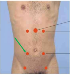
***Located 3 cm above the umbilicus + 1 stethoscope diaphragm laterally
You hear a bruit in this location. You suspect there is turbulent flow through the _____ artery.

Iliac artery
***Located diagonally from the umbilicus to the inguinal crease
You hear a bruit in this location. You suspect there is turbulent flow through the _____ artery.
Femoral artery
***Located in the inguinal crease
You hear a bruit in this location. You suspect there is turbulent flow through the _____ artery.

Right renal artery
You hear a bruit that is located 3 cm above the umbilicus and and 1 diaphragm width to the right of the midline. What artery do you think this is in?
Lungs --> Resonant
Liver --> Dull/flat
Bowel ---> Tympanic (slightly higher pitched than resonant)
When measuring the liver span, what sounds are produced when percussing over these organs?
Lungs ---> _____ sound
Liver ---> _____ sound
Bowel ---> _____ sound
13 cm
What is a normal liver span by ultrasound?
Midclavicular line
When measuring a liver span, you want to percuss down along the _____ line (anatomic line) while listening for the transition from a resonant to a dull to a tympanic sound. (lungs --> liver --> bowel)
Ultrasound machine
***Even among experienced clinicians, liver span measurements by percussion vary an average of 8 cm on the same patient. Measurement by ultrasound is much more accurate..
Who would you trust most to give you an accurate liver span measurement: Dr. Miller, Dr. Havens, Dr. Merando or an ultrasound machine?
Resonant (because you are percussing the lungs)
What percussion sound would you expect to hear superior to the upper border of the liver?
Tympany (because you are percussing the air-filled bowel)
What percussion sound would you expect to hear inferior to the lower border of the liver?
Normal - most spleens can only be palpated below the left costal margin if they are enlarged
You are unable to feel the spleen below the left costal margin. Is this normal or abnormal?
Abnormal - a spleen is enlarged if it can be palpated below the left costal margin
You are able to feel the spleen 1 cm below the left costal margin. Is this normal or abnormal?
EBV (mononucleosis)
Feature | EBV |
|---|---|
Virus | Human herpesvirus 4 (HHV-4) |
Genome | Double-stranded, linear DNA |
Envelope | Enveloped |
Family | Herpesviridae |
Transmission | Saliva (kissing) |
Target cells | B cells (CD21 receptor) and epithelial cells |
Latency | B cells |
Oncogene | EBV latent proteins (e.g., LMP1) |
Clinical associations
Disease | Classic presentation |
|---|---|
Infectious mononucleosis | Fever, pharyngitis, posterior cervical lymphadenopathy, fatigue |
Burkitt lymphoma | African endemic form; jaw mass; starry-sky appearance |
Hodgkin lymphoma | Particularly mixed cellularity subtype |
Nasopharyngeal carcinoma | Nonkeratinizing carcinoma |
Gastric carcinoma | Subset of cases |
Oral hairy leukoplakia | White, corrugated tongue lesions; immunocompromised patients |
Post-transplant lymphoproliferative disorder | Immunosuppression → uncontrolled B-cell proliferation |
Diagnosis
Test | Finding |
|---|---|
Heterophile antibody (Monospot) | Positive |
Anti-VCA IgM | Acute infection |
Anti-VCA IgG | Current or past infection |
Anti-EBNA IgG | Past infection |
Peripheral smear | Atypical lymphocytes (reactive CD8+ T cells) |
What viral infection is a common cause of splenomegaly in otherwise healthy adults?
EBV (mononucleosis)
A 18 yo female presents with a sore throat, cervical lymphadenopathy, and abdominal pain. You are able to palpate a spleen 1 cm below the left costal margin. What infection do you suspect?
Dullness
If you were to percuss the spleen, what sound would you expect?
1. Shifting dullness
2. Fluid wave
What are two tier 2 maneuvers that can detect ascites?
No - these maneuvers are only helpful for detecting large amounts of ascites
Are tier 2 maneuvers such as shifting dullness and fluid waves able to detect small amounts of ascites?
Tympany ---> dull ---> tympany
***When lying on the back, the air-filled bowel will rise to the top and will make the midline of the abdomen tympanic. The ascitic fluid is located below, and will produce a dull sound, which will become apparent as you percuss laterally from the midline.
***When lying on the opposite side, the ascitic fluid travel downwards with gravity while the air-filled bowel travels upwards, so the area that was previously dull due to the presence of ascitic fluid will now be tympanic
In the shifting dullness maneuver, when the patient is lying on their back, percussion of the midline of the abdomen would produce a _____ sound. As you percuss laterally, that will transition to a _____ sound (where fluid is present). When the patient lies on their opposite side, percussion in that same lateral area will produce a ____ sound.

30 seconds
***If you percuss too early after the patient has rolled onto their side, the ascitic fluid hasn't had enough time to travel downwards
When performing the shifting dullness maneuver, you must wait _____ seconds after the patient has rolled over before re-percussing the abdomen.

Fluid wave maneuver - used to detect large volume ascites.
What maneuver is being performed here? What is this detecting?

Involuntary guarding
***It is impossible to voluntarily contract only one side of the abdominal musculature when guarding. Unilateral guarding is always involuntary
***This is a sign of peritonitis
A patient presents to the ED with abdominal pain. When you palpate the LLQ, you notice that only the left-sided abdominal muscles contract. Is this voluntary or involuntary guarding?
Involuntary guarding
***In voluntary guarding, the abdominal contraction will usually diminish with deep breathing
A patient presents to the ED with abdominal pain. You notice that their abdominal muscles contract continuously throughout the respiratory cycle. Is this consistent with voluntary or involuntary guarding?
Voluntary guarding
***Involuntary guarding will not change with deep breathing (ie abdomen will remain rigid)
A patient presents to the ED with abdominal pain. You notice that their abdomen is rigid on examination, but when you have them take a deep breath, their abdominal muscles are able to relax and allow for palpation. Is this more consistent with voluntary or involuntary guarding?
- Have them flex their knees and hips (causes rectus abdominis relaxation)
- You can also have them take deep breaths
What is a maneuver you can do to keep a ticklish patient from contracting their abdominal muscles during abdominal palpation?
Rebound tenderness
***Expansion of the peritoneum after withdrawing the pressure causes more pain than
A patient presents to the ED with abdominal pain. You palpate deeply on their RLQ and then let go. If severe pain occurs primarily when you let go, this is an example of _____.
Peritonitis
Rebound tenderness is a classic sign that a patient has ____.
Iliopsoas test
***Test for appendicitis
***Contraction of the right psoas muscles will rub up against the appendix and cause pain
The _____ test involves having the patient flex their right hip and seeing if that causes RLQ pain.

Appendicitis
The iliopsoas test is used to test for the presence of ____.
Obturator test
***Used to test for appendicitis
***This movement causes flexion of obturator internus, which can then rub up against the appendix and cause pain
The _____ test involves having the patient flex and rotate their right hip and seeing if that causes RLQ pain.

Appendicitis
The obturator test is used to test for ____
Murphy's sign, acute cholecystitis.
***During inhalation, diaphragm expands downward, causing the inflamed gallbladder and liver come in contact with the examiner's hands. This causes immediate tenderness, which prompts the patient to stop inhaling ("inspiratory arrest")
A patient presents to the ED with abdominal pain. You have the patient exhale, then you place your fingers at the right costal margin. You then have the patient inhale. If they suddenly develop pain and then abruptly stop inhaling, the patient has a positive _____, which indicates that they may have acute _____.
Ultrasonographic Murphy's sign
A patient is having a RUQ ultrasound. The technician finds the gallbladder, presses down, and then asks the patient to take a deep breath in. If this causes tenderness and inspiratory arrest, the patient has a _____.
Acute cholecystitis
Murphy's sign is frequently seen in patients with ____.
Courvoisier's sign, malignancy
***A gallbladder should not normally be palpable (if it is palpable, it is enlarged). Since it is nontender, it is more likely to be malignancy instead of gallstones
A patient presents to the ED with jaundice. When you are finding the liver's edge, you are able to palpate the gallbladder, but this doesn't cause the patient any pain. This is called _____ sign, which is concerning for ____.
Jaundice + non-tender gallbladder
***Highly concerning for malignancy
Courvoiser's sign is an exam finding in which the patient has _____ (skin finding) as well as palpable, non-tender ____.
Malignancy
***Jaundice + non-tender gallbladder
A positive Courvoisier's sign is concerning for ____.
Umbilicus --> RLQ (specifically McBurney's point)
Pain from appendicitis often starts in the ______ region then moves to the _____ region
Back
Pain from retroperitoneal organs (pancreas/aorta, etc) often radiates to the _____.
Acute pancreatitis
A patient presents with epigastric pain radiating to the back. This is a classic presentation of ____.
1. Obstruction of a hollow tube (ie SBO)
2. Stretch/irritation of a visceral capsule (ie liver capsule)
3. Irritation of a serosal surface (ie peritonitis)
4. Vascular obstruction or lesion (ie mesenteric ischemia)
5. Abdominal wall injury (ie stab wound)
What are the 5 physiologic causes of pain that originates in the abdominal pain?
1. Obstruction of a _____
2. Stretch/irritation of a _____
3. Irritation of a _____ surface
4. _____ obstruction of lesion
5. _____ injury
(Dr. Miller abdominal pain slide #8)
Colicky
***Colicky pain = sharp pain that comes in waves. When there is an obstruction, the body responds by stimulating waves of contractions in order to get the obstruction to pass. This causes sharp waves of pain.
Patients with abdominal pain caused by obstruction of a hollow tube (SBO, appendicitis, kidney stone) will often have pain that is described as "_____"
Colicky pain
"______" pain is sharp pain that occurs in waves.
Glisson's capsule
What is the fancy name for the capsule around the liver?
Liver, spleen, kidney
What are three organs in the abdomen that are surrounded by a visceral capsule?
Stretching of the hepatic capsule (Glisson's capsule)
***Right heart failure causes blood to back up into the IVC and hepatic veins, causing congestion and swelling of the liver. The rapid swelling of the liver stretches the liver capsule, which causes pain
`
***The liver parenchyma does not have pain fibers, so the damage to the liver tissue is not the cause of the pain, it is the stretching of the surrounding capsule that causes the pain
A patient with known right heart failure presents with acute lower extremity and abdominal edema and RUQ pain. If this RUQ pain is a direct result of his heart failure, then what is the exact cause of his pain?
Liver capsule
***The liver capsule is the only part of the liver that has pain fibers. Damage to the rest of the liver tissue will not cause pain
Which is innervated by C-fibers: liver parenchyma (ie hepatocytes) or the liver capsule?
No - only acute stretching of capsules (liver, kidney, spleen) will cause pain
***This patient's liver grew so slowly that the capsule was able to slowly stretch and accommodate the growth of the liver without causing pain
A patient with non-alcoholic steatohepatitis has a liver that has slowly enlarged over several years. Would you expect this patient to have RUQ due to stretching of the liver capsule (Glisson's capsule)?
Absent bowel sounds
***Stomach and intestines will often stop moving in the setting of peritonitis or other severe abdominal injury
A patient has peritonitis from ruptured appendicitis. What type of bowel sounds would you expect to hear?
Guarding
***(this would specifically be voluntary guarding)
A patient presents to the ED with abdominal pain. When you go to press on her abdomen, she swats your hand away. This behavior is known as ____.
d. Lactic acid
***Since this patient has ischemic bowel, the lactic acid would go up due to the shift from aerobic metabolism to anaerobic metabolism.
***Abdominal ultrasound would not be able to show the embolism/plaque in the mesenteric arteries.
Btw, this is taken directly from Dr. Miller's powerpoint
You suspect that a patient has acute mesenteric ischemia. Which of these tests would be most helpful for confirming this diagnosis?
a. Serum lipase
b. Abdominal ultrasound
c. CBC
d. Lactic acid
e. LFTs
Women, diabetics
What two patient populations are more likely to have abdominal manifestations of a myocardial infarction?
Left lower lobe pneumonia.
***Pneumonia in the lower lobes often refers to the upper abdomen
A patient presents with a fever, productive cough with green sputum, shortness of breath and LUQ pain. Abdominal exam is normal except for some mild LUQ tenderness. What do you think is the most likely cause of his LUQ pain?
Myocardial infarction
A 77 yo female with a history of HTN and type II diabetes presents with acute epigastric pain, nausea, shortness of breath, and pain behind her scapula. What potentially life-threatening, extra-abdominal cause of her pain must be ruled out before obtaining an abdominal CT?
PM BAD LUNCH
***
Porphyria
Mediterranean Fever
Black widow spider bite
Addison's disease
Diabetic ketoacidosis
Lead poisoning
Uremia
Narrow angle glaucoma
Calcium (hypercalcemia)
Hereditary angioedema
What mnemonic is used to remember the metabolic and other unusual causes of abdominal pain?
Familial Mediterranean Fever
(Mediterranean Fever = "M" of PM BAD LUNCH) mnemonic for rare causes of abdominal pain
______ is a genetic condition characterized by mutations in the pyrin protein, which leads to episodes of recurrent fevers and inflammation/pain of the chest, abdomen, and joints.
Colchicine
Abdominal pain caused by familial Mediterranean fever responds well to treatment with _____.
Black widow
(B of PM BAD LUNCH mnemonic)
What type of spider bite is known to cause abdominal pain if the venom reaches systemic circulation?
Addison's disease (adrenal insufficiency)
(A of PM BAD LUNCH mnemonic)
______ is a more rare cause of abdominal pain caused by decreased production of cortisol.
Diabetic ketoacidosis (DKA)
(D of PM BAD LUNCH mnemonic)
What complication of diabetes is notorious for causing abdominal pain?
Uremia
(U of PM BAD LUNCH mnemonic)
______ is a metabolic cause of abdominal pain and confusion that is caused by renal failure.
Narrow angle glaucoma
(N of PM BAD LUNCH mnemonic)
***Mechanism is weird: In response to the build up of pressure in the eye, the pupil constricts in order to decrease some of the pressure in the canal of Schlemm (which hopefully increases some drainage of aqueous humor --> decreased intraocular pressure). This pupillary constriction is mediated by the parasympathetic nervous system. The stimulation of the parasympathetic nervous system causes overactivation of the Vagus nerve --> overstimulation of the GI tract --> abdominal pain.
_____ is an eye condition that is known to be a rare, weird cause of abdominal pain.
Hypercalcemia
(C = calcium part of PM BAD LUNCH mneumonic)
***Stones, groans, and psychiatric overtones
What electrolyte abnormality is a rare cause of abdominal pain.
Hereditary angioedema
(H of PM BAD LUNCH mnemonic)
***These patients usually have a mutation in the C1 esterase enzyme, which normally prevents overactivation of the complement system and regulates the production of kallikrein. Mutation causes overproduction of kallikrein, which leads to overproduction of bradykinin --> edema)
A patient reports that they have had recurrent episodes of severe, crampy abdominal pain, nausea, and vomiting for the past several years. They have also noted random episodes of diffuse swelling of the eyes, lips, and extremities after minor trauma or infections. What underlying genetic condition involving the complement system do you think might be contributing to this patient's symptoms?
Heme oxygenase
Biliverdin reductase
Heme is converted to biliverdin by the enzyme _____
Biliverdin is converted to bilirubin by the enzyme _____.
Albumin
***Unconjugated bilirubin is hydrophobic --> needs to bind to albumin to be transported in blood
What plasma protein does unconjugated bilirubin bind to?
1. Uptake
2. Conjugation
3. Excretion
***Excretion is the rate limiting step
What are the three steps of bilirubin metabolism in the liver?
Excretion
What is the rate limiting step of bilirubin metabolism in the liver? (uptake, conjugation, or excretion)
No
***Jaundice in hemolytic anemia is caused by overproduction of unconjugated bilirubin from the breakdown of hemoglobin. Unconjugated bilirubin is hydrophobic, so it is bound to albumin, which means that it cannot be filtered through the glomerulus and end up in urine.
urine urobilogene will increase
A patient presents with jaundice due to hemolytic anemia. Do you expect his urinalysis to be positive for bilirubin?
Conjugated bilirubin
***(this is the form that is not bound to albumin, so it can be filtered from the blood into the glomerulus and end up in urine)
Which form of bilirubin can enter the urine if it is present in the blood stream: unconjugated bilirubin or conjugated bilirubin?
Haptoglobin: Low
LDH: High
Reticulocyte count: High
***Haptoglobin: Destruction of RBCs --> release of free Hgb into plasma. Haptoglobin binds to the free hemoglobin, forming Hgb:haptoglobin complexes. This uses up haptoglobin in the plasma --> low levels
LDH = enzyme within RBCs. When RBCs are destroyed, this enzyme is released into plasma --> high
Reticulocyte count is high because the bone marrow is responding to the anemia by increasing the synthesis of RBCs, which causes increased release of immature reticulocytes into the blood
A patient has hemolytic anemia. What changes would you expect with these labs (high vs low):
Haptoglobin: ____
Lactate Dehydrogenase: ____
Reticulocyte count: ____
Unconjugated hyperbilirubinemia
***Transfusion mismatch --> hemolysis --> increased production of unconjugated bilirubin
A patient with type A blood is transfused 1 unit of type B blood. Would this cause conjugated or unconjugated hyperbilirubinemia?
Unconjugated bilirubin.
***Both of these conditions cause microangiopathic hemolytic anemia. Hemolysis of any type would cause unconjugated hyperbilirubinemia
Would conditions such as TTP and DIC cause conjugated or unconjugated hyperbilirubinemia?
Unconjugated bilirubinemia
***Both of these conditions involve mutations/deficiencies of enzymes involved with bilirubin conjugation. If bilirubin cannot be conjugated, there is a build up of unconjugated bilirubin
Would genetic conditions such as Gilbert Syndrome and Crigler-Najjar syndrome cause conjugated or unconjugated hyperbilirubinemia?
Total bilirubin > 2.0
*** In order to get these physical exam findings, the total bilirubin must be at least:
Scleral icterus t.bili > 2.0
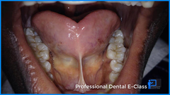
Sublingual icterus: t. bili > 5.0
Tympanic membrane icterus: t. bili > 10
Based on this physical exam finding, you suspect that the patient has a total bilirubin of at least _____.

Scleral icterus: > 2.0
Sublingual icterus: > 5.0
Tympanic membrane icterus: > 10.0
In order for a patient with jaundice to exhibit these physical exam findings, their total bilirubin must be at least:
Scleral icterus: T. bili > ____
Sublingual icterus: T. bili > ____
Tympanic membrane icterus: T. bili > ____
> 5.0
***Scleral icterus t.bili > 2.0
Sublingual icterus: t. bili > 5.0
Tympanic membrane icterus: t. bili > 10
In order for a patient with hyperbilirubinemia to exhibit this physical exam finding, they must have a total bilirubin of at least ____.
> 10.0
Scleral icterus t.bili > 2.0
Sublingual icterus: t. bili > 5.0
Tympanic membrane icterus: t. bili > 10
In order for a patient with hyperbilirubinemia to have tympanic membrane icterus, they must have a total bilirubin of at least ____.
1. Rotor Syndrome
2. Dubin Johnson syndrome
Feature | Rotor syndrome | Dubin-Johnson syndrome |
|---|---|---|
Bilirubin | Increased conjugated bilirubin | Increased conjugated bilirubin |
Inheritance | Autosomal recessive | Autosomal recessive |
Defect | Impaired hepatic bilirubin uptake/storage | Defective MRP2 canalicular transporter |
Gene | SLCO1B1 / SLCO1B3 | ABCC2 |
Mechanism | Decreased hepatic reuptake of conjugated bilirubin | Impaired secretion of conjugated bilirubin into bile |
Liver appearance | Normal | Black pigmented liver |
Symptoms | Usually asymptomatic, mild jaundice | Usually asymptomatic, mild jaundice |
Prognosis | Benign | Benign |
What are two genetic conditions that cause isolated conjugated hyperbilirubinemia with normal LFTs?
Conjugated hyperbilirubinemia with normal LFTs
***Rotor's syndrome = mutation in proteins that transport conjugated bilirubin into bile. Causes build up of conjugated bilirubin, but no hepatocyte death (no AST/ALT elevation)
***Rotor's syndrome does not cause a build up of pigment within hepatocytes, so the liver does not turn black (like it does in Dubin Johnson syndrome)
If a patient has Rotor's syndrome, would you expect them to have conjugated or unconjugated hyperbilirubinemia? What would happen to their LFTs?
Conjugated hyperbilirubinemia with normal LFTs.
***Dubin Johnson syndrome = mutation in proteins that transport conjugated bilirubin into bile. Causes build up of conjugated bilirubin, but no hepatocyte death (no AST/ALT elevation). Unlike Rotor's syndrome, this causes a build up of pigments in hepatocytes, causing the liver to be black.
If a patient has Dubin Johnson syndrome, would you expect them to have conjugated or unconjugated hyperbilirubinemia? What would happen to their LFTs?