Discovery and Fundamental properties of X-Rays, Radiological Effects and Theory of Risk Management
 Call Kai
 Learn
Learn Practice Test
 Spaced Repetition
 Match
Match Flashcards
 Knowt Play
1/139

There's no tags or description
Looks like no tags are added yet.
Name | Mastery | Learn | Test | Matching | Spaced | Call with Kai | Chat |
|---|
No analytics yet
Send a link to your students to track their progress
140 Terms
Who discovered X-rays and in what year?
Wilhelm Röntgen in November 1895.

What material fluoresced in Röntgen's initial X-ray experiment?
Barium platinocyanide-coated paper.

What is the propagation speed of X-rays in a vacuum?
The speed of light (3 × 10⁸ m/s).

Why can X-rays not be focused by optical lenses?
They are high-energy photons that do not refract like visible light.
What electric charge do X-ray photons carry?
They are electrically neutral (zero charge).

What determines the maximum energy in a polyenergetic X-ray beam?
The peak kilovoltage (kVp) selected.
What two mechanisms account for X-ray beam attenuation?
Absorption and scattering.
How does X-ray scatter affect photographic film or detectors?
It causes fogging and reduces image contrast.
Who was the first person known to die from X-ray exposure?
Clarence Dally in 1904.
Which scientist won a Nobel Prize for discovering X-ray genetic effects?
H.J. Muller in 1927.
What are the complementary DNA base pairs?
Adenine with Thymine, and Cytosine with Guanine.

What happens if a cell fails to repair significant DNA damage?
It undergoes apoptosis (programmed cell death).

What are deterministic radiation effects?
Effects caused by cell death with a dose threshold; severity increases with dose.
What are stochastic radiation effects?
Probabilistic effects without a threshold; chance increases with dose, but severity does not.
Which health outcome is an example of a stochastic radiation effect?
Radiation-induced cancer.
What does LD₅₀ signify in radiation biology?
The radiation dose that is lethal to 50% of a population.

What are the radiation thresholds for the three main Acute Radiation Syndrome (ARS) forms?
Hematopoietic (1 Gy), Gastrointestinal (6 Gy), and Neurovascular (30 Gy).

Which human cell types have the lowest radiosensitivity?
Nerve cells and muscle cells.
Which human cell types have high radiosensitivity?
Blood cells, thyroid cells, eye lens cells, and gonadal cells.

What do DICOM and PACS standards facilitate in radiology?
Standardized storage, management, and transmission of medical images.
How is Risk conceptually calculated?
Risk = likelihood × severity.
What is Risk Management in clinical imaging?
The systematic process of identifying, assessing, and controlling risks to minimize harm.
What are the 5 steps of Risk Management?
1. Identify hazards 2. Assess risks 3. Control risks 4. Record 5. Review.
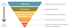
What is the Hierarchy of Control from most to least effective?
Eliminate hazard, substitute, engineering controls, administrative controls, PPE (last resort).
What does ALARP stand for in radiation safety?
As Low As Reasonably Practicable.
What does 'reasonably practicable' mean in the ALARP principle?
Further risk reduction is not required if effort/cost is grossly disproportionate to the benefit.
Which legislation principle is supported by ALARP in radiography?
The IR(ME)R principle of dose optimization.
What incident reporting software is commonly used in the NHS?
Datix.
What equipment was Röntgen experimenting with when he noticed the platinobarium screen glowing?
A cathode ray tube.
What was the result when Röntgen placed his hand between the tube and the coated screen?
He realized he could see the bones.
What other items did Röntgen explore with his rays?
A spool of wire inside a case and a compass in an enclosed box.
Who placed her hand on the photographic plate to produce the first x-ray?
Anna Bertha Lüdwig-Röntgen, Röntgen's wife.
How quickly did x-rays take the world by storm after their discovery?
Within a month.
In what year were x-rays first used to identify a Colles fracture and in early radiotherapy?
1896.
Who discovered the electron in 1897?
Thomson.
Who discovered radioactivity in 1896?
Bacquerel.
Who discovered radium and polonium in 1898?
The Curies.
X-ray travel path
Straight line; precise positioning is essential to prevent distortion, magnification, or missed anatomy.
X-ray penetration
Depends on incident photon energy and the density/thickness of the penetrated object.
Impact of incorrect kVp levels
Too low results in insufficient detector signal; too high causes over-exposure.
Three interactions of X-rays with matter
Absorption, scatter, and transmission.
Role of absorption in X-ray imaging
Provides adequate contrast as bone, soft tissue, and air absorb different amounts of radiation.
Effect of X-ray scatter
Creates noise and increases radiation exposure, which practitioners attempt to reduce.
X-ray ionising nature
Interacts with detectors and biological tissue to produce images but carries risks that require regulation.
Mass and charge of X-rays
X-rays have no mass or charge and are not influenced by electromagnetic fields.
Polyenergetic nature of X-rays
The beam contains photons of a variety of energies and must be managed for dose reduction and image quality.
Manipulation of X-ray beams
Cannot be focused by optical lenses due to high energy and short wavelength, requiring mechanical manipulation.
Fluorescence and biological changes from X-rays
Causes materials to fluoresce (used in imaging) and causes biological changes (used in therapy with side effects).
X-ray safety indicators
Signs and warning lights must indicate active X-rays because the beam is invisible to the human eye.
Clinical Governance Pillars
Consists of 7 pillars that guide healthcare quality, safety, and professional standards.
HCPC Student Guidelines on Risk
Take steps to limit harm, follow risk policies, avoid danger, and seek support if health affects performance.
Clinical Relevance in Radiography
Protects patients and staff from harm, maintains public trust, and promotes legal and professional compliance.
Benefits of Risk Management
Proactively identifies risks, ensures consistent standards, reassures patients, and builds a positive safety culture.
Five Steps of Risk Management
1. Identify risks 2. Assess likelihood 3. Implement controls 4. Record changes 5. Review and monitor.
Radiology Hazard Examples
Radiation exposure, patient falls, equipment failure, manual handling injuries, and documentation errors.
Controlling Risks in Radiology
Using lead screens, appropriate manual handling training, and regular equipment maintenance.
Recording and Reviewing Findings
Completing Datix reports, conducting audits on risk and control measures, and using errors as learning points.
Logarithmic Scales
Increments increase by multiples of 10, allowing pattern identification across a wide range of data.
Radiographer's Role in Safety
Following policies, reporting hazards and near-misses, participating in audits, and reflecting on incidents.
Rectangular collimation
Restricts and confines the X-ray beam to minimise radiation exposure.
Focus-to-skin distance
At least 20cm, but preferably 30cm.
Use of film holders
Allows for precise positioning of the film.
Digital receptors vs conventional film
Digital receptors typically require lower radiation doses.
Minimising image number
Reduce the number of images to the minimum required for diagnosis.
Limiting CBCT usage
Limit the use of cone beam computed tomography.
Radiograph justification recording
Must always be recorded in patient notes, as required under IR(ME)R.
Harmful effects of early X-rays
erythema (skin reddening) and burns; early reports of injury—previously thought nonexistent—arose from patients, tube manufacturers, and predominantly those delivering X-rays to patients, with Clarence Dally becoming likely the first fatality.