M114 part 1
 Call Kai
 Learn
 Practice Test
 Spaced Repetition
 Match
 Flashcards
 Knowt Play
 Podcast
1/105

There's no tags or description
Looks like no tags are added yet.
106 Terms
Phi and Psi backbone
stabilized by backbone hydrogen bonds (180 degrees beta), Phi (between C-N), Psi(between C-C), rigid and can only rotate

Cis and trans peptide bonds
They got to stay away in trans structure, in cis structure it has double bond(proline) becomes 2-D makes a u-turn

Types of secondary structure
Repetitive: intera peptide H bond helics(3_10—>a—>Pi), Interpeptide H bond helices structure: B strands, antiparallel or parallel B sheets
Non Repetitive: random, happen to be there
Beta Turn
2 hydrogen bonds, stabilize it, minimum 4 amino acids, secondary structure
Helics with intra peptide H bonds (folds H bond within)
3_10(1AA+AA4) distorted, alpha(AA1+AA4) NEAR PERFECT, Pi(AA1+AA5)distorted

Alpha Helix
very stable
number of H bonds-total aa-4
180 degrees for every H-bond
favorable delta S
positive at NH termibak end
negative at CO terminal end
5.4 A per turn, 3.6 residues per turn
upside down Xmas tree
NPPN
Beta sheets
1. Extended, pleated structure
2. H-bonds between adjacent β-strands
3. Parallel = same N→C direction
4. Antiparallel = opposite N→C directions
5. Antiparallel H-bonds more linear/stronger
6. Side chains alternate above/below sheet
7. ~3.5 Å rise per residue
8. Prefer V, I, C, Y, W, F, T
9. β-sheets usually right-handed twist
10. Parallel = angled H-bonds; antiparallel = straighter H-bonds
Ramachandran plot

Protein Structures

Barrel vs twist
Similar secondary structure elements, comp etely different tertiary structures

Tertiary structure
Tertiary Structure: Stablized by Interior Packing

Stablizing the Protein Fold
Add: reversable :disulfide bridge cofactor binding glycosylation phosphorylation acylation ADP-ribosylation carbamylation N-acetylation
Stablization Strategy
By disulfide bond bovine pancreatic trypsin inhibitor or By metal coordinate covalent bonds Bacterial subtilisin

Stablization Strategies
Cofactor Binding: NADP or NADPH bind to active site and change tertiary structure

Intermediate Levels of Protein Structure
Primary • Secondary • Domain — A compact assembly of secondary structures that fold into a stable entity. • Tertiary: it can consist of multiple domains • Quaternary
Motif
Definition: — Characteristics in structure or sequence, may be a whole domain or an arrangement of secondary structure element
A Motif can be as little as three amino acids (The Catalytic Triad of protease, D-H-S)
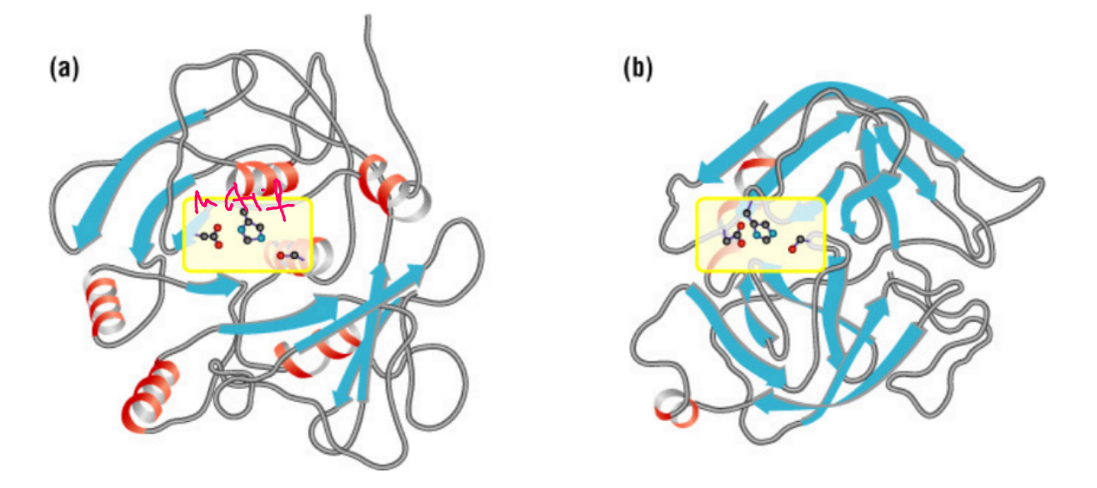
motif can consist of a few secondary structures
Helix turn helix, DNA binding motif, Major groove

Homeo-domain:
helix- turn-helix motif that binds DNA and determines our development
2 major alpha domain motifs
cytochrome(A.S top and bottom)
Globin fold( A.S inside near top)

The Hairpin Motif
No active site

Up and Down motif
A.S at top or bottom and 2 domains
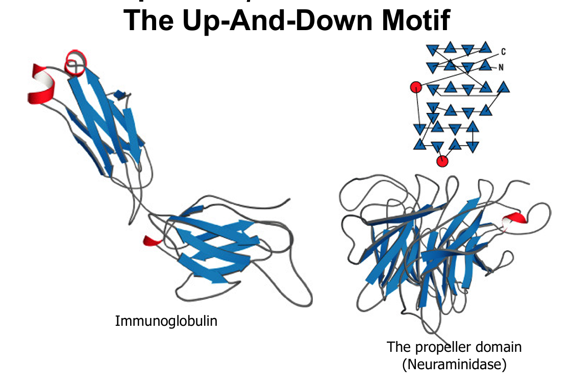
Plain b Barrel
active site in the middle

The Greek Key Motif
top or bottom A.S

The Jelly Roll
A.S top or bottom

Anti-parallel a/b Domain Motif The Saddle
A.S under beta sheet

Anti-Parallel a/b Domain Motif
Metal Ions and Disulfide Cross Links

The Zinc Finger Motif
a helix + antiparallel b 2 His + 2 Cys to one Zinc Binds DNA major groove

a+b Domains: Parallel b Strands Connected by a helices or loops

The Parallel Beta Sheet
TIM barrel or twist

The TIM Barrel
only 8 strands to have sufficient twist so that the end strand can roll back on itself and close.

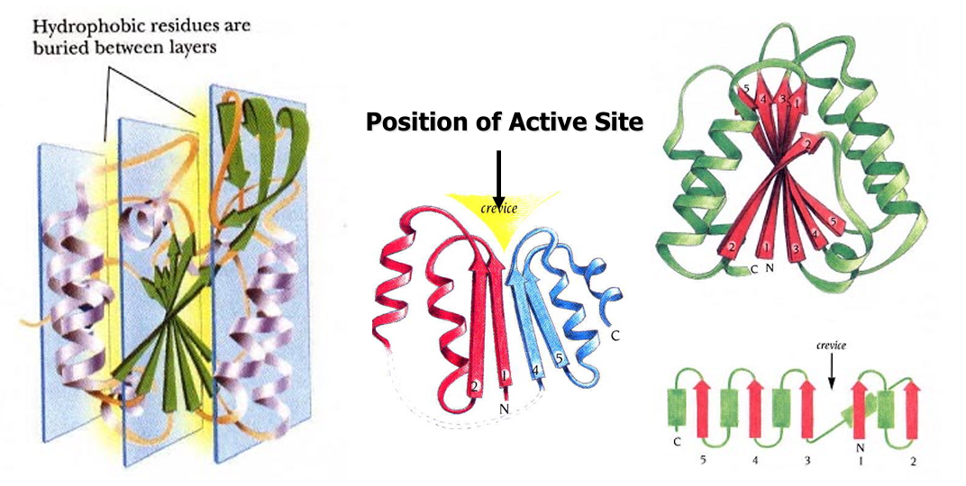
The b Twist
The alternating a / b sheet, usually < 7 b Common for nucleotide binding proteins (NADP, ATP or GTP)
Quaternary Structure
Quatemary Structure Definition — Quaternary structure refers to the number, type, and arrangement of subunits, which self-assemble into an oligomer in solution under physiological conditions. • Subunit Definition — Subunits are non-covalently associated polypeptide chains that are each folded into a structure that is a stable entity by itself. Subunits are independent units with respect to structure but not function.
Surface complementation is key to quaternary structure formation
One single mutation chan ed the quaternary structure of
In beta subunits change from ASN to VAL or Lue to Val (hydrophobic)
Motion Found in Proteins
Fluctuations, collective motions, triggered conformational changes
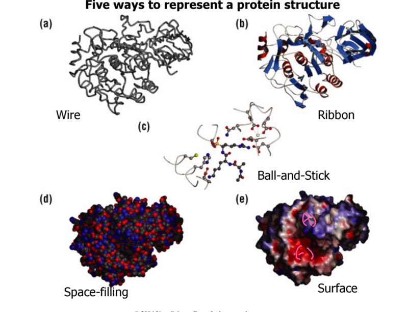
Five ways to represent a protein structure
wire, ribbon, ball and stick, space filling, surface
Mutiple confirmations
Purified protien—> NMR spec—> resonance assignment—> protien structure
Four major steps in crystallization
Obtain large amounts of pure protein samples • Choose a protein buffer in which the protein is both soluble and stable • Bring protein solution to supersaturation where spontaneous nucleation can take place • Crystal growth now begins
Sitting drop vapor diffusion
slowly evaporating away the water so protien concentration goes up and salt concentration goes up

How to Judge a Crystal Structure
Resolution (better than 3 A) • Rmerge of data (< 10% overall, <50% highest resolution shell) • Rfree of model (<25%) • Ramanchadran plot (no disallowed region { SUM UP TO 100%}
Membrane Proteins: all-alpha or all-beta

Trans-membrane proteins
All-beta (polyto ic): porin, transporter • All-alpha: — Rhodopsin — G-protein coupled receptor (GPCR) — Channel — Transporter/pump, e.g. ATP Binding Cassette (ABC) — Photosynthesis system — Protease, ATPase, oxidoreductase, respiratory enzymes — Toxins
Matching the membrane thickness with a helix
1 residue rise in helix: 1.5 To span the membrane, we need 30/1.5 = 20 aa
Matching the membrane thickness with ß strand
It has to be anti-parallel Inter-residue distance in ß stra porin : 3.5 A span the memb ne, 30/3.5 = 8 aa
The Calcium ATP se, El state
4 domains: 1st transmembrane(connects Ca²+ charged ions), 2nd A domain, 3rd P(phosphate) domain, 4th N domain (ATP bond)

The Calcium ATPase E2 state
ATP bind E1—>E2 thru phosphorylation, close to active sit (p) change of confirmation hairpin(N) and at bottom of N is ATP bind

Pumping Ca

The selectivity filter

How did K+ Channel prefer K+, not Na+
K+-protein interaction > desolvation penalty —> Go through channel
Na+-protein interaction < desolvation penalty —> Blocked

Transported by Repulsion
Selection filter can bind four K+ Charge-charge repulsion: push K+ outwar

What is Drug Design?
A multi-step process that combines many scientific disciplines to create chemical compounds that can benefit our health
Structure-Based Drug Design:
Docking & Scoring.: 1) A protein structure, 2) A small molecule database, 3) a reliable software

Criteria for Druggable Pockets
shape—> cave or minimal depression
hydrophobic interior—> higher than avg of surface exposed hydrophobic area
Size= usually 200 to 600 A
Fill the cavity w/ binding
Once hit 500 things dont work
binding specificity: alcohol dehydrogenase (blue, not used to increase amount of alcohol pieces)—> isozyme: red high alcohol processing—> isozyme binds to active site (5 H-bond 10^5 between drug and enzyme)
blue: isophone 2= lost 2H-bonds [3H bonds 10^4 pocket= drug doesnt work)

Fragment based method
compound almost not exactly like drug lead
separate drug—> chemiotaxis—>into 3= fit in enzume binding pocket
Benzyne—> delta G—> carbon group=then choose what docks in cavity[chemiotaxis]
[FlexX—> join in LUDI—> DOCK]

HIV protease:HIV Protease: Dimeric, Acid Protease
RNA virus= HIV protease—> breaksdown peptide bond of protein
protease inhibitor—> unbound—> less energy to adapt to binding site—> H-binding—>active site—>H-bond w/backbone—>improved binding—>mutation= weaken binding
Homodimer—>top(beta hairpin—>confirmational change=open for drug to enter/bind)
Asp(binding site), flap (conf.change), red color acid over Nose (active site)
tetrahedral

Two elements of a good drug
enzyme recruits own substrait= protein (polypeptide) memic
transition state analog (TSA)
Specificity pocket
every protease has a favored amino acid [Trypsin—> cuts after positivily charged residues, chymotrypsin—> cuts after larg hydrophobic residues, Elastas—> cuts after small hydrophobic residues] (lys, arg, asp, benzene ring)

Break scissile bond
between p1 and p1’= create procket to accomedate

good drug—> transition state (look alike tetrahedral)
enzyme binds tightest to transition state (high)=lower than 100,000 fold w/ enzyme
The drug needs to be tetrahedral
HIV protease #1 factor
pocket for Phe group —>and Pro group—> drug need Pro and Phe(p1 and p1’)
Combine drugs
cocktail to confuse HIV protease
COVID Protease
3 CL6^pro, M^pro
homo dimer—> 2 active sites
SARS- COV2: P1—>Q, P1’—> S
N3 p1 and p1’ (glu, sre)—> cavity phe ring
well defined tightly bond to active site of enzyme
Substrate pockets
fully different only N3 and SARS-COV2 can bind togther
virtual screening: IC—> invitiro assay
1:1 suicide inhibitor
drug vs protien
kill 1 of each other
Mpro Substrate Specificity
cut between Q and S
Q= Glutamine, S(serine), p2 = Leucine
P1’= T, S or A

What is a suicide inhibitor?
An inhibitor that forms covalent bond(s) with the active site Cys or Ser. In this case, a Michael Addition • First one is penicillin
. Explain, in one sentence, how sodium channel would bind only sodium but not potassium
Na solvent penalty < Na-channel interaction, while K-solvent penalty > K-sodium channel interaction
Backbone Hydrogen Bonds
α-helix: H-bonds = n − 4
50 aa → 50 − 4 = 46 H-bonds
Antiparallel β-sheet: 8 strands × 8 aa → 49 H-bonds
Secondary Structure Prediction
α-helix: E, Q, A, L, M, K, R
β-strand: V, I, C, Y, W, F, T
Loop/turn: G, P, N, S, D
VSVSVSVS → antiparallel β (professor's key)
GPGGP → loop (Gly/Pro favor turns)
Hydrophobic Effect
Nonpolar exposed to H₂O → ordered water → ΔSsolvent ↓ → ΔG ↑ (positive) → exposure unfavorable.
ΔG → Binding Constant
ΔG = −RT ln K
R = 0.001987 kcal/mol·K; T = 298 K
Fold change = e(ΔΔG/RT)
Loss of 10 H-bonds (+14 kcal/mol) → K decreases ≈ 10¹⁰-fold
cDNA → Protein Molecular Weight
MW (kDa) = [(bp ÷ 3) × 110] ÷ 1000
40 kb → 40,000 ÷ 3 × 110 ÷ 1000 ≈ 1,467 kDa
Shortcut: kb × 36.7 ≈ kDa
β-sheets vs. α-helices
α-helix H-bonds form within the same continuous helix (residue \(i\) to \(i+4\)).
β-sheet H-bonds form between neighboring strands, which can be far apart in the primary sequence.
Parallel vs. Antiparallel β-Sheets
Feature | Parallel | Antiparallel |
Direction | Same → → | Opposite → ← |
H-bonds | Angled | More linear |
Stability of H-bonds | Generally less optimal geometry | Generally more favorable geometry |
Connections | Often longer crossover loops | Can form short β-hairpins |

The Four Strategies of Signal Transduction:
Specificity, Amplification, Adaption, Integration
Six Mechanisms of Signal Transduction
GPCR, receptor tyrosine kinase, guanylyl cyclase, adhesion receptor, gated ion channel, nuclear receptor (no membrane bound)
Kinase inhibitor as cancer drug design:
ATP mimic
Lock kinase in an inactive conformation

SH3 Domain: binding Pro-rich helix
no cavity, SH3 bind proline, antiparallel beta sheets
SH2 Domain: binding P-Tyr
SH2—> PHOSPHORALATED TYROSIN
An protein. Mixed: electrostatic + H-bonds + hydrophobic
PTB
Binds to P-Tyrosin, ß-barrel with a helical cap Mixed: electrostatic + H-bonds + hydrophobic
PH: binding IP3 (membrane)
binding IP3 (membrane)
Two layers Of anti-parallel II-sheets with a helical cap.
Diabetes
Why did Mario Puzo's eyesight decayed? diabetes affecting eyesight
• What may aggravate de res ion of Hemingway? toxic iron deposit and hemochromtosis
• Think of the above in terms of our body organs that mainly use glycolysis (aka the breakdown of glucose) for energy? eyes, brains and kidneys
INSULIN
Insulin receptor gets Tyr phosphorylated IRS Grab2, SOS Then comes Ras the G protein Raf MeK, Erk, the MAP kinase
The auto-phosphorylation Of insulin receptor
Insulin binds to alpha subunit
beta receptrs are autophosporalted to kinase
and active site protected by facing the interior of protein (hydrophobic) with 3 tyrosines (-6 charges)
IRS
Has PTB domain and binds P-Tyr
Grb2 is the adaptor
1.SH2 binds to IRS1, SH3 binds to SOS
SOS is RAS GTP exchange factor(sos activates ras basically)
pro-rich to bind SH3 in Grb2 —> switch1: activated by cdc25 and switch2 activiated by REM domain
both switch1/2 need to face in = interacts with Raf 1[ if GTP becomes GDP then switch out and no raf1 interaction]aaaaaaaaaaaaaaaaaaaaaa
aadrugs targeting at the Ras protien?
Cancer, SADDR (sotorasib, adagrasib, divarasib, daraxonrasib, rmc-6236
Ras-GTP: allows Ser-259 P-free
RAF activated by GTP—> MEK—> ERK

INFLUENCE OF INSULIN
ACTIVATED: active transport, glycolysis, glycogen synthesis, lipid synthesis, protein synthesis
INHIBITED: gluconeogenesis, lipid breakdown
Why can’t DNA hydrolyzed by RNase.
Bec it doesn’t have 2 2’OH which is then impossinle to form ring and hydrolysis rxn
Why are vanadates good RNase inhibitor
enzyme inhibotor, trnasition metal and rnase inhibitor
RNA BREAKS EASILY BY HIS AND FIVE MEMBER RING
Self-splicing RNA. NO amino acids for acid-base catalysis so how does it work?
domain chape(loop to alpha helix)—> 1 phosphadiaster bond broken and rna catalyzes the rxn
The Proposed Transition:
Ribozyme, ribonucleoprotein enzyme, protein enzyme with nucleotide coenzyme
Metal Ion Catalysis
General acid base= HO-H + Mg, Lewis acids(O+Mg), Electrophilic catalysis (P+ Mg)

Ribozyme Pharmaceuticals, INC
Angiozyme, Hepatozyme, Herzyme
LUPTAKE LAB
he used TSA (best binder to an enzyme)
Ribosome race results
Tom Steitz won by 2.4 A but Ada Yonath first to crystalize
The ribosome has 4 binding sites
E, P, A and mRNA not found it is i the small ribosomal subunit
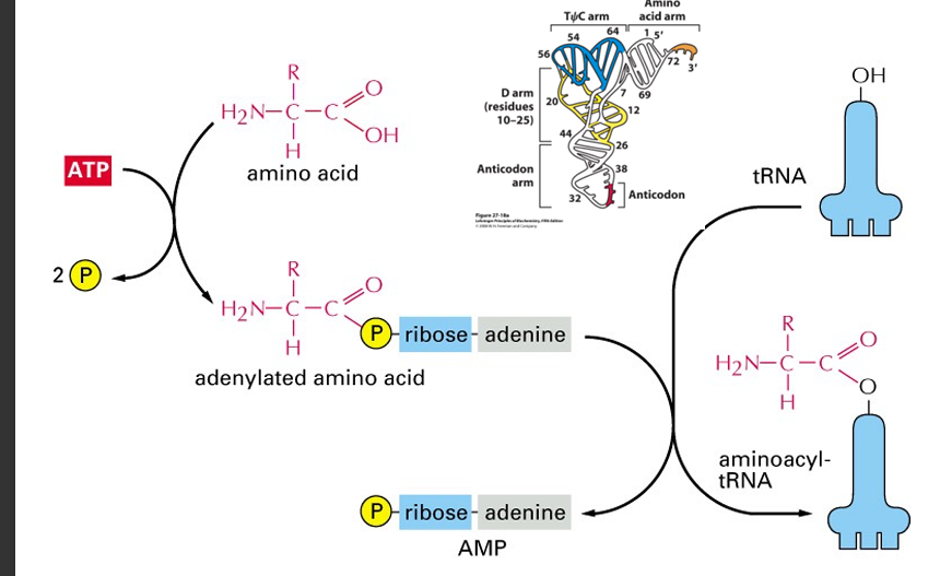
1. aa+ATP+tRNA—>aa-tRNA+AMP
ATP activate, and adenylated AA to make ribose to adenine