Bones & Skeletal system: Part 1
 Call Kai
 Learn
Learn Practice Test
 Spaced Repetition
 Match
Match Flashcards
 Knowt Play
1/32

There's no tags or description
Looks like no tags are added yet.
Name | Mastery | Learn | Test | Matching | Spaced | Call with Kai | Chat |
|---|
No analytics yet
Send a link to your students to track their progress
33 Terms
Cartilage
-Provides support, framework and attachments
-structural model for many developing bones
-Chondrocytes in Lacuna
-Abundant extracellular matrix, mostly collagen fibers, and gel-like ground substance:protein-polysaccharide(chondromucoprotein)
-surrounded by perichondrium (dense irregular connective tissue)
-Cartilage is avascular, nutrients provided by blood vessels in perichondrium
-Three types: Hyaline, Elastic, and fibrocartilage
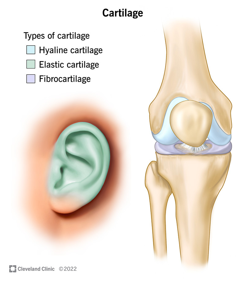
Hyaline cartilage
-Flexibility, reduce friction, absorb shock
-Most abundant type of cartilage
-Weakest cartilage
-Very fine collagen fibers in extracellular matrix (glass like appearance)
-joints, respiratory system, nose, embryonic model for bone

Elastic cartilage
-Maintain shape of organs
-Threadlike network of elastic fibers within the matrix
-More flexible than hyaline
-External ear, larynx

Fibrocartilage
-Strength and rigidity, compression, torsion-strongest
-Bundles of collagen fibers in extracellular matrix
-Found in knee joint, pubic symphysis, intervertebral discs

Skeletal system
-Bones are the organs of the skeletal system
-Composed of many tissues: bone, cartilage, dense connective, blood, epithelial, nervous
-206 bones in human body, babies have more bones(unfused), some adults have more bones
-Each bone is an organ

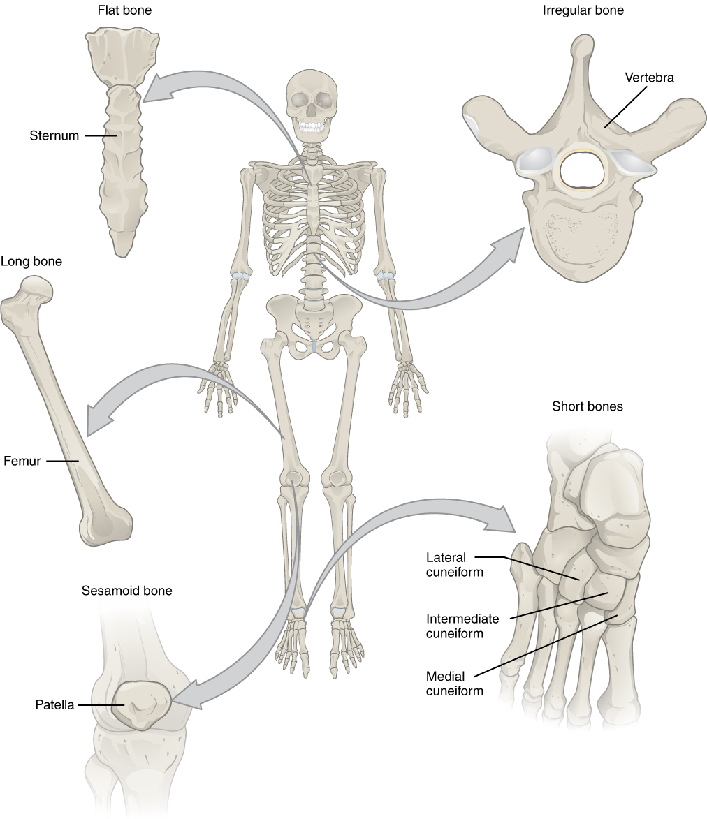
Bone classification
-Long bone(humerus), long slender
-Sutural Bones, Between skull bones
-Short bone(wrist bone), small box-shaped
-Flat bone(sternum), thin, large surface area for attachments
-Irregular bone(vertebra), Complex shapes
-Sesamoid bone(patella), small, flat, sesame seed-like
Projections
where muscles, tendons, or ligaments attach, or where bones form joins

Openings and depressions
sites where blood vessels or nerves lie alongside or penetrate the bone

Diaphysis
Tubular shaft of the bone, consists of compact bones-dense and solid, a layer of protection around a medullary cavity-central space

Epiphysis
The end parts of the long bone, consists of spongy (trabecular) bone-opening network of struts and plates resembling a lattice

Metaphysis
The region between the diaphysis and epiphysis

Epiphyseal plate/line
area of linear growth/remnant from growth

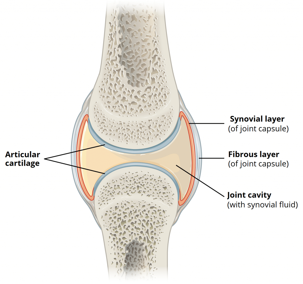
Articular Cartilage
hyaline cartilage covering joints

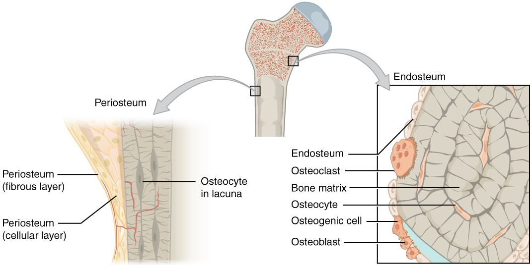
Periosteum
membrane covering outside of bone

Outer fibrous layer
dense connective tissue
Inner cellular layer
Osteoprogenitor cells, fibroblasts-like cells that differentiate into osteoblasts

Endosteum
membrane covering inside of bone, trabeculae and central canal; thinner than periosteum, reticular fibers and osteoprogenitor cells
Sharpey’s fibers
Collagen fibers from the periosteum that penetrate the bone matrix for attachment, particularly at tendons and ligaments

Red marrow
Blood cell production

Yellow marrow
lipid storage

Flat bones
Thin layer of compact bone on the outside with a core of trabecular bone

Osteoprogenitor(osteogenic) cells
progenitor cells in membranes that give rise to osteoblasts as necessary

Osteoblasts
Bone builders, are non-mitotic, secrete osteoid (organic) matrix, the matrix binds calcium and other minerals from the blood

Osteocytes
mature osteoblasts that are entrapped in matrix, maintain the protein and mineral composition of the matrix

Osteoclasts
Multi-nucleated, derived from monocytes (macrophages)and break down or resorb matrix

Matrix of bone
1/3 collagen fibers and other proteins, strength comes from calcium phosphate to form hydroxyapatite

Osteons
cylinders of circular lamellae that run parallel to the long axis of the bone (direction of max force)

Nutrient vessels
supply blood to vessels in the medulla and nerves

Central(Haversian) canals
contain endosteum, nerves and blood vessels

Perforating (Volkmann’s) canals
connect blood supply from periosteum to central canal and medullary vessels, nerves as well

Location of osteocytes
In the lacunae between the lamellae, they have fine projections that connect osteocytes via small openings called canaliculi

Location of osteoblasts and osteoclasts
found in the endosteum or periosteum where they are either building or eroding bone

Spongy (cancellous, trabecular) bone
composed of trabeculae (not osteons)
covered by endosteum
forms the core of most bones
Spaced between trabeculae is filled with red marrow-the site of hemopoiesis (blood cell production)
