L27 - Laboratory assessment of Bone marrow Pt.1
 Call Kai
 Learn
 Practice Test
 Spaced Repetition
 Match
 Flashcards
 Knowt Play
1/37

There's no tags or description
Looks like no tags are added yet.
38 Terms
What is Hematopoiesis, and Where Does It Occur?
Blood cell formation
Primarily in bone marrow in mammals and birds
What is Extramedullary Hematopoiesis?
Blood cell production OUTSIDE marrow, primarily in the spleen and liver
Which Cell Lineages are Myeloid versus Lymphoid?
Myeloid:
Granulocytes
Monocytes
RBCs
Megakaryocytes
Lymphoid:
B cells
T cells
Plasma cells
QUESTION: When should a bone marrow evaluation be performed?
ANSWER → When clinical findings and CBC abnormalities suggest a hematologic disorder not fully explained by peripheral blood
Which Unexplained Cytopenias Indicate Marrow Evaluation?
Persistent nonregenerative anemia
Neutropenia without left shift
Thrombocytopenia
Bicytopenia
Pancytopenia
Why May Horses Require Marrow Evaluation for Anemia?
Horses release few circulating reticulocytes →making erythroid regeneration difficult to assess by CBC alone
Why is Neutropenia without a Left Shift Concerning?
It raises concern for inadequate marrow production
A left shift more often suggests increased peripheral demand
When Do Increased Blood Cell Counts Indicate Marrow Evaluation?
Persistent, unexplained, or extreme elevations of WBCs, RBCs, or platelets after excluding common causes

When Do Atypical Circulating Cells Indicate Marrow Evaluation?
When unclassifiable cells are present, especially without enlarged lymph nodes or spleen to sample.

Why is Marrow Evaluated in Suspected Primary Leukemia?
To confirm leukemia, characterize cell lineage/phenotype, and assess remaining normal hematopoiesis

Which Neoplasms May Require Marrow Evaluation for Staging?
Marrow involvement helps determine disease extent and prognosis
Lymphoma(prognosis worse if marrow involvement)
Mast cell neoplasia

Which Other Conditions May Indicate Marrow Evaluation?
Unexplained hypercalcemia
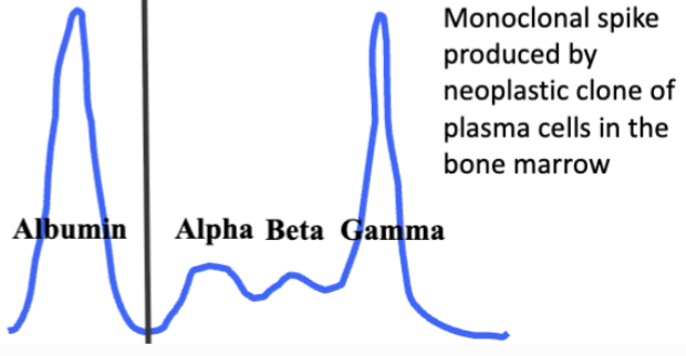
Unexplained hyperglobulinemia (monoclonal gammopathy)
Selected infections
Storage diseases
Iron deficiency
Which Infections May Be Identified in Bone Marrow?
Especially histoplasmosis and leishmaniasis
Which Findings are Generally NOT Indications for Marrow Evaluation?
Regenerative anemia
Isolated lymphopenia
Non-extreme neutrophilia
Erythrocytosis
QUESTION: Why is Lymphopenia NOT a Routine Marrow Indication?
Lymphocyte counts depend mainly on secondary lymphoid tissues and lymphocyte distribution/kinetics
Why Must Marrow Findings Be Compared with a Current CBC?
Marrow production & maturation must be interpreted alongside the peripheral blood abnormalities

What are the Advantages and Disadvantages of Marrow Aspiration?
Advantages:
Best individual-cell detail and maturation
Rapid
Inexpensive
Disadvantages:
May miss focal disease or misrepresent cellularity

What are the Advantages and Disadvantages of Core Biopsy?
Advantages:
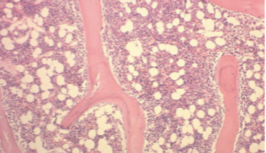
Better assessment of overall cellularity and tissue architecture
Better for diagnosis of fibrosis, necrosis, focal infiltrative disease
Disadvantages:
Slower
Costlier
Less individual-cell detail
What Does a Clinical Pathologist Assess in Marrow?
Sample adequacy
Overall marrow cellularity
Megakaryocytes
Adequacy, maturation and morphology of major cell lineages
Granulocytic to Erythroid (G:E) ratio
Other cells
Iron stores

QUESTION: Are unit particles present?
ANSWER → Adequate marrow aspirates should contain recognizable marrow particles (spicules), indicating that marrow tissue was collected
What Does Marrow Cellularity Mean?
Proportion of hematopoietic cells versus fat
Adult ~50:50
Young ~75:25
Old ~25:75

How Do Hypercellularity, Hypocellularity, and Aplasia Differ?
Hypercellular: more cells
Hypocellular: fewer cells
Aplasia: near-absence of hematopoietic cells

How Do Hyperplasia, Hypoplasia, and Neoplasia Differ?
Hyperplasia: increased normal cells
Hypoplasia: decreased production
Neoplasia: abnormal proliferating cells

What Does Megakaryocytic Hyperplasia Suggest in Thrombocytopenia?
Marrow response to peripheral platelet destruction or consumption
Severe cases in dogs suggest ITP (immune mediated thrombocytopenia)

QUESTION: Why esp. ITP ??
ANSWER → Severe thrombocytopenia with increased megakaryocytes suggests peripheral platelet destruction
ITP is an important cause of extremely low platelet counts in dogs.
What is the G:E Ratio, and What is Normal?
Granulocytic-to-erythroid precursor ratio
Normally about 1–1.5:1
Interpret with cellularity and CBC.
What Does a Hypercellular Marrow with a High G:E Ratio Suggest?
Possible granulocytic hyperplasia and/or erythroid hypoplasia
Assess each lineage separately.
What Do Maturation Arrest, Left Shift, and Dysplasia Suggest?
Arrest: neoplasia/viral disease
Left shift: demand or late-stage cell loss
Dysplasia: drugs, viruses, neoplasia
What are Potential Causes of Increased Marrow Lymphocytes or Plasma Cells?
Lymphocytes: infection, immune disease, neoplasia
Plasma cells: age, immune stimulation, plasma-cell neoplasia
QUESTION Why do plasma cells increase with age?
ANSWER → Long-lived plasma cells accumulate in bone marrow over time as a result of previous immune responses
What is Aplastic Pancytopenia, and What Can Cause It?
Pancytopenia with marrow aplasia
Causes include:
Immune disease
Viruses
Drugs
Estrogen
Ehrlichia
Radiation
Toxins
Why Do Cytopenias Develop in a Particular Order with Aplasia?
Neutropenia → Thrombocytopenia → Anemia
Reflecting progressively longer circulating cell lifespans
How Do Necrosis, Myelofibrosis, and Myelophthisis Differ?
Necrosis: marrow cell death
Myelofibrosis: fibrous replacement
Myelophthisis: displacement by abnormal cells/tissue

TABLE 1. Indications for Bone Marrow Evaluation
QUESTION: When should you consider evaluating the bone marrow?
CBC / Clinical Finding | Why Evaluate Bone Marrow? |
Persistent nonregenerative anemia | Investigate inadequate RBC production |
Neutropenia without left shift | Investigate inadequate neutrophil production |
Unexplained thrombocytopenia | Evaluate platelet production |
Bicytopenia / pancytopenia | Investigate failure or replacement of multiple marrow lineages |
Extreme or unexplained leukocytosis | Investigate possible leukemia |
Persistent unexplained erythrocytosis | Investigate excessive RBC production |
Extreme or unexplained thrombocytosis | Investigate abnormal platelet production |
Unclassifiable circulating cells | Investigate possible hematopoietic neoplasia |
Lymphoma / mast cell neoplasia | Evaluate marrow involvement for staging |
Unexplained hypercalcemia / monoclonal gammopathy | Investigate possible marrow-associated neoplasia |
Selected infections / storage diseases | Identify organisms or abnormal cellular accumulation |
Generally NOT indications:
Regenerative anemia
Isolated lymphopenia
Non-extreme neutrophilia or erythrocytosis
Exam tip: Persistent, unexplained cytopenias are particularly important because they can indicate inadequate bone marrow production
TABLE 2. Bone Marrow Aspirate vs. Core Biopsy
QUESTION: Which method is better for evaluating different marrow abnormalities?
Feature | Aspirate | Core Biopsy |
Evaluates | Individual cell morphology | Tissue architecture |
Cell maturation | Excellent | Limited |
Cellularity | Less reliable | More reliable |
Fibrosis | Difficult to assess | Better |
Necrosis | May be missed | Better |
Focal lesions | May be missed | Better |
Speed | Faster | Slower |
Cost | Less expensive | More expensive |
Remember:
Aspirate = What do the individual cells look like?
Biopsy = What does the marrow tissue look like?
They are complementary rather than interchangeable
TABLE 3. Bone Marrow Terminology
QUESTION: What do the clinical pathologist's descriptions actually mean?
Term | Meaning |
Hypercellularity | Increased proportion of hematopoietic cells |
Hypocellularity | Decreased proportion of hematopoietic cells |
Aplasia | Near absence of hematopoietic cells |
Hyperplasia | Increased number of cells in a lineage |
Hypoplasia | Decreased number of cells in a lineage |
Neoplasia | Abnormal, uncontrolled cellular proliferation |
Maturation arrest | Failure of cells to mature beyond a particular stage |
Dysplasia | Abnormal cellular development or morphology |
Myelofibrosis | Replacement of marrow with fibrous tissue |
Myelophthisis | Displacement of normal marrow by abnormal cells or tissue |
Necrosis | Death of marrow cells or tissue |
Important distinction: Cellularity describes the marrow overall, while hyperplasia or hypoplasia may describe a particular lineage.
A marrow can be hypercellular overall but have erythroid hypoplasia if another cell population has increased substantially.
TABLE 4. Interpreting Marrow Findings and Differential Diagnoses
QUESTION: If the pathologist reports an abnormal marrow finding, what conditions should you consider?
Bone Marrow Finding | Potential Causes / Interpretation |
Hypercellularity | Increased production in response to cell loss, destruction, or consumption; neoplastic infiltration |
Hypocellularity | Toxicity, immune-mediated disease, infections, drugs |
Aplasia | Aplastic pancytopenia, necrosis, myelophthisis |
Megakaryocytic hyperplasia + thrombocytopenia | Peripheral platelet destruction or consumption, including ITP |
Maturation arrest | Neoplasia, viral disease |
Marrow left shift | Increased peripheral demand or consumption; loss of later-stage cells |
Dysplasia | Drugs, viral disease, neoplasia |
Increased lymphocytes | Infection, immune-mediated disease, neoplasia |
Increased plasma cells | Age, immune stimulation, plasma-cell neoplasia |
Myelofibrosis | Fibrous tissue replacing marrow |
Myelophthisis | Abnormal cells or tissue displacing normal hematopoiesis |
Remember: A marrow finding is not always a diagnosis. For example, hypercellularity can represent either an appropriate regenerative response or neoplastic infiltration.
TABLE 5. Interpreting Cellularity and the G:E Ratio
QUESTION: How do you interpret the marrow's overall cellularity and individual lineages?
Measurement | Normal / Expected Finding | Interpretation |
Cellularity — young animals | ~75% cells : 25% fat | More hematopoietic tissue |
Cellularity — adults | ~50% cells : 50% fat | Typical adult marrow |
Cellularity — older animals | ~25% cells : 75% fat | More fatty tissue |
G:E ratio | Approximately 1–1.5:1 | Granulocytic to erythroid precursors |
Increased G:E ratio | More granulocytic relative to erythroid cells | Granulocytic hyperplasia and/or erythroid hypoplasia |
Decreased G:E ratio | More erythroid relative to granulocytic cells | Erythroid hyperplasia and/or granulocytic hypoplasia |