Bone and Muscle Biology
 Call Kai
 Learn
 Practice Test
 Spaced Repetition
 Match
 Flashcards
 Knowt Play
1/55

There's no tags or description
Looks like no tags are added yet.
Name | Mastery | Learn | Test | Matching | Spaced | Call with Kai | Chat |
|---|
No analytics yet
Send a link to your students to track their progress
56 Terms

Periosteum
thin tissue covering the outer surface of bones during life; covers all bone surfaces not covered by cartilage

Endosteum
poorly defined membrane that lines the inner surface of bones
Osteoblasts
Bone-forming cells
§ Deposit osteoid (unmineralized bone) that forms the bone
bone forming cells
Deposit osteoid (unmineralized bone) that forms the bone matrix
Blast = build

Osteoclasts
bone reabsorbing cells

Osteocytes
living bone cells that no longer secrete osteoid but maintain bone tissue

Osteoprogenitor cell
genitor = producing
Lacunae
Osteocytes live within cavities called lacunae within the lamellae
canaliculi
Osteocytes are connected to each other via channels called canaliculi
= bone is increased where needed and resorbed where not
Wolff’s Law
bone is increased where needed and reabsorbed where not needed
bones are remodeled during life to meet mechanical demands
Bone Biomechanics
§ Compression
§ Tension
§ Shear
§ Torsion
§ Response depends on:
§ Duration
§ Frequency
§ Number of repetitions
§ Age
§ Gender
§ Use/disuse
Trabecular Bone and Loading
Trabecular Bone : spongy, porous bone found inside bones, such as the vertebrae
(loading = force applied to bone)
Slow loading: The bone has more time to deform under pressure.
Fast loading: The bone behaves more stiffly, meaning it resists deformation more strongly under compression.
Viscoelastic: ~ the response to force: deformation curve ,is dependent on the rate of loading
Trabecular bone becomes stiffer in compression the faster it is loaded
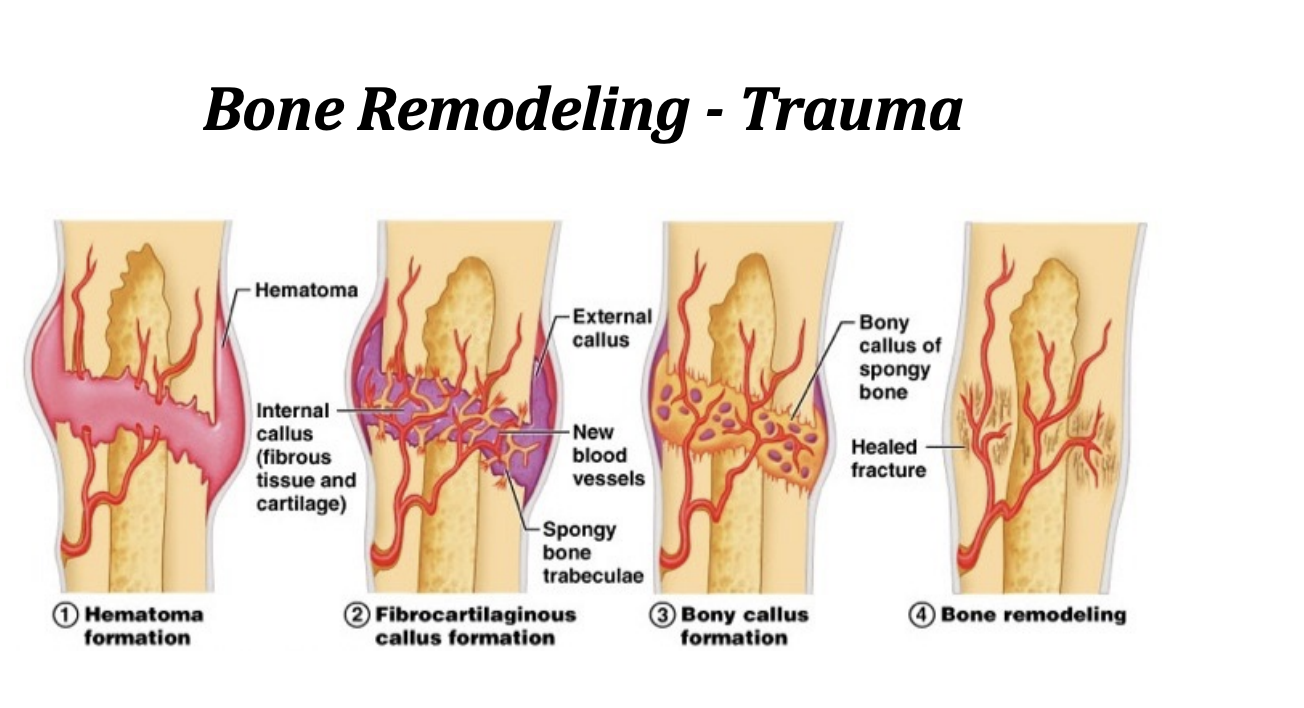
Bone Remodeling/Trauma
Woven
found during growth and repair (e.g. from a fracture or disease)
Disorganized
Not a lot of structure or stability
Primary
laid down on woven bone
Secondary (Lamellar bone):
Formed during bone remodeling
Most resistant to fractures
Bone development (ossification) occurs in 2 general sites
Intramembranous ossification
Endochondral ossification
Intramembranous Ossification
bones ossify by apposition on tissue within an embryonic connective tissue membrane (e.g., skull bones)

Endochondral Ossification
bones preceded by cartilage which ossifies over time (e.g., most bones in the body)

Intramembranous Ossification
Flat bones
From mesenchymal (embryonic) tissue
Center of ossification
location in the membrane where ossification begins
Fontanels
large membrane covered spaces between developing skull bones (unossified)

Endochondral Ossification
Long bones
Cartilage model precursor

Osteogenesis
Bone Growth/Ossification
Thin membrane called the perichondrium surrounds the cartilage model of the long bone.
Osteoblasts just beneath the perichondrium deposit bone
Once bone is deposited, the membrane becomes the periosteum
Bone Growth/Ossification
More bone is deposited layer by layer, increasing the diameter of the shaft
Appositional growth = growth in diameter
Second center of ossification is located between the metaphysis and epiphysis
Allows bones to lengthen = growth plate
Bone Growth/Ossification
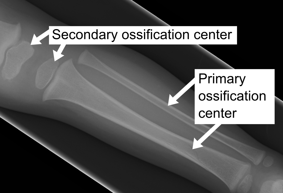
Usually around 450 ossification centers at birth
“Primary” ossification centers usually appear before birth
“Secondary” ossification centers usually appear after birth
206 bones in an adult skeleton
Bone Growth/Ossification
Usually around 450 ossification centers at birth
Some major bones fuse at predictable ages through growth
Ages of epiphyseal fusion are used to provide age estimates on immature skeletons
Long bones
Epiphysis: end part of long bone (pl: epiphyses)
Diaphysis: shaft of long bone
Metaphysis: where the epiphysis meets the diaphysis (pl: metaphyses)
Musculoskeletal system
system of bony levers operated by muscles
Joint
connection between different bony elements
Articulation
Where different bones join or touch is called an articulation
Ligaments and cartilage are often found at articulation points
Tendons
closely packed parallel bundles of collagen fibers; attach muscles to bones
Movement at joints is controlled by the shapes of the articular surfaces and the ligaments that bind the joints together and prevent dislocation
3 major types of Joints
§ Synovial § Cartilaginous § Fibrous
Synovial joint
freely moving joints composed of a joint capsule containing a joint cavity lined with hyaline cartilage and synovial fluid
Very moveable

Joint capsule
Articular capsule
Surrounds the synovial cavity (joint cavity)
Synovial fluid

Cartilaginous joint
articulating bones are united by cartilage which allows very little movement
2 types of cartilaginous joints depending on type of cartilage between the bones :
Synchondrosis ⟶ hyaline cartilage EX. EPIPHYSEAL PLATE
Symphysis ⟶ fibrocartilage EX : PUBIC SYMPHYSIS
Fibrous joint
joint between bones filled with fibrous tissue only and allowing no movement
3 Types of Fibrous joints:
Suture
Syndesmosis
Gomphosis
Cranial sutures ( Types of Fibrous Joints)
fibrous joints of the skull bones; united by fibrous tissue
Syndesmosis
tight, inflexible fibrous joint between bones and united by dense fibrous tissue such as membranes ligaments (e.g., tibia-fibula)
Gomphosis
joint between the roots of the teeth and the jaw bones
Fibrous joints
immovable

Skeletal movement
Movement of the skeleton generally takes place at synovial joint
Movement generally caused by muscles working across joints
Muscles connected to bones by tendons
Ligaments
Ligaments
connective tissue extending between adjoining bones
3 types of muscle tissue:
Striated (skeletal) muscle
Smooth muscle
Cardiac muscle
Skeletal Muscle
Attached to bone
Allows movement (voluntary)
Striated
Cardiac Muscle
Heart muscle
Involuntary
Striated
Smooth Muscle
Non-striated
Found in walls of hollow internal structures
Involuntary
Basic Muscle Biology
Muscles are composed of fiber bundles
Fibers made up of fibrils (myofibrils)
Fibrils (myofibrils) made up of myofilaments
Myofilaments are banded

Basic Muscle Biology
Bands within each myofilament are known as A bands (darker) and I bands (lighter in color)
Z-lines ⟶ thin dark strips within the I bands)
Unit between two Z-lines called a sarcomere ⟶ smallest functional unit of the myofilament

Basic Muscle Biology
Structure within a sarcomere
Central section of thick myosin positioned between thin filaments of actin
When muscle contracts, thick section slides between the thin filaments and reduces the distance between Z-lines
Basic Muscle Biology
Muscles are innervated by nerves
Nerve impulse depolarizes muscle fibers and results in an interaction of the actin and myosin molecules which results in the muscle contracting
Nerve and the group of muscle fibers that it innervates ⟶ motor unit

Work Formula
Work performed by a muscle = F*d
Work ⟶ Force x distance
Therefore, length of the muscle is related to how much work can be performed
Two muscles exerting the same force but of different length….which can perform more work?
The longer muscle, because it can contract over a greater distance. Work = Force × Distance.
Origin
in general, site that stays stable during contraction; usually closest to the trunk (proximal)
Insertion
in general, the site that is moved during contraction; usually farthest from the trunk (distal)
Antagonists
Many muscles perform opposite motions
These muscle pairs are called antagonists
E.g., flexors and extensors