US Guided Procedures (copy) 7%
 Call Kai
 Learn
 Practice Test
 Spaced Repetition
 Match
 Flashcards
 Knowt Play
1/55

There's no tags or description
Looks like no tags are added yet.
56 Terms
What is the preferred patient position during catheter retraction in an endovenous laser ablation treatment?
A. reverse Trendelenburg
B. seated with legs dangling
C. Trendelenburg
D. standing
C. Trendelenburg
When the catheter is inserted at the start of the procedure, the semi-erect or reverse Trendelenburg position is helpful to better visualize the advancement of the catheter. Once the laser is activated and the catheter is retracted, the patient is placed in the Trendelenburg position to reduce the blood volume in the veins of the leg and facilitate the closure of the vein during the procedure. The laser essentially damages the walls of the veins causing a reduced lumen diameter and thrombus formation. The less blood that's in the vein, the easier it will be to successfully treat the vein.
If the peak velocity in a transjugular intrahepatic portosystemic shunt at the portal vein anastomosis is 50cm/s and the mid graft velocity is 25cm/s, what is indicated?
A. The mid graft velocity is very low and indicates impending failure of the shunt.
B. There is most likely an occlusion in the graft at the proximal end.
C. There is approximately a 50% stenosis at the portal anastomosis.
D. The velocities indicate normal flow within the shunt.
A. The mid graft velocity is very low and indicates impending failure of the shunt.
Flow velocity <50cm/s in the intrahepatic shunt indicates impending failure.
A patient presents for a pre-procedure scan for placement of a central venous catheter in the internal jugular vein. In order to avoid potential hematoma or AV fistula formation, the relationship of the vein to the _______ must be documented on today's exam.
A. internal carotid artery
B. sternocleidomastoid muscle
C. common carotid artery
D. external jugular vein
C. common carotid artery
A patient presents for a pre-procedure scan for placement of a central venous catheter in the internal jugular vein. In order to avoid potential hematoma or AV fistula formation, the relationship of the vein to the common carotid artery must be documented on today's exam. If the CCA is punctured, serious complications can occur.
Prior to any invasive procedure, the patient should be educated about the procedure, expected outcomes and any possible complications. The patient will be required to sign a document stating their understanding of these topics in order to proceed with the procedure. This form is called:
A. health insurance waiver
B. informed consent form
C. litigation waiver
D. patient acceptance form
B. informed consent form
Prior to any invasive procedure, the patient should be educated about the procedure, expected outcomes and any possible complications. The patient will be required to sign a document stating their understanding of these topics in order to proceed with the procedure. This form is called an informed consent.
Which of the following procedures allows patients to continue their normal course of anticoagulant therapy before and after the procedure?
A. compression therapy of pseudoaneurysm
B. percutaneous transluminal angioplasty
C. ascending venography
D. carotid endarterectomy
C. ascending venography
Venography is performed using a venous puncture. Surgical procedures and invasive procedures that require an arterial puncture have an increased risk of hemorrhage when the blood is thinned by anticoagulants. Patients having these procedures must discontinue their anticoagulant therapy before the procedure. Thinned blood will also inhibit the clot formation in a pseudoaneurysm.
In most cases, a peripherally inserted central catheter (PICC) line is inserted in the basilic vein exam and advanced until the tip reaches:
A. brachiocephalic vein
B. atriocaval junction
C. axillary vein
D. proximal subclavian vein
B. atriocaval junction
The terminal tip of the venous catheter is positioned in a central vein, most commonly the distal 1/3 of the SVC (atriocaval junction). High blood flow volume in this location allows infusates to be rapidly distributed.
A patient is undergoing balloon angioplasty of the left common femoral artery. The physician just completed the first expansion of the balloon and asks you to check the flow with Doppler. Where is the preferred location to evaluate flow?
A. dorsalis pedis or posterior tibial artery
B. proximal superficial femoral artery
C. popliteal artery
D. distal superficial femoral artery
A. dorsalis pedis or posterior tibial artery
The physician is trying to increase flow distal to the obstruction. It is most important to evaluate flow in the most distal areas to evaluate improvement in overall perfusion of the leg.
Which of the following describes a difference between endovenous ablation of the great saphenous vein and of the small saphenous vein?
A. the catheter is inserted at the groin for the treatment of the GSV and at the dorsum of the foot for the treatment of the SSV
B. at the start of the procedure, the patient is placed in the supine position for the GSV treatment and in the prone position for the SSV treatment
C. the catheter is inserted at the groin for the treatment of the GSV and at the popliteal fossa for the treatment of the SSV
D. The SSV cannot be treated by endovenous ablation techniques
B. at the start of the procedure, the patient is placed in the supine position for the GSV treatment and in the prone position for the SSV treatment
The patient is placed in the supine position for the GSV treatment and in the prone position for the SSV treatment. The catheter is inserted in the distal GSV and advanced to a position 2cm from the SFJ. The catheter is inserted in the distal SSV and advanced to a position distal to the SPJ. The table position will be adjusted to Trendelenburg position after the catheter is inserted to drain excess blood from the veins before device activation. Once activated the catheter is pulled down the vein and out the puncture site.
Where is the most common site of stenosis in a transjugular intrahepatic portosystemic shunt?
A. hepatic vein anastomosis
B. portal vein anastomosis
C. biliary anastomosis
D. hepatic artery anastomosis
A. hepatic vein anastomosis
The hepatic vein anastomosis is the most common site of stenosis in a transjugular intrahepatic portosystemic shunt.
Which upper extremity vein is most commonly evaluated for use as an arterial bypass graft?
A. radial vein
B. cephalic vein
C. median cubital vein
D. brachial vein
B. cephalic vein
The cephalic vein is most commonly harvested for use as a bypass.
What is the best Sonographic technique to use to evaluate the status of a pseudoaneurysm after 10 minutes of compression therapy is applied?
A. PW Doppler
B. CW Doppler
C. Tissue Doppler
D. Color Doppler
D. Color Doppler
Color Doppler is the best for identifying the presence of the stalk within the tissues. PW and CW Doppler can be used to evaluate the stalk once it has been located with color but cannot reliably locate the stalk without color guidance.
Which of the following statements best describes the Doppler tracing found in the stalk of a pseudoaneurysm?
A. phasic forward flow
B. high resistance to and fro flow
C. continuous forward flow
D. low resistance to and fro flow
B. high resistance to and fro flow
The body of the pseudo aneurysm will demonstrate a low resistance "swirling" of flow on the Doppler tracing. The neck/stalk of the pseudo aneurysm is normally very narrow and will demonstrate a high resistance, biphasic waveform.

Which of the following is an advantage of intraoperative vascular ultrasound compared to intraoperative angiography?
A. Technique does not require vessel puncture to obtain flow information
B. Can assess flow in all branches of the intracerebral vessels
C. Can be performed during the surgical procedure
D. Provides real time flow evaluation
A. Technique does not require vessel puncture to obtain flow information
Angiography is an invasive procedure that requires a vessel puncture to administer contrast. Intraoperative US is performed by placing the transducer on the artery, no puncture required to obtain flow information. Both ultrasound and angiography can provide real time blood flow information because the contrast flow is evaluated live with fluoroscopy on angiography. Ultrasound does not evaluate all branches of the intracerebral vessels while angiography can visualize all patent branches in the FOV. Both techniques can be performed during the surgery.
What is an absolute contraindication for sclerotherapy?
A. vein location above the inguinal ligament
B. patient allergy to injectate
C. polycythemia vera
D. vein location immediately over a bony prominence
B. patient allergy to injectate
Absolute contraindications are situations which make a particular treatment or procedure absolutely inadvisable. The risk of complications caused by the patient allergy to the injected substance (sclerosant) far exceed the benefits of the procedure. The patient will be treated using other methods.
Which of the following statements is correct regarding intraoperative duplex evaluation during a carotid endarterectomy?
A. The technique is used to assess the immediate flow changes within the vessel undergoing the endarterectomy.
B. intraoperative duplex evaluation cannot be performed during a carotid endarterectomy.
C. Due to the increased attenuation of sound caused by air in the extracranial vessels, the lowest probe frequency available is preferred for the exam.
D. Sterile gel is required because a sterile sheath is not used due to potential acoustic artifacts.
A. The technique is used to assess the immediate flow changes within the vessel undergoing the endarterectomy.
The technique is used to assess the immediate flow changes within the vessel undergoing the endarterectomy. The highest probe frequency available is preferred for intraoperative duplex evaluation. The probe is placed in direct contact on the vessel that is being evaluated, so the sterile technique is required, including sterile gel with a sterile sheath protecting the transducer. The vessel should not contain air because it would become a cerebral embolism.
At the start of an endovenous ablation procedure for the great saphenous vein, the catheter is inserted _____ and advanced to a position _____ before activating the device
A. into the GSV at the groin, 2cm below the knee
B. into the distal GSV , 2cm distal to the saphenopopliteal junction
C. into the distal GSV, 2cm distal to the saphenofemoral junction
D. into the GSV at the groin, at the most distal point of reflux
C. into the distal GSV, 2cm distal to the saphenofemoral junction
At the start of an endovenous ablation procedure for the GSV, the catheter is inserted into the distal vein and advanced to a position 2cm distal to the SFJ before the device is activated. Once activated, the catheter is slowly withdrawn down the leg and out through the puncture site. This should cause fibrosis of the vein in the treated area. The catheter tip must be 2cm or more from the SFJ to prevent damage to the common femoral vein.
Which of the following is an absolute contraindication to an endovenous ablation?
A. daily aspirin use
B. history of diabetes mellitus
C. current antibiotic use
D. uncooperative patient
D. uncooperative patient
An absolute contraindication is situation which makes a particular treatment or procedure absolutely inadvisable. These include a known allergy to the injected material (ablation, steroid injection), lack of appropriate equipment or skill to complete the procedure, uncooperative patient (uncontrolled movement during the procedure can result in injury, laceration or hemorrhage)
A relative contraindication is a condition which makes a particular treatment or procedure possibly inadvisable. Changes can be made to make the procedure safer or easier to perform. These include coagulopathy and the use of blood thinners, aspirin or antibiotics. If permitted by their physician, the patient can discontinue their blood thinner medication until their PT and INR values return to normal levels and then proceed with the invasive procedure. Warfarin, Coumadin, Jantoven, Xarelto, Pradaxa or Lovenox, Aspirin, Ibuprofen, NSAIDs should be stopped at least 7 days before a procedure and resumed 7 days after the procedure.
Other relative contraindications include:
Unsafe access route - must avoid large arteries and adjacent nerves
Local infection, rash, or skin breakdown
Which of the following measurements should be obtained to evaluate the success of a percutaneous angioplasty performed on the left proximal superficial femoral artery?
A. femoral artery diameters proximal to the treated area, the treated area and distal to the treated area
B. acceleration time of the distal superficial femoral artery
C. evaluate the ABI value for the expected decrease
D. obtain waveforms at the PTLA site and measure the peak systolic velocity and pulsatility index
D. obtain waveforms at the PTLA site and measure the peak systolic velocity and pulsatility index
Evaluating a patient post-angioplasty of the lower extremity:
ABl assessment - greater than 0.9 and/or an increase of at least 0.15 in the ABI from pre-procedure levels
Evaluate inflow artery for damping - measure acceleration time <180ms is normal (AT is used to evaluate INFLOW)
Evaluate the PTLA site for residual plaque or thrombus
Record waveforms at the PTLA site and proximal to it - measure the velocity and pulsatility index
Calculate the velocity ratio - less than 2 is normal
Which of the following correctly describes proper behavior related to a sterile field?
A. Sterile gloves must be used to open the procedure tray
B. Sterile individuals should only touch other sterile items
C. Everyone in the procedure room should be in sterile PPE
D. Sterile individuals should pass by each other while facing each other
B. Sterile individuals should only touch other sterile items
Everyone in the procedure room cannot be in sterile PPE. Opening non-sterile packaging for sterile items should be done by someone not dressed in sterile attire/gloves. Touching a non-sterile item with sterile gloves renders you "contaminated". Sterile packages can be opened with non-sterile gloves, but if any items inside the tray will be touched, sterile gloves must be used. Sterile gloves must be used to remove items for the tray to prepare them (lidocaine, needle, syringe, biopsy instrument). Sterile individuals should pass back to back with other sterile individuals to prevent contamination.
Which of the following correctly describes sclerotherapy procedures?
A. Physicians may inject saline into the superficial veins of the extremity in order to cause increased viscosity and promote the formation of thrombus, which in turn reduces varicosity size.
B. Physicians may inject saline into the superficial veins of the extremity in order to improve circulation by thinning the blood, which in turn reduces varicosity size.
C. Physicians may inject saline into the superficial veins of the extremity in order to cause the vessel to contract and cause fibrosis, which in turn reduces varicosity size.
D. Physicians may inject saline into the deep veins of the extremity in order to increase viscosity and promote the formation of thrombus, which in turn reduces varicosity size.
C. Physicians may inject saline into the superficial veins of the extremity in order to cause the vessel to contract and cause fibrosis, which in turn reduces varicosity size.
Which of the following is a treatment for a pseudoaneurysm?
A. Compression stockings
B. Thrombin injection
C. Coumadin
D. IV Heparin
B. Thrombin injection
A Thrombin injection is used to treat pseudoaneurysms with large stalks. Ultrasound guidance is used to make the injection that promotes thrombosis of the stalk and body of the pseudoaneurysm.
Coumadin is a blood thinner that would decrease the ability of clot formation in the pseudoaneurysm. IV Heparin is used to treat DVT.
If the physician is having trouble visualizing the needle on the ultrasound screen during a thrombin injection, how can you help to improve the visualization of the needle?
A. increase the output power to the maximum setting
B. use a lower frequency probe
C. use electronic steering to increase the incident angle between the US beam and the long axis needle to a value closer to 90 degrees
D. use electronic steering to reduce the incident angle between the US beam and the long axis needle to a value closer to 0 degrees
C. use electronic steering to increase the incident angle between the US beam and the long axis needle to a value closer to 90 degrees
The more perpendicular the US beam is to the needle, the better the reflection properties and visibility on the screen.

A patient is scheduled for a stab phlebectomy. What is being treated for this patient?
A. carotid stenosis
B. DVT removal
C. pseudoaneurysm
D. varicose veins
D. varicose veins
Stab Phlebectomy
This method removes superficial varicose veins that are not amendable to endovenous ablation. Under local anesthesia, tiny punctures, or microincisions are made. Through these small incisions, phlebectomy hooks extract the varicose veins. The incisions are so small that no stitches are required. Ultrasound guidance can be used to guide the procedure. This procedure is well tolerated by patients and produces excellent cosmetic results.
Which of the following is part of the Sonographer's responsibilities when assisting with an endovenous ablation?
A. review patient records and ultrasound images to confirm the diagnosis and the leg that will be treated
B. inject the area around the saphenofemoral junction with anesthetic
C. performing the groin puncture and inserting the proximal end of the catheter into the vein that will be treated by the physician
D. place the patient in the Trendelenburg position to prepare for catheter insertion
A. review patient records and ultrasound images to confirm the diagnosis and the leg that will be treated
Puncturing the vein and performing the catheter insertion fall outside the scope of practice of a Sonographer. The ablation procedure must be performed by the physician.
Sonographer responsibilities include:
review patient records and ultrasound exam to confirm the diagnosis and the leg that will be treated
confirm patient understanding of procedure and the leg and vein to be treated
explain the procedure to the patient
provide the informed consent form to the patient to read (ultimately the patient should sign the form after the patient has had the chance to ask the physician any related questions about the form)
place the patient in the reverse Trendelenburg position and perform any necessary preliminary images
map the position of vein
determine area of catheter access and mark set up the supplies for the procedure cover appropriate transducer with a sterile probe cover
operate the ultrasound machine controls outside the sterile field, as directed by the physician
apply dressing to the access point
confirm that the patient understands the aftercare instructions given by the physician
disinfect the room and the ultrasound equipment after the procedure
discard all used needles and catheters into the biohazard container
What effect does a warm exam room have on a vein mapping procedure performed for coronary bypass surgery?
A. increase in tissue edema and reduced image resolution
B. contracted veins with reduced diameters
C. reduced compressibility of the superficial veins
D. normal venous diameters will be documented
D. normal venous diameters will be documented
When performing a vein mapping, the exam room should be kept warm in order to limit vasoconstriction. During the exam, cover the rest of the patient and the foot/hand to keep warm. If the veins demonstrate thickened walls and reduced diameter, warm the leg or evaluate a different vein.
Which of the following is true regarding the initial post-op examination of a transjugular intrahepatic portosystemic shunt placement?
A. It should be performed within 30 days following the procedure.
B. CTA is most commonly performed for the initial evaluation of a TIPS procedure.
C. It helps to establish baseline velocities for upcoming follow up exams.
D. Marked ascites is a common finding for the initial post op exam on a TIPS patient.
C. It helps to establish baseline velocities for upcoming follow up exams.
When performing serial evaluations of a transjugular intrahepatic portosystemic shunt, it is important to establish a baseline for the patient very soon after the shunt placement. The initial evaluation should be performed with Doppler ultrasound within 24hrs of shunt placement.
A common complication of cardiac catheterization is the formation of:
A. DVT
B. Pseudoaneurysm
C. Fusiform aneurysm
D. Arterial dissection
B. Pseudoaneurysm
During the catheterization the superficial femoral artery is punctured by the catheter as it enters the arterial system. Once the catheter is removed, there is a risk that blood will escape the artery into the surrounding tissues to form a pseudoaneurysm.
Which of the following is a responsibility of a Sonographer when assisting with a radiofrequency ablation of the great saphenous vein?
A. to advance the ultrasound catheter during the procedure
B. to send the ultrasound catheter for sterilization after the procedure
C. to perform initial puncture for the catheter prior to the vascular surgeon entering the room
D. to operate the ultrasound machine controls and/or the probe, as directed by the physician
D. to operate the ultrasound machine controls and/or the probe, as directed by the physician
The ultrasound catheter cannot be sterilized, it is disposable. The procedure is performed by the physician while the Sonographer assists with the ultrasound machine.
Which of the following techniques cannot be used to monitor distal flow in the leg while compression therapy for a pseudoaneurysm is performed?
A. PPG sensor on the ankle
B. Pressure monitor on the great toe
C. Doppler evaluation of the PTA or DPA
D. PPG sensor on the great toe
A. PPG sensor on the ankle
When PPG is used to evaluate arterial flow, the sensors are placed on the great toes. When PPG is used to evaluate venous reflux, the sensor is placed on the lower calf near the ankle. It is contraindicated to obtain an ankle pressure in a patient with an acute pseudoaneurysm. A toe monitor can be used because the inflation of the cuff has little to no effect on pressure in the femoral artery.
Which of the following is an expected finding with an AV fistula caused by an interventional procedure?
A. continuous venous outflow
B. dilated venous collaterals adjacent to the fistula
C. high resistance arterial inflow
D. direct connection between a single artery and vein
D. direct connection between a single artery and vein
An AVM is a congenital malformation with multiple connections between the arterioles and venules without a capillary bed in between. Venous collaterals will develop and dilate over time.
An AV is a direct connection of an artery or vein caused by trauma or interventional procedure. Due to the acute formation of the connection, there are no collaterals present. The arterial inflow and venous outflow will demonstrate the same hemodynamic characteristics in an AVM and AVF, increased arterial flow volume, low resistance arterial inflow and pulsatile, turbulent venous outflow.
A patient presents for a follow up exam after a recent endarterectomy that was performed 3 days ago. The intraoperative report indicates a possible shelf lesion in the ICA. How will this be demonstrated on today's exam?
A. >3mm intimal flap that causes turbulence and increased velocity
B. <3mm intimal flap demonstrates a false lumen with turbulent flow
C. Hyperechoic plaque in the vessel lumen with an abrupt edge
D. <3mm intimal flap that causes turbulence and increased velocity
C. Hyperechoic plaque in the vessel lumen with an abrupt edge
The carotid artery is lacerated and the atheroma formation is scraped for removal in an endarterectomy.
Complications include:
intimal flap
dissection
shelf lesion - residual hyperechoic plaque in the lumen that has an abrupt edge
platelet aggregate - Hypochoic/anechoic material adjacent to vessel wall that causes increased flow velocity

Why did the technologist steer the 2D image while scanning during the thrombin injection?
A. to improve the color flow demonstration
B. to make the US beam more perpendicular to the long axis of the needle
C. to make the US beam more parallel to the long axis of the needle
D. to provide a better view of the posterior enhancement from the pseudoaneurysm
B. to make the US beam more perpendicular to the long axis of the needle
The more perpendicular the US beam is to the long axis of the needle, the easier it is to visualize due to increased reflection.
A patient is referred to the vascular lab to evaluate the placement of a Greenfield filter. Where will you look for this?
A. IVC
B. subclavian or internal jugular vein
C. right or left groin
D. abdominal aorta
A. IVC
The filter is used to prevent pulmonary embolism from the lower extremities. The filter is placed into the IVC below the level of the renal veins.

Which of the following correctly describes how to prepare for an intraoperative vascular ultrasound?
A. Cover a low frequency transducer with a sterile sheath that contains gel
B. Gather extra packets of sterile gel to be used on the outside of the sterile sheath
C. Use sterile gloves to open the packaging for supplies used in the procedure
D. Cover a high frequency transducer with a sterile sheath that contains gel
D. Cover a high frequency transducer with a sterile sheath that contains gel
Intraoperative ultrasound procedures require a high frequency transducer because the transducer will usually be placed directly on the vessel being evaluated (no other tissue to penetrate). Sterile gel is placed inside the sheath, but not on the vessel or inside the body. Sterile gloves are used to touch any of the sterile instruments, but opening packaging does not require sterile gloves.
Which of the following requires the use of sterile technique?
A. Vein mapping
B. Laser ablation for telangiectasia
C. Radiofrequency venous ablation
D. Sclerotherapy
C. Radiofrequency venous ablation
Radiofrequency ablation requires the sterile technique because the procedure requires the puncture of the saphenous vein to insert a catheter into the length of the vein. The other choices do not require the sterile technique. Telangiectasia refers to dilated very small superficial veins that are treated with external application of laser treatment. Sclerotherapy refers to the injection of a solution into spider veins to cause them to atrophy and requires aseptic technique. Vein mapping is performed with ultrasound.

A patient presents for ultrasound evaluation of a PICC line. The findings or the image are most suggestive of:
A. normal catheter demonstrated in the right internal jugular vein
B. thrombus formation around the catheter in the left internal jugular vein
C. thrombus formation around the catheter in the right internal jugular cein
D. thrombus formation around the catheter in the right subclavian vein
C. thrombus formation around the catheter in the right internal jugular cein
The image demonstrates the longitudinal view of the subclavian vein on the posterior image. The transverse view of the IJV demonstrates a hypochoic mass surrounding the catheter. The CCA is medial to the IJV = right side.
Which of the following describes the sonographic appearance of a transjugular intrahepatic portosystemic shunt?
A. requires color Doppler evaluation to be able to visualize the graft within the liver
B. anechoic tube, without distinctive wall reflection, that connects the right hepatic vein to the right portal vein
C. strongly reflective curved structure connecting the right portal vein and right hepatic vein
D. shunts are not easily evaluated Sonographically and CTA is the preferred method for evaluation
C. strongly reflective curved structure connecting the right portal vein and right hepatic vein
The shunt is constructed of metallic substances with increased reflectivity in order to allow for better Sonographic visualization. The sonographic appearance of a transjugular intrahepatic portosystemic shunt is a strongly reflective curved structure connecting the right portal vein and right hepatic vein.

Which of the following is true regarding compression techniques used to treat a pseudoaneurysm of the femoral artery in the groin?
A. Thrombin injection is more effective than compression techniques in pseudoaneurysms with large diameter stalks.
B. Pressure equivalent to 100mmHg should be performed in three ten-minute intervals and if this does not close the stalk, alternative treatment must be considered
C. Compression should be performed in 30-second intervals with color Doppler evaluation in between compressions.
D. Blood pressure evaluation cannot be performed anywhere on the legs until the stalk to the pseudoaneurysm is closed.
A. Thrombin injection is more effective than compression techniques in pseudoaneurysms with large diameter stalks.
Thrombin injection is more effective than compression techniques in pseudoaneurysms with large diameter stalks. Ankle pressure should be monitored during compression techniques to avoid changes in distal flow from extrinsic compression. Compression should be performed in 10 one-minute intervals with color Doppler evaluation in between compressions.
Which of the following statements correctly describes an endovenous laser ablation of the GSV?
A. The procedure is much more easily performed if the patient already has DVT
B. Intravascular ultrasound is performed after the procedure to confirm success
C. The procedure is considered a success if thrombus fills the GSV from the ankle to the SFJ
D. The physician injects a halo of anesthesia medicine around the GSV
D. The physician injects a halo of anesthesia medicine around the GSV
Physician injects anesthesia around the vein being treated = tumescent anesthesia. A 1cm ring of anesthesia medicine should surround the vein for the appropriate level of patient comfort during the procedure. The catheter is inserted in the distal vein and advanced to the most proximal point that will be treated. The laser is turned on and the physician slowly retracts the catheter until it reaches the insertion point. A follow up ultrasound exam is performed in a week or two to evaluate the success of the procedure. The deep system should be evaluated for thrombus, the treated vein should be evaluated for diameter measurements and with color flow. The distance between the SFJ and most proximal point of closure of the GSV should be measured. If thrombus extends into the deep system, the GSV must be ligated. DVT is a contraindication for the procedure.
A patient presents for follow up after a mesenteric stent placement procedure for a 75% SMA stenosis. Which of the following describes the findings in the SMA that indicate a successful procedure?
A. Decreased resistance and increased diastolic flow
B. Increased resistance and decreased diastolic flow
C. Change from monophasic to peaked triphasic
D. Change from peaked triphasic to monophasic
B. Increased resistance and decreased diastolic flow
Normal pre-prandial flow in the SMA is high resistance with low levels of diastolic flow (monophasic). 75% SMA stenosis would cause a low resistance waveform with increased systolic and diastolic velocities. The resistance would increase in the mesenteric vascular beds as normal flow levels resume after angioplasty. Diastolic flow levels would reduce because the mesentery is now receiving enough blood during systole. Peak systolic and end diastolic velocities are reduced.
Which of the following is a contraindication for endovenous laser ablation of the great saphenous vein?
A. valvular incompetence in the great saphenous vein
B. chronic obstruction of the peroneal veins
C. Baker cyst in the ipsilateral popliteal fossa
D. chronic obstruction of the femoral vein
D. chronic obstruction of the femoral vein
IT IS VERY IMPORTANT to evaluate the venous outflow system of a patient prior to removal or closure of the saphenous vein. If the deep system is obstructed and the superficial system is the main outflow for the leg, the saphenous vein cannot be removed/closed.
Which of the following is NOT a part of the information required on an informed consent form for an interventional vascular procedure?
A. description of the procedure
B. possible complications that the patient may experience
C. percentage of patients that have experienced complications after the same procedure at the facility
D. signature line to indicate patient consent
C. percentage of patients that have experienced complications after the same procedure at the facility
The informed consent must include a description of the procedure and list the possible complications that the patient may experience. The patient must be of sound mind when giving consent by signature.
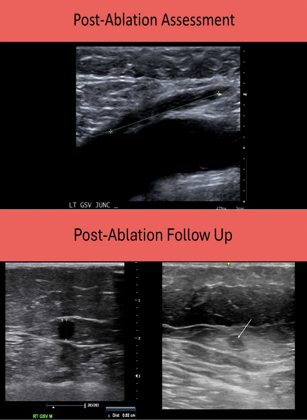
Why is the distance from the most proximal point of the contracted GSV to the saphenofemoral junction measured in a post-ablation exam?
A. to document the lack of thrombus propagation into the deep system
B. to assess the length of the segment that still requires treatment
C. to document that the procedure was unsuccessful
D. to assess the risk of future reflux in that area
A. to document the lack of thrombus propagation into the deep system
The distance between the SFJ and most proximal point of fibrosis/thrombosis of the GSV should be measured. If thrombus extends into the deep system, the GSV must be ligated because of the increased risk of embolization.

When evaluating the left arm for potential placement of a central venous catheter, you identify multiple small venous collaterals surrounding the subclavian vein, What should you do next?
A. evaluate the venous system in the right arm
B. measure the largest collateral for potential pathway for the catheter
C. evaluate the extremity arteries instead for potential catheterization
D. tell the physician the patient is not a candidate for catheter placement
A. evaluate the venous system in the right arm
If venous collaterals are identified on the pre-procedure scan for central venous catheter placement, there is a stenosis or occlusion of the adjacent major vein. The patency and size of the collaterals are insignificant when deciding not to use that extremity for the catheter. The mere presence of the collaterals indicates an issue with that venous pathway.
Normal veins are preferred for catheter placement. These findings are usually going to change the access point or placement of the central venous catheter to another vein/arm, but the Sonographer does not advise the physician on that decision. The opposite arm should be evaluated for an alternate location for catheter placement. Arteries cannot be used for the same purpose as veins for catheter delivery of medications.
You are evaluating a patient that had a balloon angioplasty on the proximal superficial femoral artery 3 years ago. The acceleration time in the common femoral artery is 230ms. This finding is most suggestive of:
A. normal arterial inflow
B. iliac artery stenosis
C. recurrence of stenosis in the proximal superficial artery
D. obstruction in the distal superficial femoral artery
B. iliac artery stenosis
Increased AT = longer time to reach peak velocity = lower resistance = proximal obstruction
Decreased AT = less time to reach peak velocity = higher resistance = distal obstruction
Increased acceleration time indicates a proximal stenosis. There is probably an iliac artery stenosis and the arteries proximal to the CFA should be evaluated.
If the AT in the CFA was shorter or decreased, this indicates a stenosis distally. In this scenario the femoral artery may have restenosis or the distal SFA could have stenosis.
Which of the following indicates a balloon angioplasty of an 80% stenosis of the superficial femoral artery was a success?
A. increased damping in the inflow artery
B. velocity ratio greater than 2.0
C. an increase in the ABI of at least 0.15
D. decrease in the pulsatility index distal to the treated segment
C. an increase in the ABI of at least 0.15
Resolution of patient symptoms, an ABI increase >0.15 and <50% diameter stenosis on duplex ultrasound indicate a successful PTLA.
Angioplasty failure = damped, low-velocity waveforms in the distal arterial tree, no change in the post-procedure ABI.
In most patients with a central venous catheter the tip of the catheter is positioned in the:
A. proximal subclavian vein
B. internal jugular vein
C. distal 1/3 of the superior vena cava
D. distal subclavian vein
C. distal 1/3 of the superior vena cava
The terminal tip of the venous catheter is positioned in a central vein, most commonly the distal 1/3 of the SVC (atriocaval junction). High blood flow volume in this location allows infusates to be rapidly distributed.
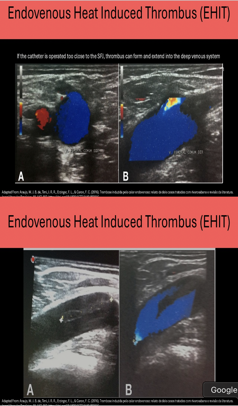
Endovenous heat-induced thrombosis is a complication of:
A. microphlebectomy
B. sclerotherapy
C. radiofrequency ablation
D. chemical ablation
C. radiofrequency ablation
Endovenous heat-induced thrombosis (EHIT) is a complication of radiofrequency ablation. The thrombus that forms in the treated vein can extend into unintended vessels in the deep system. About 7 days after the procedure the patient should return to be evaluated for procedural success and complications like DVT or EHIT.
At ______ post-injection, ______ should be performed to determine the success of a Thrombin injection for pseudoaneurysm treatment.
A. 24-48 hours, color Doppler and ABI evaluation
B. 1 hour, CW Doppler evaluation
C. 20 minutes, color Doppler and ABI evaluation
D. 5 minutes, PPG and ABI evaluation
C. 20 minutes, color Doppler and ABI evaluation
At 20 minutes post-injection, color Doppler and ABl evaluation should be performed to determine the success of a Thrombin injection for pseudoaneurysm treatment. The PSA should be filled with echogenic thrombus and no color flow should be present within it. ABI assessment is performed before and after the procedure to confirm the arterial circulation was unaffected. There should be no change in the ABI from the baseline assessment.
If the great saphenous vein is being used to create an in situ vein graft,
A. the valves will remain in place and provide little resistance to arterial flow
B. all perforator veins must be evaluated for patency because the graft will not function properly with obstructed perforator veins
C. all perforator veins must be marked on the vein mapping so they can be closed
D. the graft diameter will be greatest at the distal end of the graft
C. all perforator veins must be marked on the vein mapping so they can be closed
You must mark all perforating veins if the GSV will be used as an in situ graft or an AV fistula will be created when the graft is arterialized.
The valves must be removed with a valvulotome.
The graft diameter will be greatest at the proximal end.
Which of the following correctly describes a pre-procedure vein mapping?
A. Keep the transducer pressure constant at a medium level
B. The diameter of the vein is usually written on the arm/leg at the location it was taken
C. Veins should maintain a uniform elliptical shape during measurements
D. Larger amounts of gel will aid in visualization of the superficial vessels
B. The diameter of the vein is usually written on the arm/leg at the location it was taken
To protect the transducer from staining from the marker, cover it with a nonsterile probe cover. Remember to use limited gel to allow for easier and more permanent skin marking. The marks need to remain until the procedure is performed.
A permanent marker or carbol fuchsin stain (used in radiation therapy) is used to create the markings.
Keep the transducer perpendicular to the skin surface and use very light transducer pressure. If the vein appears elliptical, then too much transducer pressure is being applied or the transducer is off axis.
A small mark is placed in front of the ultrasound transducer along the narrow edge of the transducer. If a transverse approach is used, the vein should appear circular and centered on the ultrasound screen. A skin mark is then placed exactly at the center of the long face of the transducer.
Once the initial skin mark is placed, the transducer is moved slightly distally toward the foot while keeping the rounded vein in the center of view. A new mark is placed on the skin every 2 to 3 cm.
A straw could be used to make an indentation in the skin and then these marks are connected by lines drawn with a permanent marker after the gel is removed. The diameter of the vein is usually written on the arm/leg at the location it was taken.
When Thrombin is injected into a pseudoaneurysm, the clotting should begin:
A. after 1 minute of manual compression following the injection
B. within 24 hours
C. within 5-10 minutes post- injection
D. within 5-10 seconds post- injection
D. within 5-10 seconds post- injection
Thrombin administration is performed by injecting the clotting agent into a pseudoaneurysm to close the opening and clot the blood that has escaped. Clotting should occur immediately after injection (5-10 seconds). About 20 minutes after the procedure, a follow up exam should be performed to document the pseudoaneurysm is fully thrombosed.
Which of the following is a contraindication for harvesting of the great saphenous vein?
A. valvular incompetence in the great saphenous vein
B. Baker's cyst in the ipsilateral popliteal fossa
C. chronic obstruction of the femoral vein
D. chronic obstruction of the peroneal veins
C. chronic obstruction of the femoral vein
IT IS VERY IMPORTANT to evaluate the venous outflow system of a patient prior to removal or closure of the saphenous vein. If the deep system is obstructed and the superficial system is the main outflow for the leg, the saphenous vein cannot be removed/closed.
A patient presents for a follow up exam after radiofrequency ablation of the GSV. What is the purpose of the exam?
A. To evaluate thrombus formation in the GSV
B. To confirm the patency of the GSV and thrombosis of its branches
C. To confirm the contraction of the GSV and absence of thrombus in the deep system
D. To assess the valvular motion in the GSV
C. To confirm the contraction of the GSV and absence of thrombus in the deep system
A follow up exam after radiofrequency ablation of the GSV should be performed 2 weeks after the procedure to confirm contraction/occlusion of the GSV and absence of thrombus in the deep system. Measuring the distance from the treated GSV segment to the SFJ is important to document. The fibrosis/thrombosis of the treated vein should not extend into the deep veins. Compression views of the treated veins and Doppler demonstration of the absence of flow should also be documented.
Which of the following is a contraindication for US guided compression therapy for a pseudoaneurysm at the groin?
A. Infection at the puncture site
B. Patient that stopped taking Coumadin 3 days ago
C. A pseudoaneurysm that has been present for more than 72hrs
D. A pseudoaneurysm that measures 3.2cm in diameter
A. Infection at the puncture site
Contraindications for US guided compression therapy for a pseudoaneurysm:
Patient unable to tolerate the procedure due to pain Infection at the puncture site and surrounding tissues
Diameter >4cm
Stalk diameter >5mm
Present for more than 1 month
Patients on anticoagulant therapy (must be stopped before compression)
When preparing the sterile tray for an invasive procedure, the cover on the tray should be opened:
A. while wearing sterile gloves
B. no more than 2 hours before the procedure begins
C. by pulling the far corner of the wrapper toward you
D. by the radiologist
C. by pulling the far corner of the wrapper toward you
The Sonographer should prepare the supplies for an ultrasound guided procedure. The tray should not be opened until just before the procedure begins. The cover can be opened without wearing gloves but should be pulled toward you to expose the products facing away from you. This prevents your arm from hovering over the supplies to prevent any "debris from your person from falling into the tray. Sterile gloves must be used to remove items for the tray to prepare them (lidocaine, needle and syringe).