Biochem ch 2-6
 Call Kai
 Learn
 Practice Test
 Spaced Repetition
 Match
 Flashcards
 Knowt Play
1/117

Earn XP
Description and Tags
water, Proteins, 3D Proteins, Protein fucntions, Enzymes Catalysis
118 Terms
Ionic (Coulombic) Interactions
Electrostatic interactions between permanently charged species,
or between the ion and a permanent dipole
Dipole Interactions
Electrostatic interactions between uncharged, but polar molecules
Van der Waals Interactions
Weak interactions between all atoms, regardless of polarity
– Attractive (dispersion) and repulsive (steric) component
Hydrophobic Effect
Complex phenomenon associated with the ordering of water molecules around
non-polar substances
Noncovalent Forces and Interactions
Hydrogen bonds
• Ion-Ion 1/r
• Ion-dipole 1/r2
• Dipole-dipole 1/r3
• Dipole - Induced
dipole - 1/r5
• ID – ID (Van der
Waals) - 1/r6
• Hydrophobic
Hydrogen Bonds
Strong dipole-dipole or charge-dipole interaction that arises
between an acid (proton donor) and a base (proton acceptor)
• Typically involves two electronegative atoms (frequently
nitrogen and oxygen)
• Hydrogen bonds are strongest when
the bonded molecules are oriented to
maximize electrostatic interaction.
Van der Waals Interactions components
two components:
– Attractive force (London
dispersion) Depends on
the polarizability
– Repulsive force (Steric
repulsion) Depends on
the size of atoms
Van der Waals Interactions significance
Weak individually
–Easily broken, reversible
• Universal:
–Occur between any two atoms that are near each other
• Importance
– determines steric complementarity
– stabilizes biological macromolecules (stacking in DNA)
– facilitates binding of polarizable ligand
Water solvent interaction
Good- charged and polar such as amino, peptides, smal alcohol, carbohydrates
Poor- nonpolar such as nonpolar gases, aromatic, aliphatic chains
Water interaction to salts
high dielectric constant shields oppositely charged ions. Entropy increases as orded crystals lattice is dissolved
Ice
hexagonal ice forms regular lattice, low entrophy
Hydryphobic effect
association or folding of non-polar molecules aq. solutions, DOES NOT arise because of some attractice direct force between two non-polar molecules
hydrophobic main factors behind:
protein folding, protein-protein association, formation of lipids micelles, beding of steroid hormones to their receptors
Reasons low solubility hydrophobic solute
disruption of H-bonded H2O network, cavity formation in a medium with high surface tension Lipid molecules disperse in the
solution; nonpolar tail of each lipid
molecule is surrounded by ordered
water molecules
• Lipid aggregates – Water released,
surface area reduced
Colligative Properties
boiling point, melting point, and osmolarity
Effect of Extracellular Osmolarity
isotonic, hypertonic and hypotonic
Proton Hydration
immediately hydrated to form hydronium (oxonium) ions, associated with one of the non-bonding electron pairs
Ionization of Water:
Quantitative Treatmen
Concentrations of participating species in an equilibrium process
are not independent but are related via the equilibrium constan
pH formula
pH=-log[H+]
pKa measures?
acidity, string acids → larger Ka→ small pKa
Buffers
mixture weak acids + anions, resist change of pH, Buffering capacity of acid/anion system is greatest at pH = pKa
Henderson–Hasselbalch Equation

3 elements primarly involve in hydrogen bonding
Hydrogen, Oxygen, Nitrogen
examples weak acids, balanced chem reaction with water, describe how different than strong acid
CH3COOH+H20 →H3O+CH3COO
Weak acids only partially ionize in water, meaning the majority of the molecules remain un-dissociated. Strong acids CH3COOH or HCl ionize completely into their ions in aqueous solution. a weak acid solution has a higher pH (less acidic) and reacts more slowly with bases or metals compared to a strong acid because of the lower concentration of free H3O ions.
Amino Acids
Building blocks, protein are heteropolymers of alpha aminos, chiral

Amino acids fucntions
Capacity to polymerize
– Useful acid-base properties
– Varied physical properties
– Varied chemical functionality
Aliphatic aminos
Hydrophobc side chain


Alanine/Ala/A

Valine/Val/V

Isoleucine/Ile/I

Leucine/Leu/L

Methionine/Met/M
Aromatics aminos
Ring


Phenylalanine/Phe/F

Tyrosine/Tyr/Y
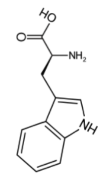
Tryptophan/Trp/W
Charged aminos
electricaly charged side chain, positive: Arg,His,Lys. Negative: Asp, Glu

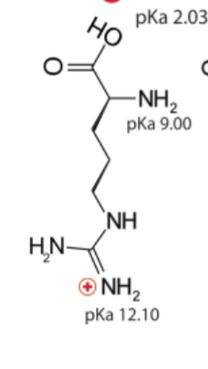
Arginine/Arg/R
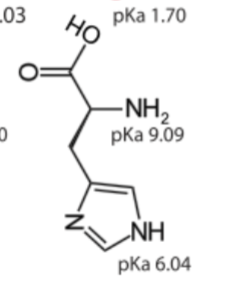
Histidine/His/H

Lysine/Lys/K

Aspartic acid/Asp/D
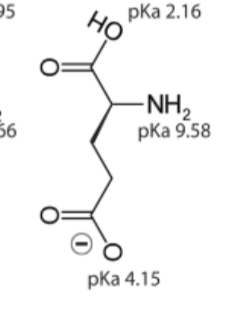
Glutamic acid/Glu/E
Polar aminos
polar uncharged side chain

Serine/Ser/S
Threonine/Thr/T
Asparagine/Asn/N
Glutamine/Gln/Q
Special aminos
Glycine, Proline, Cysteine, Selenocysteine


Glycine/Gly/G

Proline/Pro/P

Cysteine/Cys/C
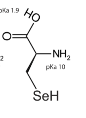
Selenocysteine/Sec/U
Ionization of aminos
At acidic pH, the carboxylgroup is protonated and the amino acid is in the cationic form
At neutral pH, the carboxyl group is deprotonated but the amino group is protonated. The netcharge is zero; such ions are called Zwitterions
At alkaline pH, the amino group is neutral –NH2 and the amino acid is in the anionic form

aminos charge 0
amino acid without ionizable side chains, the Isoelectric
Point (equivalence point, pI), the net charge is zero

Structure of Proteins
This structure is able to fulfill a specific biological
function
• This structure is called the native fold
• The native fold has a large number of favorable
interactions within the protein
• There is a cost in conformational entropy of folding
the protein into one specific native fold
Favorable Interactions in Proteins
Hydrophobic effect
– Release of water molecules from the structured solvation layer
around the molecule as protein folds increases the net entropy
• Hydrogen bonds
– Interaction of N-H and C=O of the peptide bond leads to local
regular structures such as -helixes and -sheets
• London dispersion
– Medium-range weak attraction between all atoms contributes
significantly to the stability in the interior of the protein
• Electrostatic interactions
– Long-range strong interactions between permanently charged
groups
– Salt-bridges, esp. buried in the hydrophobic environment strongly
stabilize the protein
Structure of the Peptide Bond
The resonance causes the peptide bonds
– be less reactive compared to e.g. esters
– be quite rigid and nearly planarRotation around bonds connected to the alpha
carbon is permitted
• (phi): angle around the alpha-carbon—amide
nitrogen bond
•(psi): angle around the beta-carbon—carbonyl
carbon bond
Ramachandran plot significance
Ramachandran plot shows the distribution of psi and phi
dihedral angles that are found in a protein
• shows the common secondary structure elements
• reveals regions with unusual backbone structure

Secondary Structures
alpha helix: h-bonds between nearby residues
beta sheets: h-bondds between adjacent segments not nearby
Random coil: Irregular arrangement of the polypeptide chain
alpha helix
Helical backbone is held
together by hydrogen bonds
between the nearby
backbone amides
• Right-handed helix with 3.6
residues (5.4 Å) per turn
• Peptide bonds are aligned
roughly parallel with the
helical axis
• Side chains point out and
are roughly perpendicular
with the helical axis
interactions of aminos + alpha helix
Not all polypeptide
sequences adopt alpha-helical
structures
• Small hydrophobic residues
such as Ala and Leu are
strong helix formers
• Pro acts as a helix breaker
because the rotation around
the N-Ca bond is impossible
• Gly acts as a helix breaker
because the tiny R-group
supports other
conformation
Helix Macro-Dipol
Peptide bond has a strong
dipole moment
– Carbonyl O negative
– Amide H positive
• All peptide bonds in the
helix have a similar
orientation
• The helix has a large
macroscopic dipole
moment
• Negatively charged
residues often occur near the positive end of the
helix dipole
beta sheets
The planarity of the peptide
bond and tetrahedral geometry
of the alpha-carbon create a
pleated sheet-like structure
• Sheet-like arrangement of
backbone is held together by
hydrogen bonds between the
more distal backbone amides
• Side chains protrude from the
sheet alternating in up and
down direction
parallel b-sheets
H-bonded strands run
in the same direction

antiparallel b-sheets
H-bonded strands
run in opposite directions

Beta turns (hairpins)
change the direction
• The 180° turn is accomplished over four amino acids
• The turn is stabilized by a hydrogen bond from a carbonyl oxygen
to amide proton three residues down the sequence
• Proline in position 2 or glycine in position 3 are common in beta-turns
Proline isomers
For peptide bonds involving proline, about 6-20%
can be in the cis configuration
• Proline isomerization is catalyzed by proline
isomerases
Protein Tertiary Structure
fibrous proteins
globular proteins
Fibrous proteins
typically insoluble; made from a single secondary structure
strcture to function
alpha-keratin, collagen
globular proteins
water-soluble globular proteins
lipid-soluble membraneous proteins
Quaternary structure
ormed by spontaneous
assembly of individual polypeptides into a larger
functional cluster
Chaperones
prevent misfolding, facilitate,
function of globular protein
Storage of ions and molecules
– myoglobin, ferritin
• Transport of ions and
molecules
– hemoglobin, serotonin
transporter
• Defense against pathogens
– antibodies, cytokines
• Muscle contraction
– actin, myosin
• Biological catalysis
– chymotrypsin, lysozyme
ligand
molecule that binds at binding site via non-covalent forces, which
enables the interactions to be transient
Binding: Quantitative Description
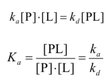
association rate constant ka
the dissociation rate constant kd
After some time, the process will reach the
equilibrium where the association and
dissociation rates are equal
• The equilibrium composition is characterized
by the the equilibrium constant Ka
Analysis in Terms of the
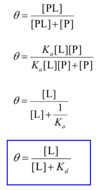
Bound Fraction
fraction of occupied binding sites, In terms of the more commonly used
equilibrium dissociation constant
Specificity: Lock-and-Key Model
Proteins typically have high specificity: only certain ligands bind
• High specificity can be explained by the complementary of the
binding site and the ligand.
•Complementarity in
– size,
– shape,
– charge,
– or hydrophobic / hydrophilic character
•“Lock and Key” model by Emil Fisher (1894) assumes that
complementary surfaces are preformed
Specificity: Induced Fit
Conformational changes may occur upon ligand binding
(Daniel Koshland in 1958).
– This adaptation is called the induced fit.
– Induced fit allows for tighter binding of the ligand
– Induced fit can increase the affinity of the protein for a
second ligand
• Both the ligand and the protein can change their
conformations
function myoglobin
Need to store oxygen for metabolism
• Protein side-chains lack affinity for O2
• Some transition metals bind O2 well but would generate
free radicals if free in solution
• Organometallic compounds such as heme are more
suitable but Fe2+ in free heme could be oxidized to Fe3+
if two heme molecules share an oxygen molecule
• Solution:
– Capture the oxygen molecule with heme that is
sequestered inside the protein
– In mammals, myoglobin is the main oxygen storage
protein

Structures of Porphyrin and Heme
Binding of Carbon Monoxide
CO has similar size and shape to O2; it can fit to the same
binding site
• CO binds over 20,000 times better than O2 because the
carbon in CO has a filled lone electron pair that can be
donated to vacant d-orbitals on the Fe2+
• Protein pocket decreases affinity for CO, but is still binds
about 250 times better than oxygen
• CO is highly toxic as it competes with oxygen. It blocks the
function of myoglobin, hemoglobin, and mitochondrial
cytochromes that are involved in oxidative phosphorylation
How Can Affinity to Oxygen Change
Like This?
Must be a protein with multiple binding sites
• Binding sites must be able to interact with each other
• This phenomenon is called cooperativity
– positive cooperativity: first binding event
increases affinity at remaining sites
– negative cooperativity: first binding event
reduces affinity at remaining sites
Positive cooperativity can be
recognized by sigmoidal binding
curves
Hill equation

What is the term that scientists use to compare the binding affinity of ligands? Give 2 (non-numerical) examples of a ligand binding to a protein.
The term scientist use to compare the binding affinity of ligads is the dissociation constant K_d.
1) oxygen binding to hemoglobin
2) enzyme binding
Myoglobin contains the _____________ prosthetic group and the ________ ion that allow for reversible to _____________ and nonreversible binding to _________________.
Myoglobin contains the heme prothetic group and the iron ion that allows for reversible to Oxygen and nonreversible binding to Carbon Monoxide
Hemoglobin contains 2 _______ subunits and 2 ________ subunits and exists in either the ___________state or the _______ state.
Briefly describe the O2 binding cooperativity in Hemoglobin.
Hemoglobin contains 2 alpha helixes subunit and 2 beta subunits and exist in either the Tense state or Relaxed state
alpha, beta, T(tense), R(relaxed) Increase the affinity of remaining subunits for oxygen. Goes from a T state to R state meaning easier binding

The image below is a typical structure of an immunoglobulin G (IgG), identifying the 2 heavy chains, 2 light chains, and which region is most variable.
Light chain: A/B left, G/H right, Heavy chains: C,D, F, E, Most variable: A and H
Enzyme?
Enzymes are catalytically active biological
macromolecules
• Specific, Efficient, Active in Aqueous Solution
• Most enzymes are globular proteins,
however some RNA (ribozymes, and ribosomal RNA) also
catalyze reactions
Non-peptide Co-Factors (Metals, Vitamins, Coenzymes)
• Enzymes can be classified functionally
Biocatalysis?
Higher reaction rates
• Greater reaction specificity
• Milder reaction conditions
• Capacity for regulation
Metabolites have
many potential
pathways of
decomposition
• Enzymes make the
desired one most
favorable


enzymatic Substrate Selectivity
Kinetics and Equilibrium
Enzymes affect rates but not equilibria

Transition State Theory
Slow reactions face significant activation
barriers that must be surmounted during the
reaction
–transition state theory is applicable for
catalysis
–rate constants and free energies can be
related
Rate Acceleration
The enzyme
lowers the
activation barrier
compared to the
uncatalyzed
aqueous reaction

How to Lower changeG‡?
Enzymes organizes reactive
groups into proximity
• Uncatalyzed bimolecular reactions:
two free reactants single restricted transition state
conversion is entropically unfavorable
• Uncatalyzed unimolecular reactions:
flexible reactant rigid transition state conversion is
entropically unfavorable for flexible reactants
• Catalyzed reactions:
Enzyme uses the binding energy of substrates to organize
the reactants to a fairly rigid ES complex
Entropy cost is paid during binding
Rigid reactant complex transition state conversion is entropically OK
How is TS Stabilization Achieved?
acid-base catalysis: give and take protons
– covalent catalysis: change reaction paths
– metal ion catalysis: use redox cofactors, pKa shifters
– electrostatic catalysis: preferential interactions with TS
acid-base catalysis
give and take protons,

metal ion catalysis
use redox cofactors, pKa shifters
electrostatic catalysis
preferential interactions with TS
Proteases and peptidases
– chymotrypsin, elastase, subtilisin
– reactive serine nucleophile
• Some aldehyde dehydrogenase
– glyceraldehyde-3phosphate dehydrogenase
– reactive thiolate nucleophile
• Aldolases and decarboxylases
– amine nucleophile
• Dehalogenases
– carboxylate nucleophile
Enzyme Kinetics
Enzymes affect rates but not equilibria
• Reactions are Complex – Simplifying
assumptions
Michaelis-Menten kinetics
Total Enzyme concentration is fixed