Sectional Anatomy, Anatomic Planes & Directions, Transducers & Image Shapes, Scanning Techniques
 Call Kai
 Learn
 Practice Test
 Spaced Repetition
 Match
 Flashcards
 Knowt Play
1/44

Earn XP
Description and Tags
Chapters 5 & 6
45 Terms
Assumes that the body is standing erect, the eyes are looking forward, and the arms are at the sides with the palms and toes directed forward.
Anatomic Position
The sonographer images, the body in multiple planes, which include but are not limited to
Transverse, Sagittal, Longitudinal, Coronal
Divides the body or any of its parts into right and left sides
Sagittal/Longitudinal
Lengthwise plane running from front to back
Sagittal/Longitudinal
Divides the body into equal halves
Midsagittal plane
Horizontal to the body; Divides the body or any of its parts into upper and lower portions
Transverse
Lengthwise plane that runs from side to side; Divides the body into anterior and posterior parts
Coronal
The top of the head is the most _______ point of the body
superior

cephalic and cranial
towards the head
The most ________ point of the body is the bottom of the feet
inferior
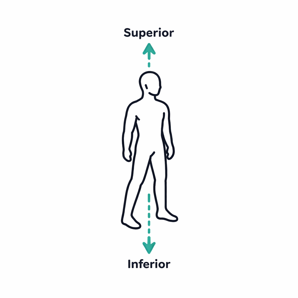
Caudal
toward the tail
The front (belly) surface of the body; also called ventral
Anterior

Back surface of the body; also called dorsal
Posterior

Patient lies on their back, face forward
Supine

Patient lies on their abdomen, face down
prone

An imaginary line from the center of the top of the head to the groin
Body Axis
Structures are those that are closer to the midline of the body than they are to another structure; The superoinferior body axis that goes right through the midline of the body
Medial

Structure toward the side of the body
Lateral

Structure is closer to the body midline or point of attachment to the trunk
Proximal

Farther from the midline or point of attachment to the trunk
Distal

Located close to the surface of the body
Superficial
Located farther inward, away from the body surface
Deep
Transducers
•Convert one form of energy to another
•Various types
•Type used dependent on many factors
Common Transducers
Curved
Linear
Phased

Linear Array
Thin, rectangular transducer
Rectangle shaped image
Used for Superficial Structures and Vascular imaging
Curved Array/Curvilinear
Transducer has a curved/convex shape
Curved sector (pie) shaped image
Used for Abdominal Scanning and OB/Gyn
Phased Array
Small rectangular or square transducer
Smaller “Footprint”
Sector (pie) shaped image
Used in mainly Echocardiography
What is the best scanning window determined by?
The area of interest—what do you need images of?
The anatomy of the patient—where is it located?
The presence/absence of bowel gas—can you see the liver from the anterior surface of the abdomen?
The patient’s position—is he/she lying supine or on his/her side?
The patient’s body habitus (their build)—how far from the skin surface is the area of interest?
What is the transducer selection determined by?
The type of exam being performed
The window available (array)
Can you use a large footprint or the does window require a small footprint
The patient’s body habitus (high or low frequency)
Patient is either sitting or standing
Erect

Patient lies on their right side w/ left arm over head (if possible)
Right Lateral Decubitus (RLD)


Patient lies on their left side with right arm over head (if possible)
Left Lateral Decubitus (LLD)
Patient lies on their back with the left side elevated between supine and lateral decubitus –maintaining hip & shoulder alignment
Right Posterior Oblique (RPO)
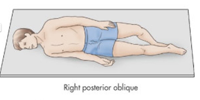
Patient lies on their back with the right side elevated between supine and lateral decubitus –maintaining hip & shoulder alignment
Left Posterior Oblique (LPO)

How should the image be labeled?
The image should be labeled in such a way that someone else could recreate the image by looking at the label. Accurate labeling of images is EXTREMELY important!
Patient ID-Each exam should include
Patient’s name
Patient ID number (MRN: medical record number)
Date of exam
Initials of sonographer
What labeling should image should include?
The patient’s position
Plane you are scanning
Organ you are imaging
A set of guidelines that tell what images are necessary to complete a specific exam
Protocol
Protocol also includes
patient preparation requirements and equipment set-up instructions
When looking for an organ/structure, the sonographer must be able to identify
neighboring structures or vasculature as landmarks
Where is the caudate?
Between the Ligamentum Venosum & IVC
What plane screen is oriented with the head (superior) to the left of the screen and the feet (inferior) to the right of the screen and the top of the screen represents the anterior (superficial) aspect of the patient and the bottom of the screen is posterior
Sagittal/Longitudinal

What plane is the probe is oriented vertically and the sonographer sweeps through the structure in the lateral-medial direction?
Longitudinal/Sagittal
What scans are oriented with the right side of the patient’s body on the left side of the screen and the left side of the patient’s body on the right side of the screen as if you are facing the patient?
Transverse

What plane is the probe is oriented horizontally and the sonographer sweeps through the structure in the superior-inferior direction?
Transverse