Chapter 3: the eye and retina
 Call Kai
 Learn
Learn Practice Test
 Spaced Repetition
 Match
Match Flashcards
 Knowt Play
1/117

There's no tags or description
Looks like no tags are added yet.
Name | Mastery | Learn | Test | Matching | Spaced | Call with Kai | Chat |
|---|
No analytics yet
Send a link to your students to track their progress
118 Terms
The sharp, colorful scene represents perception created by activation of
cone receptors in the retina.
The less focused, grey-scale scene represents perception created by activation of
rod receptors in the retina.
Larry Hester
began noticing a rapid decline in his vision. He had always had poor eyesight, but this was different;
had a genetic disorder of the eye called retinitis pigmentosa that would result in total blindness, and that there was no stopping it
had the opportunity for a bionic eye
step 1 in visual process
the distal stimulus (tree)
step 2
light is reflected from the tree and enters the eye to create the proximal stimulus on the visual receptors
step 3
receptors transform light into electrical signals
step 4
electrical signals are processed as they travel through a network of neurons
The ability to see a tree, or any other object, depends on
light being reflected from that object into the eye
Vision is based on
visible light, which is a band of energy within the electromagnetic spectrum
The electromagnetic spectrum is a
continuum of electromagnetic energy that is produced by electric charges and is radiated as waves
wavelength
For light energy, the distance between one peak of a light wave and the next peak.
the energy in the electromagnetic spectrum can be described by this.
the wavelengths in the electromagnetic spectrum range from
short wavelength gamma rays to long wavelength radio waves
visible light
The band of electromagnetic energy that activates the visual system and that, therefore, can be perceived. For humans, visible light has wavelengths between 400 and 700 nanometers.
For humans and some other animals, the wavelength of visible light is associated with the
different colors of the spectrum, with short wavelengths appearing blue, middle wavelengths green, and long wavelengths yellow, orange, and red.
eyes
The eyeball and its contents, which include focusing elements, the retina, and supporting structures.
contain the receptors for vision
Light reflected from objects in the environment enters the eye through the
pupil
cornea
The transparent focusing element of the eye that is the first structure through which light passes as it enters the eye. The cornea is the eye’s major focusing element.
lens
The transparent focusing element of the eye through which light passes after passing through the cornea and the aqueous humor. The lens’s change in shape to focus at different distances is called accommodation.
retina
A complex network of cells that covers the inside back of the eye. These cells include the receptors, which generate an electrical signal in response to light, as well as the horizontal, bipolar, amacrine, and ganglion cells.
photoreceptors
the receptors for vision
two types of photoreceptors
rods and cones
rods
A cylinder-shaped receptor in the retina that is responsible for vision at low levels of illumination.
cones
receptors in the retina that are primarily responsible for vision in high levels of illumination and for color vision and detail vision.
outer segments
Part of the rod and cone visual receptors that contains the light-sensitive visual pigment molecules
visual pigments
A light-sensitive molecule contained in the rod and cone outer segments. The reaction of this molecule to light results in the generation of an electrical response in the receptors.
Signals from the receptors flow through the network of neurons that make up the
retina, and emerge from the back of the eye in the optic nerve
optic nerve
Bundle of nerve fibers that carry impulses from the retina to the lateral geniculate nucleus and other structures. Each optic nerve contains about 1 million ganglion cell fibers.
fovea
A small area in the human retina that contains only cone receptors. The fovea is located on the line of sight, so that when a person looks at an object, the center of its image falls on the fovea
peripheral retina
The area of retina outside the fovea.
contains both rod and cones
The peripheral retina contains many more
rods than cones because there are 120 million rods and only 6 million cones in the retina
the vertical brown bar near 20 degrees indicates the place on the retina where
there are no receptors because this is where the ganglion cells leave the eye to form the optic nerve. (blind spot)
macular degeneration
A clinical condition that causes degeneration of the macula, an area of the retina that includes the fovea and a small surrounding area.
common in older people
destroys the cone-rich fovea and small area that surrounds it
creates a blind region in central vision (when a person looks directly at something they lose sight of it)

macular degeneration

retinis pigmentosa
retinitis pigmentosa
A retinal disease that causes a gradual loss of vision, beginning in the peripheral retina.
passed from one generation to the next (not always affecting everyone though)
first attacks peripheral rod receptors and results in poor vision in the peripheral visual field
in severe cases, the foveal cone receptors are also attacked resulting in complete blindness
blind spot
The small area where the optic nerve leaves the back of the eye. There are no visual receptors in this area, so small images falling directly on the blind spot cannot be seen.
Why aren’t we usually aware of the blind spot?
the blind spot is located off to the side of our visual field, where objects are not in sharp focus.
a mechanism in the brain “fills in” the place where the image disappear***
CREATES A PERCEPTION THAT MATCHES THE SURROUNDING PATTERN
How the brain can create a coherent perception of our world
example: filling in the area served by the blind spot
Light reflected from an object into the eye is focused onto the retina by a two-element optical system:
the cornea and the lens
The cornea, the transparent covering of the front of the eye, accounts for about
80 percent of the eye’s focusing power,
BUT fixed in place and cannot adjust its focus
The lens, which supplies the remaining 20 percent of the eye’s focusing power, can
change its shape to adjust the eye’s focus for objects located at different distances
achieved by the action of ciliary muscles (can increase curvature of lens)

myopia (eye relaxed)

correction of myopia
. (a) Rays of light coming from a small light source that is more than 20 feet away are approximately
parallel
focus point is on the retina
(b) Moving an object closer to the relaxed eye pushes the focus point
back
light is stopped by the back of the eye so the image on the retina is out of focus
(c) Accommodation of the eye (indicated by the fatter lens) increases the focusing power of the lens and brings the focus point
for a near object back to A on the retina, so it is in focus
caused by the action of the ciliary musscles
(d) In the myopic (nearsighted) eye, parallel rays from a distant spot of light are brought to a focus in
front of the retina, so distant objects appear blurred.
a corrective lens bends light so it is focused on the retina
accommodation
In vision, bringing objects located at different distances into focus by changing the shape of the lens.
refractive errors
Errors that can affect the ability of the cornea and/or lens to focus incoming light onto the retina.
accomodation is not foolproof
sometimes focusing the image onto the retina fails
refractive error: aging - Presbyopia
The inability of the eye to accommodate due to a hardening of the lens and a weakening of the ciliary muscles. It occurs as people get older.
struggle to see at close range
solution: reading glasses
refractive error: myopia
nearsightedness (can’t see distant objects clearly)
why: the optical system brings parallel rays of light into focus at a point in front of the retina, so the image that reaches the retina is blurred.
problem of light being focused in front of the retina (blurred)
refractive myopia, axial myopia
refractive myopia
the cornea and/or lens bends the light too much
axial myopia
the eyeball is too long
hyperopia
farsighteness
can see distant objects but struggle seeing nearby objects
why: because the focus point for parallel rays of light is located behind the retina, usually because the eyeball is too short.
Visual transduction occurs in
photoreceptors (the rods and cones) and transforms light into electricity.
Visual pigments have two parts:
opsin and retinal
opsin
a long protein
100 of times longer than retinal
retinal
smaller, light-sensitive component
when the retinal and opsin are combined, the resulting molecule
absorbs visible light

A = retinal
B = opsin
the change in shape of the retinal molecule is called isomerization, and triggers a sequence of reactions that culminates in generation of an electrical response in the receptor
When incoming light hits the retina, the first step of
transduction is initiated
the visual pigment molecule absorbs the light
causes the retinal within that molecule to change its shape from bent to STRAIGHT
Isomerization
Change in shape of the retinal part of the visual pigment molecule that occurs when the molecule absorbs a quantum of light. Isomerization triggers the enzyme cascade that results in transduction from light energy to electrical energy in the retinal receptors.
he visual pigments in these two types of photoreceptors influence two aspects of visual perception
how we adjust to darkness, and
how well we see light in different parts of the spectrum.
dark adaptation
Visual adaptation that occurs in the dark, during which the sensitivity to light increases. This increase in sensitivity is associated with regeneration of the rod and cone visual pigments.
measured by determining a dark adaptation curve
dark adaptation curve
The function that traces the time course of the increase in visual sensitivity that occurs during dark adaptation.
dark adaptation curve, which is the
function relating sensitivity to light to time in the dark, beginning when the lights are extinguished.
The first step in measuring a dark adaption curve is to have the participant
look at a small fixation point while paying attention to a flashing test light that is off to the side
image falls on the fovea
While still in the light, the participant turns a knob that adjusts the intensity of the flashing light until it can just barely be seen
the method of adjustment
This threshold for seeing the light, the minimum amount of energy necessary to just barely see the light, is then converted to
sensitivity
Because sensitivty = 1/threshold , this means that a high threshold corresponds to
low sensitivity
light-adapted sensitivity
The sensitivity of the eye when in the light-adapted state. Usually taken as the starting point for the dark adaptation curve because it is the sensitivity of the eye just before the lights are turned off.
measured in the light

the downward movement of these curves represents an increase in sensitivity.
The curves actually begin at the points indicating “light-adapted sensitivity,” but there is a slight delay between the time the lights are turned off and when measurement of the curves begins.
higher sensitivity is at the bottom (participant’s sensitivity is increasing/downward curve)
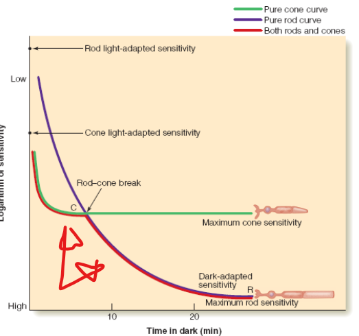
. The red line is the two-stage dark adaptation curve, with an initial cone branch and a later rod branch, which occurs when the test light is in the peripheral retina,

The green line is the cone adaptation curve, which occurs when the test light falls on the fovea.

The purple curve is the rod adaptation curve measured in a rod monochromat
The dark adaptation curve shows that as adaptation proceeds,
the participant becomes more sensitive to the light.
dark-adapted sensitivity
The sensitivity of the eye after it has completely adapted to the dark.
The sensitivity at the end of dark adaptation, labeled dark-adapted sensitivity, is about 100,000 times greater than
the light-adapted sensitivity measured before dark adaptation began.
keeping an eye in the dark triggers the process of
dark adaptation, which causes the eye to increase its sensitivity in the dark.
This curve, which measures only the activity of the cones, matches the initial phase of our original dark adaptation curve but does not include the second phase. Does this mean that the second part of the curve is due to the rods?
yes!
rod monochromats
people, who have no cones because of a rare genetic defect
in order to reveal how the sensitivity of the rods is changing at the very beginning of dark adaptation we need to measure it in a person who has no cones
rod-cone break
The point on the dark adaptation curve at which vision shifts from cone vision to rod vision.
visual pigment regeneration occurs more rapidly in the
cones than in the rods
visual pigment bleaching.
The change in the color of a visual pigment that occurs when visual pigment molecules are isomerized by exposure to light.
change in shape and separation from the opsin causes the molecule to become lighter in color,
visual pigment regeneration
Occurs after the visual pigment’s two components—opsin and retinal—have become separated due to the action of light. Regeneration, which occurs in the dark, involves a rejoining of these two components to reform the visual pigment molecule. This process depends on enzymes located in the pigment epithelium.
Our sensitivity to light depends on the
concentration of a chemical—the visual pigment.
The speed at which our sensitivity increases in the dark depends on a chemical reaction
the regeneration of the visual pigment.
detached retina
A condition in which the retina is detached from the back of the eye.
what occurs when a person’s retina becomes detached from the pigment epithelium, a layer that contains enzymes necessary for pigment regeneration
Rods and cones also differ in the way they respond to light in different parts of the
visible spectrum
spectral sensitivity
The sensitivity of visual receptors to different parts of the visible spectrum.
Spectral sensitivity is measured by determining the
spectral sensitivity curve—the relationship between wavelength and sensitivity.
monochromatic light
Light that contains only a single wavelength.
cone spectral sensitivity
A plot of visual sensitivity versus wavelength for cone vision. Often measured by presenting a small spot of light to the fovea, which contains only cones. Can also be measured when the eye is light adapted, so cones are the most sensitive receptors.
rod spectral sensitivity curve
The curve plotting visual sensitivity versus wavelength for rod vision. This function is typically measured when the eye is dark adapted by a test light presented to the peripheral retina.
Purkinje (Pur-kin’-jee) shift
The shift from cone spectral sensitivity to rod spectral sensitivity that takes place during dark adaptation
absorption spectrum
A plot of the amount of light absorbed by a visual pigment versus the wavelength of light.
neural circuits
A number of neurons that are connected by synapses.
bipolar cells
A retinal neuron that receives inputs from the visual receptors and sends signals to the retinal ganglion cells.
ganglion cells
A neuron in the retina that receives inputs from bipolar and amacrine cells. The axons of the ganglion cells are the nerve fibers that travel out of the eye in the optic nerve.