3.1.5 Investigates the visual fields of patients with all standards of acuity and analyses and interprets the results
 Call Kai
 Learn
 Practice Test
 Spaced Repetition
 Match
 Flashcards
 Knowt Play
1/62

There's no tags or description
Looks like no tags are added yet.
63 Terms
What are the characteristics of the visual field?
The binocular field has a zero zone where you achieve stereopsis. Beyond this zone is the extreme periphery, it is only governed by one eye at a time.
The normal hill of vision
Light sensitivity is greatest at the fovea
The blind spot is 5.5° wide and 7.5° high
The blind spot is located 15° degrees from the fixation point and 1.5° below the horizontal midline.

Decibels (dB)
refers to stimulus intensity
0dB = maximum brightness
51dB = minimum brightness
Visual Field Index (VFI)
Quantifies the overall status of the visual field
Useful for monitoring changes over time but not a useful in classifying the severity of vision field loss
E.g. patients could have a dense central scotoma but have a VFI of 99%
Mean Deviation (MD)
How the average of the whole field differs from agenormal findings
Approx. 0 dB in normal fields
Pattern Standard Deviation (PSD)
- The SD of the values in the total deviation plot
- An indication of focal loss as it demonstrates whether the total deviation are approximately the same or different from each other
- As focal loss increases, PSD becomes more positive
- Once PSD >12.0dB, it decreases as focal loss is so large that it begins to diffuse and be less anomalous
Glaucoma Hemifield TEST (GHT)
- Pattern deviation in 5 zones of the upper hemifield are compared to mirror-image zones in the lower hemifield (Used in 30-2 and 24-2 tests)
“Outside normal limits” - one zone pair differs by amount of found in fewer than 1% normal subjects
“Borderline” - one zone pair differs by amount found between 1-3% normal subjects
“Abnormally high sensitivity” - when best points are so high that they’re on seen by 0.5% normal subjects
“General reduction of sensitivity” - when best points are so low that they are not seen by fewer than 0.5% normal subjects
The Greyscale
- Visual representation of raw decibel sensitivity
- Not compared to normal data ranges
- Successfully shows profound visual defects or artifactual loss
Total deviation
- Threshold sensitivity is compared with age-corrected normal at each test point to produce numerical maps
- These numerical values are then images with probability agents to clearly show areas outside normal limits
Pattern deviation
Removes any generalised depression or elevation so vision loss pattern can be distinguished
Based on a “general height adjustment”
The external readings are placed in order form positive to negative
The 7th most positive value is applied to all the readings so that the 7th value becomes 0db
Means that subtle but significant variations are highlighted and easily visible to the interpreter
Stimulus parameters
SITA - Swedish Interactive Threshold Algorithm
-Compares the frequency of seeing curve at each location in a normal eye and also in the glaucomatous eye to the patients frequency of seeing curve
- Allows for short test times as the predicted values are close to the threshold
- SITA STANDARD uses 2-4dB jumps
- SITA FAST uses 4dB jumps
Stimulus parameters
Stimulus colour
- Background is always at 31.5 ASB
- Colour of stimulus is always fixed as ‘Goldman white’
Stimulus parameters
Stimulus size
- All normative data and statistical analysis is based upon the Size III stimulus
Stimulus parameters
False negatives
-Presenting 9dB brighter stimulus on a place where a stimulus was apparently seen
- If exceeds 20% then test deemed unreliable
Stimulus parameters
Stimulus parameters
- When patient clicks the button too quick that there were clearly anticipating the stimulus
- Unreliable test if error exceeds 15%
The “-2” indicates…
the stimulus offset from the horizontal and vertical midline
30-2
- Stimuli are 6º apart; the middle stimuli are 3º off the midline
- The field of examination is 27º wide as there a 9 spaces between stimuli across the wides horizontal line
24-2
- Stimuli are 6º apart; the middle stimuli are 3º off the midline
- The field of examination is 21º wide as there a 7 spaces between stimuli across the wides horizontal line
24-2c
- Modified 24-2 the incorporates 10 additional test points in central 10º of vision
10-2
- Stimuli are 2º apart; the middle stimuli are 1º off the midline
- Field is 9º wide
- Used for advanced glaucoma when patient has small visual field
Fixation stability
- Blind spot monitoring
- The patient shouldn’t see stimuli presented to the blind spot area
Gaze tracking system
Analysis of distance between centre of pupil and the corneal reflex
- Upward markings indicate fixation loss of >10º
- Downward markings indicated blinking
What is Suprathreshold perimetry (C40)
- Presents stimulus at a known luminance higher than expected threshold
- Stimulus is either seen or not seen
- Doesn’t give indication of the depth of the loss so. if the stimulus is not seen then the depth of the loss should be investigated though full threshold testing
Suprathreshold perimetry (C40)
Disadvantages
Non-quantitative
Cannot separate optical loss from neural loss
Insensitive to shallow field loss
- USELESS AT DETECTING EARLY GLAUCOMATOUS FIELD LOSS
Artefacts
Ocular factors
-Increasing cataract or media opacities
- Reduction in pupil size
Artefacts
Upper lid position
- Ptosis often impacts 30-2 tests
- May need to elevate lid with surgical tape to make sure lid doesn’t give superior defect
Artefacts
Untrained patient
- 10% patients show decreases sensitivity on first attempt
Artefacts
Incorrect date of birth
Inability to compare results with correct DoB results in unfavourable total deviation maps
Artefacts
Trial lens power and position
- ~1dB reduction per 1.00D defocus
- High plus sense cause magnification and some points will be obscured by peripheral scotoma
Artefacts
Patient fatigue or inattention
- Peripheral locations exhibit greater reduction
- May form ‘clover leaf field’
Artefacts
Trigger-happy field
Many false positive results will cause algorithm to push threshold to a non-humanly possible level
Common Glaucomatous Field Defects
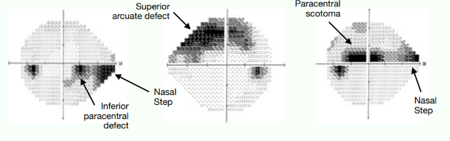
Nasal Step
Widespread involvement of fibres in all parts of the optic disc are effected
Light sensitivity in opposing hemifield may not be equal
Doesn’t cross horizontal meridian
Common Glaucomatous Field Defects
Arcuate Defect
Deep focal notch at optic disc leads to loss of all RNFL in the area corresponding to the notch
Visual loss circumnavigates around the point of fixation abiding horizontal raphe
Common Glaucomatous Field Defects
Ring scotoma
When two separate arcuate defects occur in each hemifield and potentially meet at the horizontal raphe
Common Glaucomatous Field Defects
Central scotoma
Can arise from superior and inferior fixation splitting defects are both present

Label the defect:


Label the defect:

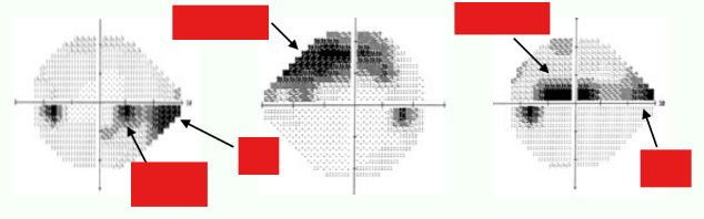
Label the defects:

Neurological Field Defects
The Optic Chiasm
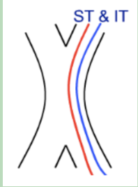
Nasal macula fibres cross over from one eye to the other through the centre of the chiasm.
Temporal fibres do not cross over
Neurological Field Defects
The Optic Chiasm
Superior / inferior temporal fibres don’t cross over, they come from the right optic nerve and carry on into the right optic tract

Neurological Field Defects
The Optic Chiasm
Even though nasal fibres cross, the path isnt direct.
Superior nasal fibres cross over at the chiasm but pass into the optic tract of the same eye before coming back and crossing over to the contralateral optic tract. “Knee of Willbrand"

Neurological Field Defects
The Optic Chiasm
Inferior nasal fibres go back into the opposite side of the optic nerve head before heading to the contralateral optic tract
Neurological Field Defects
Retinal induced defects
Retinal toxicity
Vascular damage
Age-related Macular Degeneration
Retinitis pigmentosa
Retinal induced defects
Retinal toxicity
Drugs for systemic conditions can be toxic to the retina causing photoreceptor damage
Vigabatrin is an anti-epilepsy drug where 1/3 patients give rise to permanent bilateral nasal defects.
Retinal induced defects
Vascular damage
BRVO obscures light from reaching the photoreceptors and can give rise to an arcuate defect if severe
Retinal induced defects
Age-related Macular Degeneration
Visual field loss mimics damage to the photoreceptors, resulting in pronounced and severe central field loss
Retinal induced defects
Retinitis pigmentosa
Tunnel vision due to the degeneration of photoreceptors starting at in the peripheral retina
What are the characteristics of optic nerve disease?
Only affects the affected eye
Most optic nerve diseases cause central scotoma
Early optic disc oedema may only enlarge the blind spot
What optic nerve diseases can produce visual field defects, and what are these defects?
Optic disc drusen or optic nerve head coloboma can produce arcuate defects that may be indistinguishable from glaucomatous defects
Optic neuritis can cause defects from complete inferior field loss to a small paracentral defect
Meningioma tumour can compress the optic nerve but this doesn’t have a characteristic defect
What happens to the optic nerve fibres as they pass down towards the chiasm?
- As the optic nerve fibres pass down there is a reorganisation of the fibres so by the time it reaches the chiasm, all macula fibres are in the centre of the optic nerve and there is a concentration of different nerve fibres with different functions in different aspects of the nerve.

Optic chiasm lesions

View of the chiasm

There are a variety of ways in which we can damage the optic chiasm:
1. Compressive injury (tumour)
2. Vascular origin
What are the effects of meningioma compression on the anterior optic chiasm?
Compresses the left optic nerve and the anterior knee of Willbrand.
This will cause complete loss of vision in one eye, and a scotoma in the other eye.

What are the effects of pituitary gland tumours on the visual field?
Fairly common. Tumour grows upwards, causing pressure on the chiasm. Over a period of time the pressure starts to damage fibres and kill them, leading to visual field loss.
Pituitary adenoma is the most common lesion and it is a bitemporal defect, which leads to reduced visual acuity.

What are the effects of subarachnoid haemorrhage?
This will damage the chiasm but more importantly, the brain, this is life threatening.

What causes damage to the circle of Willis, and what are the effects?
Due to atherosclerosis.
This is plaque forming, containing cholesterol, which stick to blood vessels in the aorta and in the internal carotid artery in the neck.
VF defects from this are in the form of an altitudinal defect or sometimes a diffuse visual field defect.
What are the effects of lateral geniculate nucleus (LGN) damage?
The LGN blood supply is often thin vessels so vascular damage is the most likely cause of LGN damage.
Damage to the ventral boundary of the LGN leads to an hourglass defect.

What are the effects of optic tract lesions?
Tumours in the visual pathway become rarer the further back in the pathway you are.
The most common of the rare tumours is the glioglioma.
Results in a homonymous hemianopia. There may be low symmetry as the overlap between the two eyes is poor.
What are the effects of optic radiation damage?
Compressive tumours or vascular damage can damage the optic radiations.
The types of tumours are very rare.
What are the effects of a stroke on the visual field?
Most common cause of death
Obstructing blood supply in the brain leads to oxygen starvation, giving rise to vision damage.
The most common visual field presentation is a homonymous hemianopia.
If recovery occurs it will tend to happen within 1 month of onset.
What are the effects of optic chiasm lesions on the visual field?
Damage to the optic chiasm can cause visual field defects affecting both eyes.
Pituitary adenoma is the most common lesion and it causes a bitemporal defect, which leads to reduced visual acuity.

What are the characteristics of the striate cortex?
The central 30º visual field is represented by approximately 80º of the striate cortex
Macular sparring is difficult to demonstrate clinically.
Hard to image on a 30-2 or 24-2 as the stimuli are 6º apart. However a 10-2 grid (2º apart) and Amsler grid have enough resolution to demonstrate macular sparing