A&P 1 - Lab Manual Unit 3
 Call Kai
 Learn
 Practice Test
 Spaced Repetition
 Match
 Flashcards
 Knowt Play
1/134

There's no tags or description
Looks like no tags are added yet.
135 Terms
Psoas Major
I: lesser trochanter of femur
A: flexes at hip

Iliacus
O: iliac fossa
A: flexion at hip

Piriformis (cant see on model)
O: anterolateral surface of sacrum
I: greater trochanter of femur
A: lateral rotation and abduction at hip

Gracilis
O: inferior ramus of pubis
I: medial surface of tibia
A:
knee flexion
adducts thigh
medially rotates at hip

Adductor Magnus (cant see on model)
O: inferior ramus of pubis and ischial tuberosity
I: linea aspera of femur and adductor tubercle of femur
A: adduction at hip (part can do flexion and medial rotation at hip; inferior part can produce extension and lateral rotation at hip)

Adductor Longus
O: body of pubis
I: linea aspera of femur
A:
adduction at hip
can contribute to flexion and medial rotation of hip

Gluteus Maximus
I:
(IT) tract
gluteal tuberosity of femur
A:
extension (moves leg backward)
lateral rotation at hip (turns leg outward)

Gluteus Medius (can’t see on model)
O: anterior iliac crest of ilium
I: greater trochanter of femur
A: abduction and medial rotation of hip

Tensor Fasciae Latae
O: iliac crest and (ASIS)
I: (IT) tract
A:
abduction (moves leg away from midline)
medial rotation of hip

Sartorius
O: (ASIS)
I: medial surface of tibia
A:
flexes knee
flexion
lateral rotation of hip

Rectus Femoris
O: (AIIS) and superior acetabular rim of ilium
I: tibial tuberosity via patellar ligament
A:
extension at knee
flexion at hip

Vastus Medialis
O: linea aspera of femur
I: tibial tuberosity via patellar ligament
A: extends knee

Vastus Intermedius (can’t see on model)
Origin, Insertion, Action: same as vastus medialis (linea aspera of femur; tibial tuberosity via patellar ligament; extends knee)

Vastus Lateralis
Origin, Insertion, Action: same as vastus medialis (linea aspera of femur; tibial tuberosity via patellar ligament; extends knee)

Biceps Femoris
O: ischial tuberosity and linea aspera of femur
I: head of fibula
A:
flexion at knee (bends the leg)
extension
lateral rotation at hip

Semitendinosus
O: ischial tuberosity
I: proximal and medial end of tibia
A:
flexion at knee
extension
medial rotation at hip

Semimembranosus
O: ischial tuberosity
I: medial condyle of tibia
A:
flexion at knee
extension
medial rotation at hip

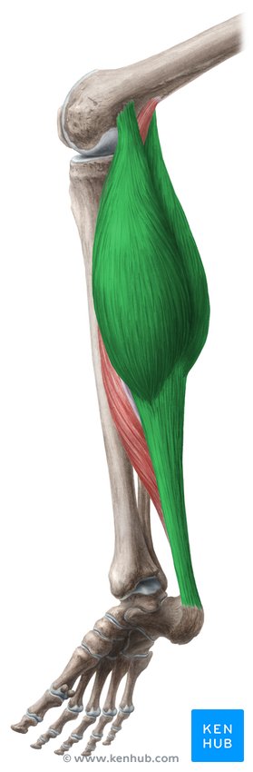
Gastrocnemius
O: femoral condyles
I: calcaneus via calcaneal tendon
A:
plantarflexion
inversion
knee flexion
Soleus
O: head and proximal shaft of fibula
I: calcaneus via calcaneal tendon
A: plantarflexion

Tibialis Anterior
O: lateral condyle and proximal shaft of tibia
I: metatarsal and cuneiform
A:
dorsiflexion
inversion of foot

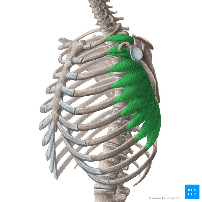
Serratus Anterior
O: ribs 1 through 9
I: vertebral border of scapula
A: protracts scapula
Pectoralis Minor
O: ribs
I: coracoid process of scapula
A: protracts and depresses scapula

Trapezius
O: occipital bone and thoracic vertebrae
I: clavicle and scapula
A: elevates scapula

Pectoralis Major
O: sternum, clavicle, costal cartilages
I: greater tubercle, intertubercular groove of humerus
Action: flexes and adducts shoulder

Latissimus Dorsi
I: intertubercular groove of humerus
A: extends and adducts arm

Deltoid
O: clavicle and scapula
I: deltoid tuberosity of humerus
A: abducts arm

Supraspinatus
G: Moves the arm; rotator cuff muscle
O: supraspinous fossa of scapula
I: greater tubercle of humerus
A: abducts arm

Infraspinatus
O: infraspinous fossa of scapula
I: greater tubercle of humerus
A: lateral rotation of shoulder

Teres Major
O: inferior angle of scapula
I: intertubercular groove of humerus
A: extends and adducts arm

Teres Minor
O: lateral border of scapula
I: greater tubercle of humerus
A: lateral rotation of shoulder

Subscapularis
G: Moves the arm; rotator cuff muscle
O: subscapular fossa of scapula
I: lesser tubercle of humerus
A: medial rotation of shoulder

Biceps Brachii
O: scapula
I: radial tuberosity
A:
flexes arm at elbow
supination

Triceps Brachii
O: humerus, infraglenoid tubercle of scapula
I: olecranon of ulna
A: extends arm at elbow

Brachialis
O: distal humerus
I: ulna
A: flexes arm at elbow

Brachioradialis
O: lateral epicondyle of humerus
I: styloid process of radius
A: flexes arm at elbow

Pronator Teres
O: medial epicondyle of humerus
I: radius
A: pronation

Flexor Carpi Radialis
O: medial epicondyle of humerus
I: metacarpals
A: flexes wrist

Flexor Carpi Ulnaris
O: medial epicondyle of humerus
I: carpals and metacarpals
A: flexes wrist

Palmaris Longus
O: medial epicondyle of humerus
I: palmar aponeurosis
A: flexes wrist

Flexor Pollicis Brevis
O: radius
I: phalanx of thumb
A: flexes thumb

Extensor Carpi Radialis
O: lateral epicondyle of humerus
I: metacarpals
A: extends wrist

Extensor Carpi Ulnaris
O: lateral epicondyle of humerus
I: metacarpals
A: extends wrist

Extensor Digitorum
O: lateral epicondyle of humerus
I: phalanges
A: extends wrist and fingers

Frontalis
O: galea aponeurotica
I: skin of eyebrows, bridge of nose
A: raises eyebrows

Orbicularis Oculi
O: medial edge of orbit
I: skin of eyelids
A: closes eye

Orbicularis Oris
O: maxilla and mandible
I: lips
A:
compresses
purses lips

Buccinator
O: alveolar processes of mandible and maxilla
I: orbicularis oris
A: compresses cheeks

Platysma
O: fascia covering the pectoralis major and deltoid muscles
I: mandible
A:
tense neck
depress mandible
lower corners of mouth (frown)

Masseter
O: zygomatic arch
I: mandibular ramus
A: elevates mandible

Temporalis
O: temporal lines on parietal bone
I: coronoid process of mandible
A: elevates mandible

Sternocleidomastoid
O: manubrium of sternum, clavicle
I: mastoid process of temporal bone
A:
together they flex the neck
separately they bend the head to one side

Rectus Abdominis
O: pubis
I: costal cartilages and xiphoid process
A: flexes vertebral column

External Oblique
O: ribs
I: linea alba and iliac crest
A: compresses abdomen, flexes spine

Internal Oblique
O: lumbodorsal fascia and iliac crest
I: ribs, xiphoid process, and linea alba
A: compresses abdomen, flexes spine

Transverse Abdominis
O: ribs, iliac crest, lumbodorsal fascia
I: linea alba and pubis
A: compresses abdomen

Splenius
Action:
together they extend the neck
separately they rotate the head side-to-side

Erector Spinae Group
The 3 muscles:
Iliocostalis
Longissimus,
Spinalis
A: extend the vertebral column

Iliocostalis
G: Erector spinae
A: extends vertebral column

Longissimus
G: Erector spinae
A: extends vertebral column

Spinalis
G: Erector spinae
A: extends vertebral column

Flexors of the Wrist/forearm
Muscles in this group:
Flexor carpi radialis
Flexor carpi ulnaris
Palmaris longus
Flexor pollicis brevis
O: medial epicondyle of humerus
A: flex the wrist (and thumb)
Extensors of the Wrist/forearm
Muscles in this group:
Extensor carpi radialis
Extensor carpi ulnaris
Extensor digitorum
O: lateral epicondyle of humerus
A: extend the wrist (and fingers)
Iliopsoas group
Muscles: Psoas major + Iliacus
Moves: the thigh
A: flexion at hip
Adductor group
Muscles on test:
Gracilis
Adductor magnus
Adductor longus
Moves: the thigh
Action: adduction at hip
Gluteal group
Muscles:
Gluteus maximus
Gluteus medius
Tensor fasciae latae
Moves: the thigh
Quadriceps femoris group
Muscles:
Rectus femoris
Vastus medialis
Vastus intermedius
Vastus lateralis
A: extend the knee
I: tibial tuberosity via patellar ligament
Hamstrings group
Muscles:
Biceps femoris
Semitendinosus
Semimembranosus
O: ischial tuberosity
A: flex the knee, extend the hip
I: sciatic nerve
Rotator cuff muscles
Supraspinatus
Infraspinatus
Teres minor
Subscapularis
Moves: the arm/shoulder
Origin
The attachment site of a muscle on a bone
Does not move during contraction
Usually proximal to the insertion
Insertion
The specific site on the bone that does move during contraction
In some cases origin and insertion may interchange if the muscle can do more than one action
Action
The specific movement that results from muscle contraction at an articulation
Must state the direction of movement and the body part moved
(e.g., "flexion of the thigh")
Prime Mover (Agonist)
The main muscle involved in a specific movement
Antagonist
Muscles that have opposite actions
Typically found on the opposite side of the body part to the agonist
Synergist
Muscles that assist the prime mover, either by providing additional force or by stabilizing a joint so the prime mover can work more effectively
Fixator
A synergist that stabilizes a joint
Tendons
Connective tissue that attaches muscle to bone
Aponeuroses
Broad, flat sheets of connective tissue that attach muscles
(e.g., palmar aponeurosis, galea aponeurotica)

Angular movement
The angle between two articulating bones changes
Rotational movement
A bone rotates around an axis
Flexion
Decreases the angle between two bones
Extension
Increases the angle between two bones
Hyperextension
Extension beyond the normal anatomical position
Abduction
Movement away from the midline
Adduction
Movement toward the midline
Protraction
Moving a body part anteriorly (forward)
Retraction
Moving a body part posteriorly (backward)
Elevation
Moving a body part superiorly (upward)
Depression
Moving a body part inferiorly (downward)
Plantarflexion
Pointing the toes downward (foot moves away from the shin)
Dorsiflexion
Pulling the toes up toward the shin
Inversion
Turning the sole of the foot medially
Eversion
Turning the sole of the foot laterally
Circumduction
Circular movement of a limb (combination of flexion, abduction, extension, and adduction)
Rotation
A bone turns around its own long axis
Pronation
Rotating the forearm so the palm faces posteriorly/down
Supination
Rotating the forearm so the palm faces anteriorly/up
Muscle naming: Location
gluteus maximus, temporalis, brachialis
Muscle naming: Relative position
external and internal obliques
Muscle naming: Shape
deltoid and trapezius
Muscle naming: Size
pectoralis major and pectoralis minor