1.Reptiles & ZOO Animals EXAM (copy)
 Call Kai
 Learn
 Practice Test
 Spaced Repetition
 Match
 Flashcards
 Knowt Play
1/25

Earn XP
Description and Tags
PART 1 - OF exam qs. NB - gets questions on treatment on different infections - på tvers av qs.
26 Terms
1.History and clinical examination in diagnostics of reptile´s diseases.
Anamnesis & Clinical examination, incl. visual inspection & physical examination.
In reptiles, cs are often subtle, majority of diseases in captivity are directly linked to environmental and husbandry mistakes. Thus, a detailed husbandry history is considered most important in dg. process.
Anamnesis (1st step)
Getting history from the owner, we include:
1)patient data - species, sex, age, origin, other animals in household, diet, reproduction state
Check sex on reptile - snake, we can use probing → count 8 or more scales = male, if 3-6 scales = female.
2)past history - vaccine, past diseases, previous therapy, result of previous tests.
3)Present history - why animal is at the clinic today, sympotms, duration of issue, other animals affected.
4)Husbandy - caging, heat, cooling sources (ask if there is thermal gradient like basking area or cooler zone) so that animal can thermoreg. by moving back and forth), temp. range, water, supplements, diet, changes in appetite, recent stressors, changes in environment, shedding etc.
Temperature & thermal gradients - reptiles are ectothermic and poikilothermic (cold-blooded) - cannot generate their own body heat - relies on external environment to survive. The history must evaluate if enclosure provides the species-specific POTR (pref. optimal temp. range)!
Dg. clue: Incorrect temp. + stress&malnourishment → mouth rot (stomatitis) & egg binding, while misuse of heat lamps or hot rocks cause severe thermal burns.
Lighting - must hvae unfiltered sunlight / full spectrum UV-B light bulbs (sith within 0.5m). Dg clue: Not enough UV-B light prevents proper Ca metabolism → rickets, anorexia…
Humidity & hydration - dg clue: low humidity → abnormal skin sheddding.
Environmental stress - cage mates/density. Enclosure with rough surfaces, lack of objects to rub against prevents normal shedding..
5)Information on diet - last meal, what kind of meal, live or dead pray, how much was eaten. Dg clue: nutritional deficiencies (low Ca/vit. D) → MBD. general malnourishment → mouth rot…
Visual inspection (2nd step)
Look at the animal before handling (because of stress).
Temp. is generally not obtained in reptiles (ectotherms), but breathing and hearth rate by doppler method is used.
NB → reptiles can hold their breath for a long time!
Normal phys. values in reptiles have a large range, varies.
Physical examination (3rd step)
Start at head → work down to tail.
Eyes, ears & oral cavity - free of discharge & debris
Thoracic cavity&coelomic cavities - free of soft tissue swelling, hard masses
In snakes - heart, gallbladder, ribcage & prey items/feces can be palpated. While in lizards - difficult to palpate organs.
Turtles - are challenging due to their shell. One or two fingers can be placed in inguinal area (femoral fossa) bw. hindlimbs and shell for deep structures.
prefemoral (axillary regions) in front of hind leg & behind the front leg. Check for fluid (ascites), bladder stones, reprod. status - retained eggs, masses…
More important to note shell quality & color or skin quality, shedding, color in other reptiles
During a systemic infection → can be petechiae and ecchymosis (blåmerker) seen
Snakes in their shedding zone should not be examined
Signs of shedding → dull, blue tinged or opaque coloration to skin + flaky areas
Mucous membranes - can be examined for dehydration status
Common sign of dehydration = dark/tacky MM w/thick mucus strands. Wrinkled skin or skin that lacks elasticitiy.
Snakes can have paler MM compared to other animals.
fecal & urine examination, hematological values, x-ray and USG - common tools to examine
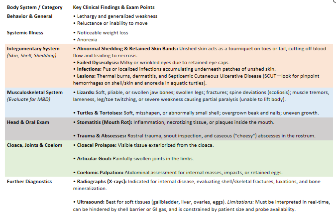
2.Diseases of reptiles caused by husbandry failures.
Captivity → challenges for reptiles, including limited space, diet, incorrect temperature, light, thermal injuries, humidity, wrong housing & stress => lead to diseases.
husk! Reptiles → poikilothermic (cold-blooded) & ectothermic - rely entiery on local surroundings to regulate body temp., absorb nutrients + maintain normal phys. functions.
Temperature
For most spp. → a focal basking light should be positioned at one end of enclosure, which gives an area with higher environmental temperature. If there is proper ventilation - rest of enclosure will be cooler. This is important so that the reptile can choose their suitable temperature.
Overall temp. of terrarium: 24-29C.
basking spot/hot zone should be: 25-39C
cold zone should be: 21-24C
Incorrect temp. → affects immune function, growth, shedding, digestion.
Dysdecdysis (Abnormal shedding)
common disease due to wrong temperature or humidity, or if reptile does not have access to water/bathing or hypocalcemia & hypovitaminosis A.
If too cold or too hot → cannot shed, unshedded skin can cause ligation of extremities, local ischemic necrosis + possible sec. osteomyelitis. Cannot grow either, if shedding does not occur.
High humidity - linked to fungal & bacterial infections. 50-70% is correct humidity, lower for desert animals.
Light
General rule: 14h of light during summer, 12h during winter
Most reptiles need UVB to activate production of vit. D, and natural direct sunlight is more recommended than UVB artificial light.
Metabolic bone disease (MBD)
Can be due to insufficient light, occurring by imbalance of Ca & P. Vit. D is important in uptake of Ca in digestive tract.
typical cs: Bowed and swollen legs, arched spine, bumps along legs, spine and tail, softening of bone
Tx: correct lighting and UVB, nutritional correction, calcitonin or Ca supplements, strict cage rest, bathe reptile 2x a day in baby bath temp.
Thermal injuries
Due to improper positioning of heat sources - as reptiles associate heat with overhead light - have poor thermal avoidance behavior on their undersides and will sit on dangerously hot surfaces without realizing they are being burned.
misuse of heating - unshielded heat lamps, basking lights placed too close to animal, or use of “hot rocks” - notes as unaccaptable!
Types of burns:
First degree - superficial epidermal reddening & blisters
2nd degree - deeper partial-thickness damage destroying the epidermis and damaging the dermis → severe pain, swelling, blistering, oozing of serum, bruise & scab-like scarring
3rd degree - destruction of entire thickness of skin - actually painless as all local pain-sensing nerves are destroyed. Tissues takes on a dry, whitish or charcoal-black charred look.
Tx: rapid cooling, local antisepsis, debridement, ATBs, supportive care (fluids)
Analgesia, sedation/anesthesia - for 2nd degree burns. (Anesthesia for doing procedures liked debridement).
Regular wound lavage, remove necrotic tissue (debridement). Topicals → cover wound with sterile occlussive dressing or wet to dry bandages or bandages soaked in silver sulfadiazine. Manuka honey.
low fomite environment (must be kept on simple, sterile substrates like paper towels).
if high humidity → incr. risk for bacteria, virus, parasites & fungi to accumulate. Thus good hygiene is essential!
3.Diseases of reptiles caused by improper feeding, basics of reptiles nourishment.
Reptiles can be classified in a number of ways, one is according to their diet - 4 main categories:
Carnivores - snakes
Insectivores / herbivores - lizards, require access to greens + insects like crickets and mealworms
Omnivores/herbi - turtles, tortoises, require access to fresh plants, legumes and veggies
Feeding:
Snakes - Size, age, and species affect how frequent they eat. Small snakes can be fed up to 2x a week, while larger can be fed 1x a month.
Chelonians & lizards - should be fed every day / every other day
Prey Preparation (Snakes): Ideally alive or sedated to maintain natural instincts. Prey should not be fully lucid or dead to avoid injury to the snake. Freezing meals can be used and eliminates potential bacteria or viruses in the prey.
Metabolism & Hydration:
Snakes have a very slow metabolism; overfeeding can lead to obstruction.
Fresh quality water must be provided.
Diseases caused by Improper feeding:
Post-Hibernation Anorexia:
Occurs in hibernating reptiles (tortoises) after using up all energy storage.
after hibernation = no energy → anorexia and dehydration due to poor nutrition prior to hibernation or failure to recover from hibernation.
Obesity:
A common problem as reptiles dont really move much in their enclosures compared to in the wild.
Can lead to gout (excess protein), soft tissue mineralization, and fatty liver syndrome.
Gout: Characterized by uric acid accumulation, which can occur due to dehydration inside the body (visceral gout).
Refeeding Syndrome:
Occurs after a period of anorexia when excess calories and proteins cause a rapid uptake of glucose into cells along with kalium and calcium.
Leads to hypokalemia and hypophosphatemia, which may result in arrhythmias.
Metabolic Bone Disease (MBD):
Caused by a lack of calcium in the diet, excess phosphorus, or a lack of vitamin D.
Clinical Signs: Bowed and swollen bones, bumps along the spine and neck, softening of bone, inactivity, osteoporosis, and tooth loss.
Treatment: Administering calcium supplements and calcitonin.
Dysdecdysis (“abnormal / incomplete shedding of the skin in reptiles”)
Can occur secondary to hypocalcemia.
4.Metabolic disorders and nutritional deficiencies in reptiles.
Metabolic disorders = up to 80% of all medical cases in reptiles.
Most Common: Gout, hypovitaminosis, hypervitaminosis, mineral imbalance, and post-hibernation anorexia.
1) Metabolic Bone Disease (Nutritional Secondary Hyperparathyroidism)
Etio: Inadequate nutrition or insufficient lighting:
Lack of vitamin D or calcium in diet.
Excess phosphorus in diet.
Insufficient UVB lighting.
Pathophys: Long-term calcium deficiency → secretion of PTH, leading to the mobilization of calcium from bones. - Affects mostly femur, humerus, jawbone (lizards) & carapace (turtle - its hard upper dorsal shell).
Clinical Signs:
Bowing and swelling of bones, softening of bones, osteoporosis, and tooth loss.
Sternal position, lethargy, and inactivity.
Lizards: Flexible jaw ("rubber jaw").
Turtles: Misshaped and soft shell/scutes.
Diagnostic: X-ray; do not use EDTA if blood is taken.
EDTA is avoided = cause severe hemolysis (rupture of RBCs) in many reptiles - thus changing the values.
Treatment:
Nutritional and husbandry correction.
Strict cage rest and fixing fractures.
Soaking reptile in baby bath temperature water twice a day.
Calcium gluconate injection: 20% (must be diluted with saline/glucose to prevent local necrosis).
2) Hypovitaminosis
Hypovitaminosis A (vit.A def)
Clinical Signs: Ear abscesses, squamous metaplasia of epithelium and skin, eyelid edema, lethargy, anorexia, and pneumonia.
Treatment: Subcutaneous injections of parenteral vitamin A once a week for 2–3 weeks.
Vitamin B1 Deficiency:
Clinical Signs: Neurological symptoms including ataxia, blindness, paresis, and torticollis.
Treatment: Addition of thiamine to the diet.
Vitamin D Deficiency: Same presentation and implications as Metabolic Bone Disease.
3) Gout
= Accumulation of uric acid (a waste product from protein metabolism).
Forms:
Articular Form: Accumulation of the uric acid in joint spaces, presenting as swelling and inflammation in joints.
Visceral Form: Forms a mineralized coating on the lining of internal organs.
Ante-Mortem: Cachexia (muscle wasting&weight loss) and dehydration.
Treatment & Management:
Fluid therapy.
Allopurinol: Helps prevent worsening of the condition. (it is a xanthine oxidase inhibitor, stopping the enzyme that is responsible for converting enzyme - xanthine into uric acid).
Analgesia: Meloxicam.
5.Post-hibernation syndrome, diagnosis, therapy and prevention.
Post-hibernation syndrome:
Occurs in hibernating species during winter months.
Pathogenesis depends on the underlying cause of the syndrome.
Causes:
Pre-existing disease prior to hibernation.
Poor nutrition leading to poor fat reservoirs prior to hibernation.
Incorrect hibernation conditions (10 weeks in winter months at 2–15°C).
Owner failing to recognize when hibernation ends.
Clinical Signs:
Inappetence and anorexia after hibernation
Respiratory tract infections.
Decreased blood glucose levels (hypoglycemia) and high levels of uric acid.
Dehydration, renal deficiency, and ketoacidosis.
Diagnosis:
Weight Monitoring: Comparing weight prior to and after hibernation (healthy individuals lose 1% of body weight per month during hibernation).
Blood Work: Shows hypoglycemia, hyperproteinemia, and hyperuricemia.
Therapy:
Intensive fluid therapy and nutritional support (feeding with a syringe using small meals).
Bathing the turtle/tortoise 3–4 times a day in water covering 50% of the individual at 40°C.
Treatment of secondary conditions if they occur.
Prevention:
Ensure the animal is healthy prior to hibernation.
Do not hibernate if the tortoise is underweight.
Perform regular checks during hibernation.
Bathing the tortoise in warm water.
Provide food and water immediately available after hibernation.
6.Clinical examination and dystocia and egg retention management in reptiles
Dystocia: Retention of follicles or eggs in female reptiles.
Common in snakes, chelonians, and lizards.
Forms of Dystocia:
Post-Ovulatory Stasis (POES):
Refers to the condition where the egg is stuck in the oviduct and cannot be laid properly.
Obstructive: Something physically blocking the oviduct (tumors, inflammation, malformed eggs).
Non-obstructive: Wrong husbandry conditions, lack of nesting material.
Other Causes: Hypocalcemia, infections, malnutrition, calculi, lack of nesting material.
Note: Female chameleons often turn black when laying eggs.
Pre-Ovulatory Stasis (POFS): (Note: not necessary to talk about POFS in this question) - Occurs when follicles are produced and enlarge but do not shed into the oviduct.Ovaries accumulate un-shed yolks over time → replaces other organs in the colemoic cavity. sign: anorexia, pressure in abd.region, elevated tail, failed attempts to lay eggs. Can be linked with long-term absence of a male.
Diagnosis:
USG: Often used in POFS (since there is no thin calcium shell, yolks appear as spherical densities).
x-ray: More often used in POES cases (VD and LL views; also CC view in chelonians).
Abdominal Palpation.
Anamnesis: ask if there is lack of a suitable nesting site, unsuitable temp., diet, matings, and dates of shedding.
Treatment:
Mild Signs: Correction of husbandry problems may be enough.
Moderate to Severe Signs:
Physical Manipulation: Using hands or a thermometer to dig out stuck eggs (high risk).
Hormonal Stimulation: Oxytocin (give calcium + glucose 30 minutes before) or arginine vasotocin, plus calcium injection (do not use during an obstruction).
Ovocentesis: Removing/aspirating egg contents through a needle.
Surgical Removal: Incision made between the first and second row of lateral scales over retained eggs; oviduct is isolated and incised to remove eggs.
7.Reproductive syndromes POES,POFS, etiologies, diagnostics and therapy in various species of reptiles. (SVARET E HEILT LIKT Q.6) - idk why dont ask me bruh
Dystocia: Retention of follicles or eggs in female reptiles.
Commonly affects snakes, chelonians, and lizards.
Forms & Etiologies:
Post-Ovulatory Stasis (POES):
Refers to condition where the egg is stuck in the oviduct and cannot be laid properly.
Obstructive: Something physically blocking the oviduct (tumors, inflammation, malformed eggs).
Non-Obstructive: Wrong husbandry conditions (decreased humidity, decreased light, lack of nesting material).
Other Causes: Hypocalcemia, infections, malnutrition, calculi, lack of nesting material.
Pre-Ovulatory Stasis (POFS):
Follicles are produced and enlarge but do not shed into the oviduct.
Ovaries accumulate un-shed yolks over time, eventually replacing other coelomic organs.
Clinical Signs: Anorexia, lethargy, apathy, pressure in abdominal region, elevated tail, failed attempts to lay eggs.
Etiology: Commonly associated with long-term absence of male, incorrect husbandry conditions like UVB light (hypocalcemia).
Diagnostics:
Ultrasonography (USG): Used more often in POFS (yolks lack a thin calcium shell, appearing as spherical densities).
Radiography (X-Ray): Used more often in POES.
Abdominal Palpation.
Anamnesis: Essential to establish lack of a suitable nesting site, unsuitable temperature, diet, matings, and dates of shedding.
Therapy:
Mild Signs: Correction of husbandry problems may be sufficient.
Moderate to Severe Signs:
Physical Manipulation: High-risk procedure to manually assist stuck eggs.
Hormonal Stimulation: Oxytocin or arginine vasotocin + calcium injection (contraindicated during obstruction).
Ovocentesis: Aspiration/removal of egg contents through a needle.
Surgical Removal: Incision made between first and second row of lateral scales over retained eggs; oviduct isolated and incised to remove eggs.
8.Viral diseases of reptiles, clinical manifestations, laboratory diagnostics and prevention.
Common in captive reptiles, but not commonly diagnosed.
Prevention: Quarantining new animals, regular checkups, good husbandry, and hygiene.
General Dx: Histology, serology, and virus isolation (more common during postmortem inspection).
General Management: Supportive and symptomatic treatment commonly used to relieve pain.
Specific Viral Pathogens:
Arenavirus (Inclusion Body Disease - IBD):
Species: Most associated with boa constrictors, but also seen in pythons.
Transmission: Body fluids.
Clinical Signs: Star gazing, anorexia, weight loss, poor wound healing, decreased WBC count as disease progresses. Chronic cases show neurological symptoms ranging from tics to seizures.
Diagnostics: History, clinical signs, blood smear (shows inclusion bodies in cytoplasm of leukocytes), and biopsy.
Treatment: No specific treatment (owners tend to choose euthanasia).
Herpesviridae (Chelonian Herpesvirus Infection):
Species: Reported in many reptiles, but common in sea turtles. Often associated with stomatitis-rhinitis complex.
Clinical Signs: Anorexia, skin infections, stomatitis, rhinitis, vomiting, nasal discharge, swelling of eyelids, pneumonia, and neurological signs.
Characteristics: Tumors/plaques growing on several body parts and organs.
Diagnostics: Serology, PCR.
Treatment: Acyclovir; tumors can be surgically removed.
Paramyxovirus:
Clinical Signs: Respiratory and neurological infections (pneumonia, neurological signs, restlessness, open-mouth breathing); may also be asymptomatic.
Diagnostics: Virus isolation, immunofluorescence, HIT, histology.
Treatment: Supportive and symptomatic therapy, nebulization (no curative treatment, prognosis is bad).
Other Notable Reptile Viruses:
Adenovirus: Liver, intestinal, and neurological disease.
Poxvirus: Skin lesions.
Flavivirus: West Nile Virus.
Iridovirus: Necrosis of liver, kidney, and spleen.
Reovirus: Respiratory symptoms and papillomatous skin lesions in lizards.
9.Parasitic diseases in reptiles, clinical signs, therapy and prevention.
Captive reptiles have an increased risk of parasite infection if husbandry conditions do not meet their needs.
Ectoparasite Location: Accumulate under scales, skinfolds, and postorbital areas.
General Signs/Complications: Anemia, skin damage, secondary infections, immunosuppression, inflammation, depression, anorexia, dysdecdysis, and death.
Specific Ectoparasites & Myiasis:
Snake Mite (Ophionyssus natricis):
Red/dark non-burrowing mite hiding under overlapping scale edges.
Signs: Irritation, pruritus, anemia, dysdecdysis; transmits bacteria and viruses.
Cloacal Mites: A problem in aquatic turtles.
Ticks (Ixodes ricinus, Ixodes hexagonus): Found around the neck inlet and in front of hind legs in tortoises.
Blowfly Myiasis:
Affects reptiles in poor conditions or those with diarrhea.
Larvae hatch within 1–2 hours in peak conditions, burrowing into the body to cause serious damage.
General Tx:
Manual removal, bathing, rinsing, soaking, and cage cleaning.
Ivermectin: Given IM or PO (not in chelonians).
Endoparasites:
Entamoeba invadens:
One of the most serious diseases in snakes, tortoises, and lizards.
Clinical Signs: Anorexia, blood or mucus in feces, vomiting, swelling in caudal abdomen.
Diagnosis: Trophozoites observed in direct fecal smear.
Treatment: Metronidazole.
Coccidia (Isospora, Eimeria):
Infect GIT and gallbladder; present as enteritis, intussusception, and anorexia.
Treatment: can try Sulfamethazine once daily for 7 days (complete recovery can be difficult; prevention is more appropriate).
Cryptosporidium:
Treatment: Spiramycin for 7 days (does not resolve infection; supportive therapy is better).
Strongyloides & Rhabdias:
Strongyloides: Causes GIT problems and respiratory stress due to migration through lungs.
Rhabdias: Inhabitant of the lungs in snakes.
Pinworms (Oxyuris):
Usually non-pathogenic; clinical signs present only in severe infestation.
Treatment: Fenbendazole.
Tapeworms: Treated with praziquantel for 2 weeks.
Flukes:
Less common (more in snakes).
Prevention/Treatment: Freezing prey eliminates flukes; treated with praziquantel or fenbendazole.
10.Bacterial diseases in reptiles, methods of drug administration
Routes of Drug Administration:
Per Os (PO): Used for critical care and administration of contrast media, for example.
Intramuscular (IM):
Turtles & Lizards: Front leg muscles.
Snakes: m. longissimus dorsi.
Subcutaneous (SC):
Turtles: Prefemoral fossa.
Lizards: Lateral side of spine.
Snakes: Loose skin lateral to spine.
Intraperitoneal / Intracoelemic.
Blood:
Sampling Sites: Ventral vein on tail in snakes and lizards; dorsal vein in turtles (or subcarapacial vein via midline puncture in turtles).
Taking a blood sample from turtles we get → hemolymph (blood mixed with lymphatic fluid).
Cell Morpho: Reptiles have blood similar to birds = nucleated erythrocytes and heterophils.
Bacterial Pathogens & Specific Diseases:
General: Bacterial diseases are common; most are opportunistic bacteria that replicate in immunosuppressed individuals.
Salmonella & Edwardsiella:
Opportunistic bacteria in many reptiles; can be asymptomatic or cause septicemia (bacterial infection of blood), pneumonia, and abscesses.
Treatment is not normal/routine in reptiles.
Mycobacterium:
linked with chronic wasting; seen as granulomatous lesions at necropsy. No successful treatment is known.
Septicemic Cutaneous Ulcerative Disease (SCUD):
Shell disease of aquatic turtles caused by Citrobacter freundii.
Clinical Signs: Pitted scutes that may slough with underlying purulent discharge, anorexia, lethargy, petechiae on shell and skin, and liver necrosis.
Treatment: Systemic antibiotics and good sanitation.
Diseases Caused by Aeromonas spp. & Pseudomonas spp.:
Ulcerative Necrotic Dermatitis (Blister Disease / Vesicular Dermatitis): Seen in snakes and lizards kept in unhygienic conditions with excessive humidity and moisture. Signs include dermal necrosis, ulceration, and exudative discharge. Treated with systemic antibiotics, topical antibiotic ointment, and good hygiene.
Pneumonia: Treated with proper husbandry conditions and systemic antibiotics.
Infectious Stomatitis: Caused by Aeromonas and Pseudomonas as common oral inhabitants. Signs include petechiae in the oral cavity and development of caseous material along the dental arcade. Treated with antiseptic irrigation, systemic antibiotics, supportive therapy, and vitamin supplements (vitamins A and C).
Abscesses:
Focal infections caused by traumatic injury, bite wounds, or poor husbandry. Etiological agents include Pseudomonas, Aeromonas, Salmonella, E. coli, etc.
Treatment: Small, localized abscesses should be completely removed surgically. Large abscesses should be marsupialized with aggressive wound treatment (lining aggressively scraped to remove necrotic tissue and promote healing). Systemic antibiotics may also be administered.
11.Supportive treatment in reptiles, convalescence patterns.
Supportive Treatment: Maintenance of normal physiological values within the individual without directly treating the primary underlying cause of disease.
→ Maintaining optimal temperature, providing appropriate nutritional support based on the reptile's diet, fluid therapy to avoid or correct dehydration, antibiotics, and analgesia.
Water Retention Capabilities: Reptiles excel at retaining water (e.g., secreting uric acid, minimal water loss through the skin).
Assessment Parameters: Skin elasticity, mucous membrane color, capillary refill time (CRT), and cornea brightness.
Dehydration Severity Levels:
3% Dehydration: Increased thirst, slightly lethargic, decreased urates.
7% Dehydration: Increased thirst, anorexia, dullness, loss of skin turgor, slightly delayed CRT.
10% Dehydration: Dull to comatose, skin remains tented, delayed CRT, dry/pale mucous membranes, dull cornea.
Fluid Therapy & Electrolyte Solutions:
Ringer-Lactate: Used for rehydration and maintenance fluid.
Glucose/Saline Combination: Used for hypoglycemia.
Hypertonic Saline: Used for acute hypovolemia.
Colloidal Fluids: Administered IV during serious anemia or blood loss.
Routes of Fluid Administration:
Oral (PO):
Advantages: Minimum stress, lower risk, rapid administration.
Disadvantages: Slow rehydration rate, potential damage to stomach or general GIT.
Subcutaneous (SC):
Advantages: Can give large volumes, rapid administration, faster uptake than oral, minimal risk of hitting internal organs.
Disadvantages: Risk of tissue trauma, slow absorption rates relative to vascular routes; hypertonic fluids cannot be injected SC.
Intracoelemic (IC):
Advantages: Accommodates large volumes, fast uptake, not painful.
Disadvantages: May exert pressure on lungs, slow absorption rates relative to vascular routes; hypertonic fluids cannot be injected IC.
Intravenous (IV):
Sites: Ventral tail vein in snakes, cephalic vein in lizards, jugular vein in chelonians.
Advantages: Rapid absorption rate, accurate delivery.
Disadvantages: Requires high technical skill, dependent on patient size.
Intraosseous (IO):
Advantages: Rapid absorption rate, accurate delivery, highly useful in small species.
Disadvantages: Contraindicated in cases of bone disease (e.g., MBD); requires sedation.
12.Anesthesia, most common surgical procedures and surgical treatment principles in reptiles.
Analgesics, sedatives and anesthetics used in reptiles
Turtle & tortoise: Alfaxolone, buprenorphine, butorphanol, ketamine + diazepam, ketamine + medetomidine, ketamine + midazolam (sedative), propofol, zolazepam. Meloxicam - NSAIDs, alfaxolone.
Snake: Propofol, ket+med, zolazepam, inhalation - isoflurane
Common surgical procedures:
Lizards: coeliotomy - opening abdominal cavity (ovariectomy + salphigectomy), castration, cystotomy - opening urinary bladder, oesophagostomy, orthopaedics - fractures.
Snakes: Colopexy (- fixing colon to body wall to avoid displacement or collapse - cloacal prolapse), coeliotomy, skin biopsy, hemipenis amputation
Chelopans: plastostomy - incision to oviduct to get access to eggs, shell repair, wound care, aural abscess removal, cloacal prolapse repair
POES and POFS - also common surgeries in reptiles, amputation of toes, Enucleation - removing eye if there is irreversible changes on eye by circular incision.
Anaesthesia
For some small procedures (like taking blood sample) - simply restraining the animal is all that is needed.
This can be better by temporary immobilization techniques such as dorsal recumbency, reduced light intensity or gentle ocular pressure (vasovagal response).
For invasive / painful procedure = General anesthesia must be used.
Preanesthetic Assessment and Stabilization
Before anesthesia: full clinical examination should be done + weight measure, reptiles should not be dehydrated or underweight.
Checking hydration status - esp. in sick or those coming out of hibernation.
Elective surgeries (like neuter): if sick or underweight - delay surgery and fix until healthy.
Emergency / non-elective: give fluids (IV / intraosseus into bone) to fix dehydration before surgery. Unstable reptiles often die during or after operation.
Fasting: fast reptiles. A full stomach can push against their lungs or cause vomiting/regurgitation during anesthesia
Premedication: Sedeatives are rarely used, but analgesics (pain relief) should be given.
Anesthetic Induction (putting them under)
IV or intraosseous - fast and controlled (propofol)
safe even if a little leaks out of the vein (low risk of vein infla.) which helps since reptile veins can be hard to hit. (red.risk of thrombophlebitis if inj. perivascularly)
IM injection: used when IV is difficult. A combo like ketamine + medetomidine / dexmed. + morphine works well in turtle/tortoise
Can be reversed by atipamezole (and naloxone if needed) to help them wake up safely.
Induction (gas via mask or chamber): works well for lizards and snakes, but induction can take 10-30min.
does not work well for turtle/tortoise/croco because they can hold their breath for long periods.
Maintenance of Anesthesia
Isoflurane / Sevoflurane = the agents used
has fast mode of action, easily controlled and gives faster recovery
Intubation of reptiles = simple, we just insert the small-gauge endotracheal tubes / catheters through the glottis caudal to tongue.
Monitoring - how to look after reptiles under anesthesia
Temp. during anesthesia is optimal. 32-35C
Check heart rate (auscultate or see/feel), breathing, temp.
Pulse oximetry (SpO2) - can place a probe in esophagus / cloaca to check HR & pulse strength. The O2 % (SpO2) are not 100% accurrate for replites, but watching whether it goes up or down can be helpful!
reflexes - corneal and palpebral (not in snakes)
check if reptile is deep asleep:
Blink reflex (palpebral / corneal reflexes) - not in snakes.
Pupil size - dont rely on this
jaw completely relaxed, body is limp, no reaction when pinching or pulling its tongue, leg or tail = fully under for surgery. + they will also lose their ability to flip themselves right-side up (righting reflex), complete muscle relax.
Towards end of surgery (WAKING IT UP): stop anesthetic gas, but keep pumping O2 / air for 5-10min to help clear gas out of their system. Switch to room air - turn off pure O2 and pump normal room air using bag valve mask (manual rescusc. bag). Pure O2 can actually supress the reptiles urge to breathe so room air encorage them to start breathing on their own again!
13.Capture specifications, Manipulation and transport in zoo animals (Anesthesia and immobilization, fixative and capture facilities).
Capture of zoo animals:
Done by immobilization (restricting an animal`s movement by using either physical or chemical methods)
Generally, handling cause stress, so avoid full anesthesia whenever possible..
Training - positive reinforcement training (ex. teaching animal to present a limb) is preferred for injections and physical exams - to avoid risk and stress.
Monitoring: Immobilized animals (by drug) require continuous clinical monitoring for complications like hypoxemia (low blood O2), respiratory depression (shallow/slow breathing), and thermoregulatory failure (overheating or hypothermia)
Capture Myopathy: Severe stress and physical exertion during capture can trigger massive myoglobin release, leading to nephrotoxicity and kidney failure.
Physical (Mechanical) Restraint
Physical restraint uses tools / barriers to limit movement for exam, sample collection or treatment.
Blindfolds (to block visual stimuli and lower stress), ear covers.
confinement: crush cages, chutes, squeeze cages, bags, towels
Capture device: drop nets, snares, ropes
Physical barriers: shields, blankets, plastic sheets, snake tubes
Traps - box traps, cage traps, pitfall traps
Chemical Restraint
Chemical restraint uses drugs to sedate, paralyze or make an animal unconscious. Risk: all immobilizing drugs are potent toxins, some like ultra-potent opioids can be fatal to humans if acc. injected / absorbed by skin/MM.
Delivery Methods:
Dart Guns: shoot Projectile darts that injects drug on impact by compressed gas / small charge.
Spring-Loaded Pole Syringes: Used for manual injection at close range.
Common Immobilizing Agents:
Etorphine Hydrochloride (Immobilon):A potent synthetic opioid used for large mammals (ungulates, elephant, rhinos).
Extremely lethal to humans; requires protective gear and the antidote (diprenorphine)
Hellabrunn Mixture: A combination of liquid ketamine (100 mg/mL) and powdered xylazine (125 mg/mL).
Transport Protocol
State of Animal: Animals must be transported awake (never under sedation/anaesthesia - risk of airway collapse/bloat).
Crate & Space: Crates should be sized with the animal not being able to turn around.Ventilation is mandatory. Keep in dark (cover eyes/ears).
Separation & Care: Adult animals must be transported separately, food and water are generally not required during short-distance transit.
Personnel: Trained staff must accompany the animal throughout the entire journey.
14.Preventative veterinary measures in Zooˋs - health control, vaccination, deworming, drug administration and quarantine conditions.
Preventive vet measures:
Why prevention? → Catching and treating wild animals can be stressful and dangerous → stress makes sick animals worse
Main goal - keeping diseases out of the zoo collection entirely so you rarely have to treat sick animals
Components of a prev. program: quarantine, parasite control (fecal exams), booster vaccines, health screening procedures, nutrition, necropsy exams, pest control program
Quarantine (Isolating new animals)
Strict isolation required for all new arrivals before joining the permanent collection.
Facility rules:
Hygiene - easy to clean and sanitize safely
pest-proof - sealed against pest that carry disease, restricted access
All-in-all-out principle (adding a new animal restarts the quarantine timer for the whole group)
Quarantine Duration:
Standard Animals: 21–30 days
Psittacines (Parrots): Up to 60 days
Primates: Up to 90 days
Procedures - what is done during quarantine: Physical exams, diagnostic testing (bloodwork), parasite treatments, enteric bacterial screening (gut bacteria tests), and vaccinations prior to release.
Routine Health check up
Zoos constantly trade and move animals between facilities / zoos, so regular health check ups are important!
zookeepers know individual animal behavior best, they are the first to spot early warning signs (lack of appetite, abnormal feces). Finding issue early = tx easier = stop outbreaks.
Preventive Treatments & Drug Administration
Vaccination: vax programs for car, primates, equids, birds etc. Common is - rabies, canine parvovirus, feline panleukopenia, distemper, leptospira, parainfluenza, measles.
Deworming & Parasite Control:
Targeted against ecto- and endoparasites using standard veterinary drugs. But vet must check for spp. senstivities (some drugs are toxic to specific species)
Rotate anthelmintics to prevent resistance, mixing drugs into feed minimize stress, control IH to reduce parasites with indirect LC.
Drug Administration:
Most medications are used extra-label (off-label). (as few are officially approved (licenced) for zoo spp.)
Oral: Minimal stress, but exact dosing is harder to guarantee.
Intramuscular (IM): Delivered via hand-held syringe or remote dart.
Intravenous (IV): Typically reserved for animals under general anesthesia.
15.Veterinary care of carnivores in Zoo
Nutrition & Feeding Types
→ nutrition and maintaining some of their natural instincts are important to maintain the mental health of the captive animals.
In the wild, carnivores hunt a variety of whole prey. In captivity, their diet changes based on age, pregnancy, and nursing.
Carcass Feeding (Whole Animals):
Pros: Very natural; provides physical exercise and mental stimulation.
Cons: Hard to source consistently; carries risks of food poisoning, infectious diseases, or nutritional deficits if animals only eat their favorite parts (selective eating).
important to be selective + finding out reason to death before giving to carnivore.
Prepared Meat (Muscle Meat): Plain, skinned muscle meat from livestock lacks bone, organs, and skin, which can lead to severe nutritional deficiencies unless vitamins and minerals are added.
Complete Commercial Diets (Kibble/Canned):
Pros: Safe from diseases, fully balanced, and prevents nutritional deficiencies.
Cons: Lacks mental stimulation and can cause dental problems because animals don't chew or tear tough meat/bones.
Nutrition-Related Health Problems
Obesity: Caused by lack of exercise (no hunting required) and overfeeding. Leads to joint pain, metabolic issues (like diabetes), and higher risks of certain cancers.
Dental Issues:
Tartar buildup: From soft commercial diets without bone scraping.
Malocclusion (misaligned bite): From lack of vigorous chewing, tearing, and crunching.
Housing & Welfare
Enclosures must meet physical, environmental, and social needs while allowing safe public display.
Flooring: Soft-footed carnivores need non-abrasive substrate (grass, soil) to prevent paw scrapes, bleeding, and foot infections.
Canids (Wolves, Wild Dogs): Usually housed in single-sex groups (except during breeding season). They need an indoor cubicle with heat during winter, plus a outdoor run.
Felids (Big/Small Cats): Housed in large, heavy-mesh outdoor enclosures with indoor heated sleeping quarters. Grouping (pairs vs. small groups) depends on species compatibility (e.g., solitary tigers vs. social lions).
Housing Hazards: Poor housing leads to physical injuries or severe psychological distress (stereotypies).
Environmental Enrichment
Enrichment keeps animals mentally active, encourages natural behaviors, gives them choices, and educates zoo visitors.
Food Enrichment:
Simulated Hunts: Zookeepers can move whole prey on zip-lines or drop items from moving vehicles to trigger hunting instincts.
Puzzle Feeders: Devices or boxes that require tearing or problem-solving to reach food rewards.
Environmental Items: Logs, boulders, climbing shelves, water features, and trees to mimic natural habitats. + toys, sticks..
Stereotypies (Stress Behaviors)
= Unnatural, repetitive, useless behaviors caused by boredom, frustration, or cramped conditions.
Most Common in Carnivores → Pacing back and forth along fence lines (locomotor stereotypy).
Vaccination
Zoo carnivores are vulnerable to deadly domestic animal viruses. Key vaccines include:
For Cats (Felids): Feline Panleukopenia (Parvovirus), Feline Rhinotracheitis (herpes), Feline Calicivirus, Rabies. Corona.
For Dogs/Bears/Mustelids (Canids, Ursids, Mustelids): Canine Distemper, Canine Parvovirus, Rabies.
16.Veterinary care of primates in Zoo
Housing & Welfare
Primates have complex physical, social, and mental needs. Good housing allows them to show natural behaviors like grooming, climbing, and foraging.
Primates are skilled climbers and spend most of their time off the ground, so enclosures must be tall, not just wide.
Social Structure: Keep natural group structures intact. Avoid separating bonded pairs or mothers and offspring, as breaking hierarchy causes severe stress. Kept in mixed groups - packs.
Environment: Enclosures require controlled temperature, humidity, and ventilation.
Nutrition
Primates are generally omnivores (eating fruits, grains, vegetables, insects, and small animals depending on the species).
Vitamin C: Primates cannot synthesize their own Vitamin C (just like humans). Their diet must include Vitamin C-rich fruits or supplements to prevent scurvy.
Vitamin D3: New World primates (e.g., marmosets, tamarins, capuchins) utilize Vitamin D poorly. If they don't have direct sunlight, they must get stabilized Vitamin D3 in their diet to prevent metabolic bone disease.
Protein: Needed for strength, but too much protein causes kidney damage.
Treats & Foraging: Foods like eggs, yogurt, nuts, insects, and fruits are great for treats or hiding around the enclosure to encourage natural hunting and gathering.
Environmental Enrichment & Training
Because primates are highly intelligent, boredom leads to severe psychological issues.
Types of Enrichment:
Foraging: Hiding food in logs, branches, and puzzle boxes.
Physical: Ropes, swings, climbing structures.
Sensory: Mirrors (visual), radios/music (auditory).
Rule: Rotate toys and puzzles constantly (to avoid boredom)
Target Training: Training primates to present limbs, open their mouth, or step on scales eliminates the need for stressful chemical anesthesia during routine health checks.
Behavioral Problems
Stereotypies (Stress Habits): Bored or stressed primates develop repetitive behaviors:
Oral: Excessive licking or pacing with mouth movements.
Locomotor: Pacing, rocking back and forth.
Grooming: Over-grooming leading to hair loss or skin self-mutilation.
Aggression: Often caused by cramped spaces, poor group dynamics, lack of enrichment, or high stress.
Vaccination & Zoonotic Risks
Because primates are closely related to humans, diseases easily jump back and forth (zoonoses) between primates, keepers, and zoo visitors.
High-Risk Pathogens: Tuberculosis (TB), Measles, Mumps, Rubella, Influenza, Polio, Rabies, Tetanus, Salmonella, and Ebola, herpes, hepatitis B
Prevention: Extensive routine screening (especially for TB) and strict vaccination programs protect both the animals and the human staff.
Contraception & Breeding Control
Primates housed in natural family groups will breed rapidly. Unwanted pregnancies are controlled via:
Females: Permanent spaying or reversible hormonal implants/injections.
Males:
Castration: Removes testicles, which lowers testosterone and reduces male aggression. However, castrating only some males in a group upsets social hierarchy and makes the intact males overly dominant.
Vasectomy: Cuts the sperm ducts without dropping testosterone. This stops reproduction while keeping natural social structures, behaviors, and rank intact.
17.Veterinary care of ruminants in Zoo
Housing & Enclosures
Outdoor Habitat: Needs large, predator-proof fenced areas with shade, trees, and shrubs for natural grazing or browsing. RU can be together independent of spp. Usually in packs, giraffes in loose groups.
Indoor Quarters: Animals must have secure outdoor pens during the day and warm, dry indoor stalls/pens for night and winter. Should be enough for animal to lie down and stand/walk around.
Nutrition & Feeding Types
Zoo herbivores are split into three main feeding groups:
Browsers (Tree/Leaf Eaters): ex. Giraffe, moose.
Grazers (Grass Eaters): Cattle, buffalo, sheep, bison, zebra, camel (note: elephants and rhinos are non-ruminants, but follow similar feeding rules).
Mixed Feeders: Goats, white-tailed deer.
Hay
Constant Supply: Hay makes up the bulk of their diet and should be available all day, not fed in small timed meals.
Legume Hay (e.g., Alfalfa/Lucerne): Richer in protein, nitrogen, and calcium; ideal for browsers.
Grass Hay (e.g., Timothy/Meadow): Lower energy and high fiber; ideal for grazers.
Feeding Height: Serve hay from racks at eye level to prevent soil contamination and mimic natural browsing (except for elephants).
Pellets
Formulated to provide extra protein, vitamins, and minerals.
High-Fiber Pellets (High NDF/ADF): Required for browsers to mimic fibrous woody plant leaves.
Moderate-Fiber Pellets: Sufficient for grazers.
Individual Feeding: Feed animals separately when possible so dominant individuals don't steal food.
Specific Mineral Considerations
Elephants: Need extra calcium supplements because grass hays are often low in calcium.
Black Rhinos: Need low-iron diets to prevent dangerous iron overload disease (hemosiderosis).
Vaccinations
Given annually (or as needed based on local risk):
Bovine Viral Diarrhea (BVD), Clostridial diseases, Leptospira, and Parainfluenza.
Common Health & Behavioral Problems
Hoof Overgrowth & Lameness: Softer zoo ground (dirt/grass) doesn't wear down hooves like wild terrain. Uncut, overgrown hooves alter stance, leading to severe lameness.
Oral Stereotypies: Stress or lack of chewing time causes repetitive tongue-rolling, bar-licking, or wind-sucking.
Seasonal Aggression: Males fight fiercely during breeding season. Prevent injury by separating males from each other or keeping them away from females.
Capture Myopathy: Severe stress, panic, or overexertion during handling ruptures muscle tissue, releasing myoglobin that damages the kidneys.
Grain Overload (Rumen Acidosis): Eating too many high-starch pellets/grain breaks down rumen fermentation, causing life-threatening acid accumulation.
Degenerative Joint Disease (Arthritis) & Obesity: Common in older or inactive animals that lack space or exercise.
Fra her - DEL B - Exotic birds
1B: History and clinical examination in diagnostics of exotic bird`s diseases
1. Taking the Anamnesis (Medical History)
Because birds hide illness, getting a complete history from the owner is essential.
Bird Details: Species, age, gender, origin, hand-reared vs. parent-reared, how long the owner has had it, and if there are other birds in the house.
Environment & Care: Diet, vitamin supplements, cage setup, cleaning, indoor air quality, and whether wings are clipped or the bird is free-flying.
Behavior & Background: Any recent behavior changes, past illnesses, previous diagnostic results, and current treatments.
Current Problem: Main symptoms, when they started, and how quickly they are progressing.
2. Clinical Examination
Step A: Visual Inspection (Hands-Off)
Always observe the bird in its cage before handling it. Birds are prey animals and instinctively hide signs of illness ("masking phenomenon").
Behavior & Posture: Attitude, mental state, sitting on a perch vs. sitting fluff-up on the cage bottom.
Breathing: Breathing rate, effort, tail bobbing (a sign of respiratory distress), or open-mouthed breathing.
General Appearance: Beak alignment, feather condition, toe grip, and how the bird interacts with its owner.
Step B: Physical Examination (Hands-On)
Restrain the bird safely using a towel to prevent bites and scratch injuries (tailor restraint strength to the species). Work quickly to minimize stress.
Body Condition Score (BCS): Palpate the pectoral muscles on both sides of the keel bone (sternum). + look at weight, subcut.fat.
Feathers & Skin: Check for clean, undamaged feathers, stress bars, missing feathers, ectoparasites, skin flaking, redness, or masses.
Head: Inspect the eyes, ears, nostrils (nares), and beak. Use a speculum to gently open the beak and examine the oral cavity.
Crop & Abdomen: Palpate the crop for food, fluid, or thickening. Gently feel the abdomen for swelling, fluid, or egg-binding.
Wings & Legs: Check wing locomotion, joint mobility, feet, and toes for lesions (pododermatitis/bumblefoot).
Cloaca: Check that it is clean and free of vent pasting, swelling, masses, or prolapsed tissue.
Hydration Status: check the refill time of the basilic vein (located over the elbow joint).
3. Common Diagnostic Tests
Fecal Analysis: Direct smears, flotation, and cytology to check for intestinal parasites, yeast, or abnormal bacteria.
Bloodwork:
Complete Blood Count (CBC) & blood smear (for infection, anemia).
Biochemistry panel (checks liver, kidney, and metabolic function).
DNA Sexing: Done via blood / endoscopy, visual inspection (polymorphism)
Microbiology: Swabs from the choana (roof of mouth) or cloaca for bacterial culture and sensitivity.
Imaging: Radiographs (X-rays) or endoscopy to evaluate internal organs, air sacs, bones, and foreign bodies. (need sedation).
Infectious Disease Screening: Specific PCR tests, especially for Psittacosis (Chlamydia psittaci) = zoonotic.
2b) Bacterial diseases in exotic birds, diagnostics, therapy and prevention
Bacterial diseases in exotic birds are commonly triggered by poor husbandry, bad diet, or environmental stress.
General symptoms: lethargy (listlessness), loss of appetite, and weight loss. But it will depend on type of bacteria, location in body and affected organs.
1) Clostridiosis (Clostridium perfringens)
An infection of the small intestine that can spread to other organs.
Transmission: Ingestion or inhalation of spores from contaminated food, water, cages, or utensils.
Mechanism & Symptoms: Occurs when normal gut flora becomes imbalanced, allowing Clostridium to overgrow and produce harmful tissue-damaging toxins → damage of intestinal wall. Leads to rapid health decline, bloody feces 🩸, undigested food in droppings, weight loss & lethargy.
Treatment: Fluid therapy, probiotics, and targeted antibiotics (e.g., amoxicillin or tetracycline).
2) Avian Chlamydiosis / Psittacosis (Chlamydia psittaci)
An obligate intracellular bacterial infection common in parrots, cockatiels, and budgerigars. Zoonotic risk (flu-like / pneumonia in man)
Clinical Signs: Can range from asymptomatic (carrier state) to severe illness. Symptoms include eye and nasal discharge, anorexia, dyspnea, severe depression, dehydration, diarrhea, and biliverdinuria (bright lime-green urates).
Diagnostics:
Bloodwork: Leukocytosis (high WBC), monocytosis, and high bile acid concentrations.
X-rays (Radiographs): May show enlarged liver (hepatomegaly), enlarged spleen (splenomegaly), or inflamed air sacs (airsacculitis).
Confirmation: PCR swabbing (most common) or serological tests (ELISA, indirect/direct fluorescent antibody, complement fixation).
Treatment: Doxycycline for 45 days. Dietary calcium must be reduced during oral treatment because calcium binds doxycycline and lowers its absorption.-
3) Other Common Bacterial Conditions
Rhinitis (Nasal Inflammation): Caused by E. coli, Pseudomonas, or Klebsiella. Hypovitaminosis A (vitamin A deficiency) is a major predisposing factor.
Sinusitis: Caused by Pseudomonas, Aeromonas, E. coli, or Mycoplasma.
Fowl Cholera (Pasteurella multocida): Causes painful swelling of the head, footpads, and joints.
Avian Tuberculosis (Mycobacterium avium): A chronic, progressive disease primarily affecting the liver and GIT.
Staphylococcosis (Staphylococcus aureus): Causes skin lesions including redness, swelling, itching (pruritus), crusting, and tissue necrosis.
General Diagnostics, Treatment & Prevention
Diagnostics:
based on cs, result of cytologic examination, culture of samples.
Gram Stain: Evaluates normal vs. abnormal gut flora, yeast, and spore-forming bacteria.
Cytology & Bacterial Culture with Sensitivity: Identifies the exact organism and determines which antibiotics will successfully eliminate it. Swabs can be taken from the respiratory, GI, urinary, or reproductive tracts.
Treatment: based on location of infection, result of culture & senstivity testing - ATBs tx. fluids, nursing care.
Prevention: quarantine, vaccine, cleaning & disinfection.
3b) Mycotic diseases in exotic birds, diagnostics, therapy and prevention
Fungal (mycotic) infections in companion birds are usually secondary infections - they “take over” when a bird's immune system is weak or when husbandry is poor.
Aspergillosis, candida & megabacteriosis - common fungal infections in birds.
1) Aspergillosis (Aspergillus fumigatus, A. flavus, A. niger)
An opportunistic infection caused by inhaling airborne spores. It thrives in warm, humid, poorly ventilated, or unsanitary environments.
NOTE: Mainly attacks the upper resp. tract, lungs & air sacs. It forms fungal masses (granulomas) and is not spread from bird-bird.
Clinical Signs:
Respiratory distress: Tail bobbing (a sign of heavy breathing), dyspnea, exercise intolerance, and voice changes.
Systemic: Weight loss, loss of appetite (anorexia), and weakness.
Diagnostics:
Bloodwork: High white blood cell count (leukocytosis) and high monocytes (monocytosis).
X-rays (Radiographs): can find air sac inflammation (airsacculitis), pulmonary disease, or granulomas.
Definitive Diagnosis: Direct visualization via endoscopy or surgery, confirmed with biopsy, cytology, or fungal culture of lesions.
Treatment - antifungals:
Nebulization w/Enilconazole or F10 solution (15–30 minutes, 2–3 times daily).
Oral: Itraconazole or Voriconazole (5–10 mg/kg PO, 1–2 times daily).
2) Candidiasis / Yeast Infection (Candida albicans)
An opportunistic yeast that naturally exists in small numbers in the gut. Overgrowth occurs when normal gut flora is disrupted by stress, immunosuppression, or poor hygiene. It most commonly affects unweaned hand-fed chicks.
Clinical Signs:
Juvenile Birds: White plaques in the mouth, crop slow-down or stoppage (crop stasis), regurgitation, loss of appetite, and rapid weight loss.
Adult Birds: Milder signs—slight weight loss, lethargy, and dull feathers.
Diagnostics: Cytology/Gram stain of fecal smears, crop flushes, or regurgitated material showing budding yeast.
Treatment:
Crop Management (Neonates): Empty and flush the crop, then feed smaller meals. Metoclopramide can be given to stimulate crop motility.
Antifungals: Nystatin (local action in GI tract) or Fluconazole (systemic).
Flock Treatment: Chlorhexidine added to drinking water (10 mL per gallon for 1–3 weeks).
3) Megabacteriosis (Macrorhabdus ornithogaster)
= Large yeast (despite its name).
Targets stomach lining - glands bw. proventriculus (true stomach) and ventriculus (gizzard) - esp. in small birds like budgies, parrots, cockatiels, lovebirds and finches.
Cs: Chronic weight loss, regurgitation, lethargy, diarrhea, and passing undigested seeds in droppings.
Asymptomatic carriers are common.
Diagnostics: Fecal Wet Mount / Staining: showing very large, rod-shaped yeast organisms. Shedding is intermittent, so a single neg. fecal test does not rule it out!
Treatment:
Oral Antifungal: Amphotericin B (100 mg/kg PO, twice daily for 30 days) is the most effective drug.
Stomach Acidification: Adding organic acids (like apple cider vinegar or Vitamin C) to water creates an acidic gut environment that suppress yeast growth.
Experimental - Sodium benzoate added to drinking water.
4b) Viral diseases in exotic birds, diagnostics, therapy and prevention
Most imp. viral diseases in birds: Avian bornavirus, psittacine circovirus & avian polyomavirus
1. Proventricular Dilatation Disease (PDD)
Cause: Avian Bornavirus (ABV). Spread via the fecal-oral route. Typ. affects macaws, conures & african gray parrots.
Mechanism: The virus attacks and destroys the nerve ganglia supplying the stomach (proventriculus) → paralysis of the digestive tract.
CS: GI - chronic weight loss (often despite huge appetite), regurgitation, passing whole undigested food in droppings.
Neuro in some: tremors, seizures, ataxia, blindness
Diagnosis: PCR testing on choanal, cloacal, or fecal swabs, or serology (ELISA). Note: Shedding is intermittent. X-rays often show a dilated proventriculus.
Treatment & Control: Supportive care with easily digestible food and NSAIDs (e.g., meloxicam) to reduce neural inflammation. Infected birds must be isolated.
2. Psittacine Beak and Feather Disease (PBFD)
Cause: Psittacine Circovirus. Targets rapidly dividing cells (feather follicles, crop, thymus/bursa).
Transmission: Inhalation or ingestion of feather dust/dander, feces, or oral secretions. Can also pass vertically (hen to egg).
Clinical Signs:
Early Sign: Loss of powder down (beak looks glossy/dirty instead of matte grey).
Feather Lesions: Clubbed, pinched, or deformed growing feathers, feather shaft bleeding, easy plucking, and color loss.
progressive: Severe immunosuppression → fatal secondary bacterial / fungal infections.
Diagnosis: CS, PCR testing (blood/swabs), and feather follicle biopsy.
Treatment: No cure exists—treatment is purely supportive. Euthanasia is often recommended. Prevention relies on strict quarantine, dust control, and viral screening.
3. Polyomavirus / Budgerigar Fledgling Disease (BFD)
Cause: Avian Polyomavirus (APV). Primarily attacks young, hand-fed neonates and fledglings.
CS: Sudden onset of severe lethargy, delayed crop emptying (crop stasis), abdominal swelling, skin hemorrhages (bruising), and acute death within 24–48 hours.
Dx: PCR DNA probes (cloacal swabs/blood) and virus neutralizing Ab test.
Tx: Supportive care only (no targeted antiviral). Prevent by hygiene, quarantine, avoid cross-contam. bw. brooders. Vaccinate.
Other viruses:
Newcastle disease (paramyxovirus g.1)
inhalation of resp. aerosols / ing. of feces/direct contact → resp. signs - nasal discharge, dyspnea + bright yellow-green diarrhea, severe neuro signs like torticollis (twisted neck).
West nile disease (west nile virus)
Vector-borne flavivirus by mosquitoes → head tremor, weight loss, ataxia, blind/seizure and sudden death.
→ No tx for these, hard to diagnose, but serology maybe.
→ Generally not recommended to do symptomatic tx due to strict reg./quarantine laws and rapid spread.
5b: The most common surgical Procedures used in exotic birds.
Most common surgical procedures used incl. Ingluviotomy, pododermatitis, cloacapexy, cloacatomy, proventriculotomy, hysterectomy, celiotomy, cysts & neoplasia.
1) Ingluviotomy (Surgical Opening of the Crop)
Done to remove FB, cleaning crop impaction or sour crop, crop biopsy, to repair traumatic injuries, burns and rupture.
method: bird is anesthesized and intubated. Head is kept elevated to prevent fluid from flowing backward into the throat (pharynx), causing fatal aspiration.
2) Surgical Treatment of Pododermatitis (Bumblefoot)
= Inflammatory + infected lesion on plantar surface of foot (bottom). Common in ex. falcons. Tx depends.
Mild cases (grade I): treated w/wraps / habitat change, conservative tx (ATB, wound care, analgesia):
Severe case: needs surgery to remove necrotic + infected tissue (debridement)
3) Cloacopexy & Cloacotomy (surgery affecting claoca)
Cloacopexy (closing/repositioning of cloaca):
To treat chronic cloacal prolapse. Done by reattaching cloaca to abd. wall, preventing it from slipping out, but recurrence is common because underlying cause is often uknown and cloacal sphincter mm is usually stretched + weak.
Cloacotomy (Surgically Opening the Cloaca): Opening the cloaca directly to inspect its walls or surgically remove internal masses, such as cloacal papillomas.
4) Proventriculotomy / Ventriculotomy
Removing FB or metallic objects (lead / Zn toxicity) from proventriculus / ventriculus when it is not possible by endoscopy / flushing. Done via a left-abd. approach (left lateral celiotomy)
Incision in the gizzard (ventriculotomy) leak easily due to mm movement. Needs fine monofil. sutures + often tissue patch (serosal patch) to seal line against digestine fluids.
5. Hysterectomy / Hysterotomy (Salpingectomy)
Removing stuck or obstructed eggs (egg binding), repairing a ruptured oviduct, or tumors/cysts.
method: A full surgical opening of the abdomen (celiotomy) is required to safely remove the egg and/or the affected portion of the oviduct. Simple ventral midline approach.
6. Celiotomy (Surgically Opening the Abdomen):Diagnostic exploration or access to internal organs. Ventral midline incision - access to liver, pancreas, intestines within hepatoperitoneal cavity. This specific compartment does not open directly into the main lateral air sacs.
7. Feather Cyst Removal
Common in small birds (like canaries) with soft feathers when a growing feather fails to break through the skin and curls inside the follicle. Cyst is excised under anesthesia, feather follicles are vascular - so strict hemorrhage control is important. Wound is left to heal naturally, if cyst reoccur in same spot → deep feather root must be removed (dermal papilla)
8. General Mass Removal - skin tumors, skin lesions, papillomas..
6b: Parasitic diseases in exotic birds, diagnostics, therapy and prevention
Parasitic diseases - less common in captive pet parrots as wild importation has stopped and indoor pet birds have minimal exposure to outdoors or wild bird vectors.
Ectoparasites include cnemidocoptes mutans / gallinae / pilae, dermanyssus gallinae, sterostoma tracheacolum, ticks.
1. Ectoparasites (External Parasites)
Scaly Face & Leg Mite (Knemidocoptes pilae)
Host: Most common in budgerigars; occasionally seen in passerines (e.g., canaries, finches).
Clinical Signs:
Budgies: White, porous, honeycomb-like crusts around the corners of the mouth, cere, beak, eyelids, legs, and vent.
Passerines: Thick crusts on the legs and toes ("scaly leg").
Dx: Microscopy - facial / skin scrapings.
Tx: Systemic antiparasitics - Ivermectin (PO/IM) or Moxidectin (Topically/PO).
Red Mite / Feather Mite (Dermanyssus gallinae)
= Blood-sucking mites that hide in cage crevices during the day and feed on birds at night. They survive for long periods in the environment without feeding.
CS: Feather plucking, intense itching (pruritus), skin irritation, and severe anemia (from blood loss).
Tx: Spray/powder environmental treatments (Pyrethrin, Carbaryl spray) / systemic Ivermectin for 2w
deep clean cage, throw away wooden nest boxes/perches.
Air Sac Mite (Sternostoma tracheacolum)
Host: Parasitizes the entire resp. tract (trachea, syrinx, lungs, + air sacs), esp. in canaries and Gouldian finches.
Clinical Signs: Heavy infestations cause high-pitched respiratory noises, clicking sounds, sneezing, tail bobbing, open-mouthed breathing, and death. Mild - asymptom.
Diagnosis: Transillumination of the trachea (shining a bright light through the skin of the neck in a dark room to see tiny dark dots inside the windpipe). Endoscopy/tracheoscopy.
Treatment: Ivermectin or Moxidectin.
Other External Parasites:Flat flies (Hippoboscids), ticks, and lice.
Endoparasites (Internal Parasites)
→ like trichomonas, giardia, coccidian, ascaridia, capillaria, syngamus trachea & cestodes
1) Trichomoniasis (Trichomonas gallinae)
Known as Canker in pigeons/doves (Columbiformes) and Frounce in birds of prey (raptors).
Cs:
Raptors/Pigeons: Thick, whitish-yellow, cheesy (caseous) plaques attached to the mouth, crop, and esophagus, causing difficulty swallowing (dysphagia), breathing trouble (dyspnea), and weight loss.
Budgies: Usually do not show visible oral plaques, but present with regurgitation and weight loss.
BUT 80% = asympt.
Dx: crop swab.
Tx: Antiprotozoals such as Metronidazole, Ronidazole
2) Giardiasis (Giardia spp.)
CS: Classic "screaming and plucking" syndrome in cockatiels (feather picking under the wings/axillary region and inner thighs + vocalization), along with diarrhea and weight loss.
Dx: Direct saline fecal smear (showing motile trophozoites), zinc sulfate flotation test, or ELISA.
Tx: Metronidazole or Carnidazole.
3) Coccidians (Eimeria spp. &Isospora spp.)
CS: Weight loss, chronic diarrhea (sometimes bloody), and lethargy. Dx by flotation.
Tx: Toltrazuril or Amprolium. sulphonamides.
4) Roundworms (Ascaridia spp. ex. Heterakis gallinarum)
Direct LC, common in birds housed outdoors w/ground access.
affects GIT (diarrhea/weight loss), Dx: eggs on fecal flotation. Tx: General anthelmintics (e.g., Fenbendazole, Ivermectin).
5) Hairworms (Capillaria )
= infects the oral cavity, esophagus, and crop. Mites burrow deep into the MM → creating blood-filled tracts and yellow membrane patches (diphtheritic lesions).
Cs: Inability/reluctance to swallow (dysphagia), anorexia, weight loss, and severe diarrhea.
dx: Detecting double-operculated eggs in feces.
tx: Difficult due to frequent anthelmintic resistance
6) Tapeworms (Cestodes - choanotaenia, amoebotenia for ex.)
Uncommon in strictly indoor domestic birds (as it needs IH like an insect or earthworm). Dx: fecal flotation or observing proglottids in droppings. Tx: praziquantel.
Prevention: good hygiene, quarantine, regular health checks and deworming / ectoparastiics (regular but not too often).
7.Endoscopy in exotic birds, patient preparation, procedure, diagnostic findings.
Endoscopy uses a tiny camera (telescope) inserted through a small body opening or incision. In birds, this procedure works exceptionally well because their unique air sac system acts as natural, transparent air-filled chambers, allowing direct visualization and sampling of internal organs with minimal trauma.
Indications, Equipment & Risks
Main Uses: Sex determination, diagnosing internal organ diseases (e.g., liver, lung, kidney, or air sac disease), collecting tissue biopsies, and performing minimally invasive surgeries.
Basic Equipment: Rigid endoscope (typically 2.7 mm diameter for most species), fiber-optic light source, camera system, and micro-biopsy forceps.
Potential Complications: Infection at the entry site, organ puncture, subcutaneous emphysema (air leaking under the skin), or fistula formation.
Key Endoscopic Procedures1. Tracheoscopy & Upper GI Endoscopy
Target Area: Throat, trachea (down to the voice box/syrinx), oral cavity, esophagus, crop, proventriculus, and gizzard.
Technique: The bird is placed under deep general anesthesia to prevent coughing. The neck is extended, and the scope is passed directly through the glottis into the windpipe or down the digestive tract.
2. Cloacoscopy
Target Area: The cloaca and its three compartments—coprodeum (digestive exit), urodeum (urinary/reproductive exit), and proctodeum.
Technique: Warm saline solution is flushed into the cloaca to dilate the space. It is the gold standard for diagnosing cloacal papillomas, assessing ureter openings, and evaluating the oviduct/shell gland in females.
3. Coelioscopy (Laparoscopy)
Target Area: The main body cavity (coelom) and its internal organs. Requires strict sterile conditions and full general anesthesia.
Primary Route (Left Approach): The standard entry point is on the left side, behind the last rib and just under the leg muscles, because female birds only have functional reproductive organs on the left side.
                  [Entry Point]
       Last Rib ───►  *  ◄── Flexor cruris medialis muscle
                     / \
    (Small skin incision into Caudal Thoracic Air Sac)
Procedure Steps:
A tiny skin incision is made over the left flank.
A sharp-tipped sleeve (trocar) is pushed gently through the body wall until a "pop" is felt, indicating entry into an air sac.
The inner sharp pin is removed, leaving the hollow sleeve in place.
The telescope camera is inserted through the sleeve to view organs directly through the clear air sac membranes.
Normal Endoscopic Anatomy & Findings
From the Cranial Thoracic Air Sac: Visualizes the lungs, heart, major blood vessels, and liver.
From the Abdominal Air Sac: Visualizes the kidneys, adrenal glands, gonads (ovary/testes), spleen, and intestines.
Endoscopic Sexing Findings
Females: The left ovary lies near the cranial tip of the kidney. In non-breeding birds, it looks flat with a "cobblestone" pattern; in active breeding birds, it expands to look like a cluster of yellow/orange grapes.
Males: Testes appear as smooth, oval/bean-shaped structures sitting over the cranial pole of the kidneys. They can swell significantly during the breeding season.