Lecture 1-4: Introduction to Amino Acids
 Call Kai
 Learn
Learn Practice Test
 Spaced Repetition
 Match
Match Flashcards
 Knowt Play
1/49

There's no tags or description
Looks like no tags are added yet.
Name | Mastery | Learn | Test | Matching | Spaced | Call with Kai | Chat |
|---|
No analytics yet
Send a link to your students to track their progress
50 Terms
Small Molecules and Macromolecules
Small molecules
Sugars, amino acids, nucleotides, carboxylic acid derivatives
Act as building blocks for macromolecules
Macromolecules
Proteins – chains of amino acids
Polysaccharides – chains of simple sugars
Nucleic acids – chains of nucleotides
Macromolecule Example
Myoglobin is a protein that stores O2 in muscle tissue

How large is a protein molecule?
Most proteins: 10,000 to 100,000 g mol-1
Protein size is expressed in kiloDaltons (kDa)
1 Dalton (Da) = 1 g mol-1 (mass of H atom)
1 kDa = 1000 g mol-1
Myoglobin is 16.5 kDa – small protein
P-glycoprotein is 170 kDa – large protein
Macromolecule Building Block Principle

What are Proteins Made Up Of?
Linear chains of amino acids
Linked by peptide bonds (type of amide bond)
Each protein has:
Unique sequence of different amino acids
A well defined size and structure
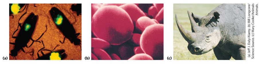
Proteins have diverse functions including:
Catalyzing reactions (enzymes)
Forming complex subcellular structures
Describe the Basic Amino Acid Structure
Each amino acid has an amino group and a carboxylate group
Each amino acid has a different side chain R
20 different amino acids are found in proteins
Note the central alpha carbon!

What form of bond links two amino acids together?
Peptide Bonds!
Condensation (Dehydration Synthesis): To join two amino acids, the carboxyl group (COO- /COOH) of the first amino acid reacts with the amino group (NH3+ /NH2-) of the second. A molecule of water (H2O) is removed (an -OH from the carboxyl end and an -H from the amino end). The resulting covalent linkage (CO-NH) is called a peptide bond (or an amide bond).
Hydrolysis (Breaking the Bond): The reverse reaction breaks the peptide bond. Adding a water molecule splits the bond and regenerates the free carboxylic acid and amino groups
NOTE: At physiological pH (~7):
Amino group (NH₃⁺): Protonated (gains/retains H⁺).
Carboxyl group (COO⁻): Deprotonated (loses H⁺).

Where is the point of weakness in the newly formed molecule?
C=O! Oxygen is significantly more electronegative than carbon:
Oxygen pulls the shared electrons strongly toward itself, giving the oxygen atom a partial negative charge
This drains electron density from the carbon atom, leaving it with a partial positive charge
Because that carbon is starved for electrons, it acts as an electrophile—essentially a target waiting for any electron-rich molecule to come along
H2O has two unshared lone pairs of electrons on its oxygen atom:
Because opposite charges attract, the electron-rich oxygen of water attacks the partially positive carbonyl carbon
Where does the unique combinations occur in a protein?
The "R" groups (or side chains) are precisely where all the diversity, individuality, and unique chemical combinations occur in a protein
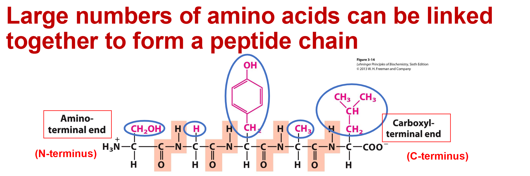
The Main Chain (Backbone): The repeating -N-Cα-C-(=O)- unit highlighted across the slide is identical in every amino acid. It simply acts as the structural scaffold holding the chain together.
The Side Chains (R1, R2, R3): These are the chemical groups attached to each central alpha-carbon (Cα).
The combination of different side chains R1, R2, R3, etc. gives each protein its unique properties.
Side Chain Structure
Carbon atoms of the amino acid core are identified by Greek letters
The α-carbon is the central backbone atom
The β- carbon is the first atom of the side chain, the y-carbon is the second, etc
Functional groups may be linked to different core atoms:
α-amino
e-amino

The 20 natural amino acids
Amino acids share a common backbone, but differ in the side chain
Amino acids can be grouped according to structures or by similar properties
How many “categories” of amino acids are there? How many belong in each group?
5 Based on Polarity Properties
6 with very non-polar side chains
5 with moderately non-polar side chains
4 with polar but uncharged side chains
3 with positively charged side chains (very polar)
2 with negatively charged side chains (very polar)

What Side Chains do the very non-polar amino acids tend to have?
The side chains are dominated by hydrocarbon, and consist only of C-C and C-H bonds
Hydrocarbon is non-polar and hydrophobic (or water avoiding)
Ala, Val, Leu, Ile, Met, Phe

Recap: Polar and Non-Polar Properties
Polarity is a consequence of atoms having different electronegativity or tendency to hold bonding electrons
Electronegativity: Electronegativity is a measure of an atom's tendency to attract shared electrons toward itself in a chemical bond
O > N > S > C = (Roughly) H
Atoms with similar electronegativity share bonding electrons equally, e.g. C-C, C-H, and are non-polar
Pairs of atoms with different electronegativity distribute bonding electrons unequally – more electronegative atoms such as O or N get greater than 50% share, and this leads to unbalanced charges and polar bonds

Moderately Non-Polar Amino Acids
Glycine has single H atom as side chain, not enough to be very non-polar
Hydrophobicity is related to the number of CH, CH2 or CH3 groups present
Cysteine contains the slightly polar SH group
Proline is unique because the side chain links to α-N as well as to α-C. The polar
N moderates the non-polar hydrocarbon.Tyrosine has a single polar group that partly offsets the very non-polar benzene ring.
Tryptophan behaves similarly.

Amino acids with polar uncharged side chains
Ser, Thr, Asn, Gln
Serine and threonine have side chains that include the polar hydroxyl group -OH (simple alcohol)
Asparagine (Asn) and Glutamine (Gln) both contain the polar amide group
These side chain groups do not gain or lose H+ in aqueous solution at pH 7, so they are uncharged
All four side chains act as good hydrogen bond donors or acceptors

Recap: Hydrogen Bonds
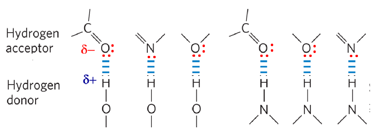
Hydrogen bonds are electrostatic attractions between a H-bond donor and an acceptor
An acceptor = electronegative atom with an available lone pair of electrons
It "accepts" the partially positive hydrogen by attracting it toward its lone pair.
A donor = hydrogen atom covalently bonded to another electronegative atom.
It "donates" its hydrogen's positive partial charge to the interaction because it pulls electron density away from that hydrogen.
The hydrogen bond is about 5-10% as strong as a covalent bond, enough to make the two molecules stick loosely to each other but not to form a permanent link
H-bonds are directional - stronger if donor and acceptor line up with one another
The Amino Acids with Positively charged side chains (very polar)
These side chains contain weak bases that gain H+ (become protonated) and so are positively charged in aqueous solution at neutral pH
Charge make them very polar, overriding the non-polar hydrocarbon chain

The Amino Acids with Negatively charged side chains (very polar)
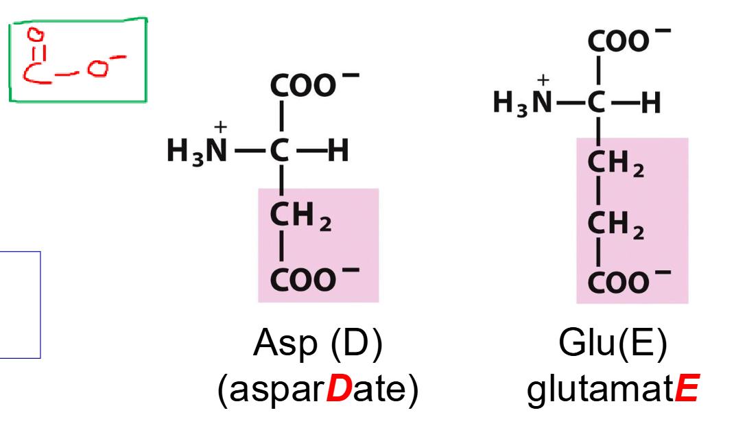
Side chains have carboxylic acid groups R-COOH that lose H+ (become deprotonated) at neutral pH
When deprotonated these are described as carboxylate groups R- COO-
Carboxylate groups are negative and also very polar
Asp side chain: -CH2-COO-
Glu side chain: -CH2-CH2-COO-
What can amino acids act as?
As both acids and bases!
Recall: HA → A- + H+
The amino and carboxylate groups of amino acids and the side chains of some amino acids can act as acids and bases
These groups will gain (protonate) or lose (deprotonate) H+ depending on availability of H+ in solution
Normal biochemical processes occur close to pH 7 (physiological pH is 7.0-7.4)

Worked Example 1
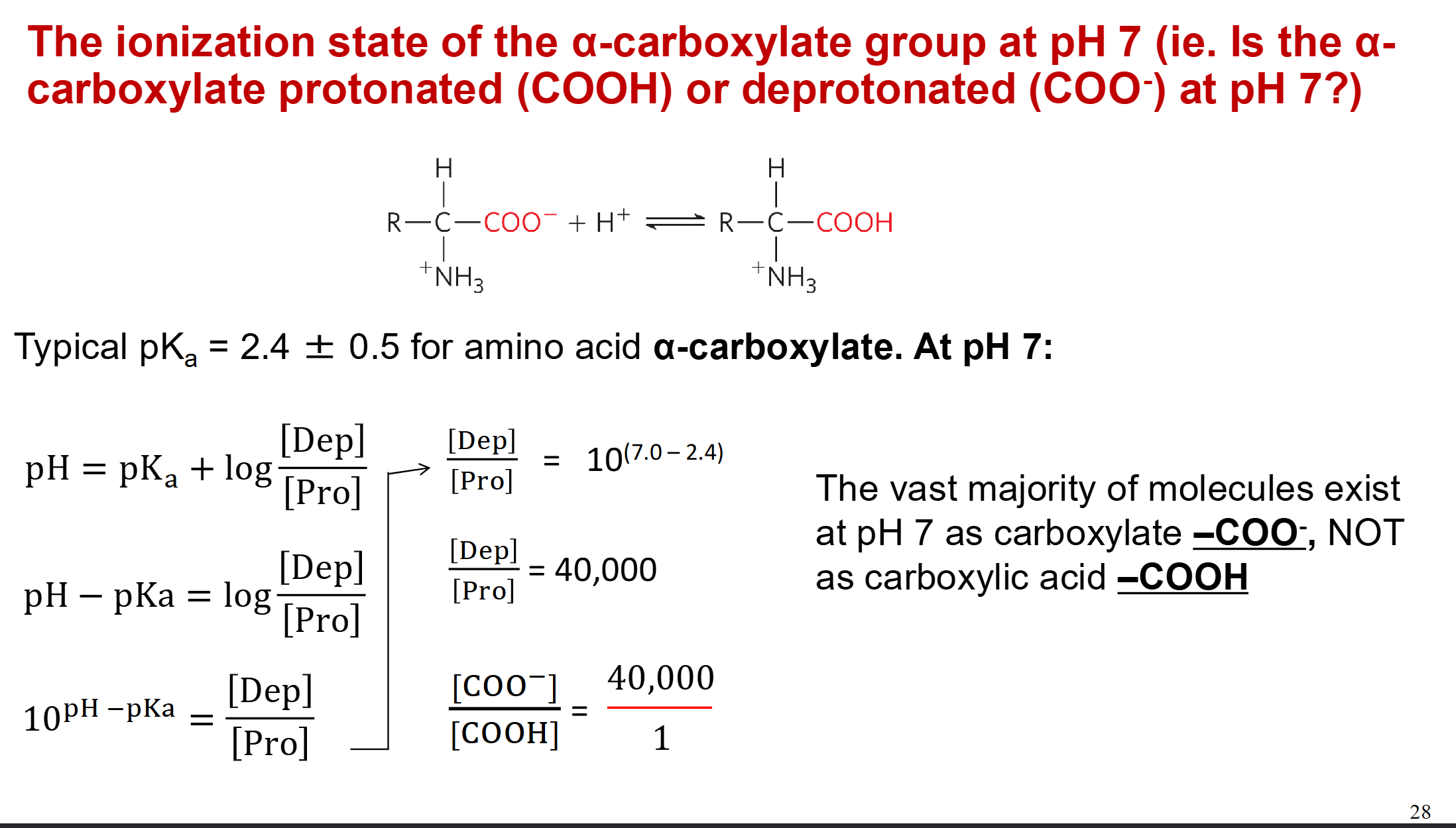
Worked Example 2

Important! What is the correct way to represent an Amino Acid at a Neutral pH?
The correct structure to represent an individual amino acid at neutral pH is NH3+ and COO-
This is also known as a Zwitterion
But when the amino acid is part of a peptide chain, the α-amino groups and α-carboxylate groups are linked as uncharged amide bonds (except for the ones at the N and C termini, which are also NH3+ and COO- at pH 7)

How many amino acids have ionizable side chains?
Seven!
Note: The polar but uncharged amino acids go from 0 to -1 charge
Note: The very polar and charged amino acids go from +1 to 0 charge
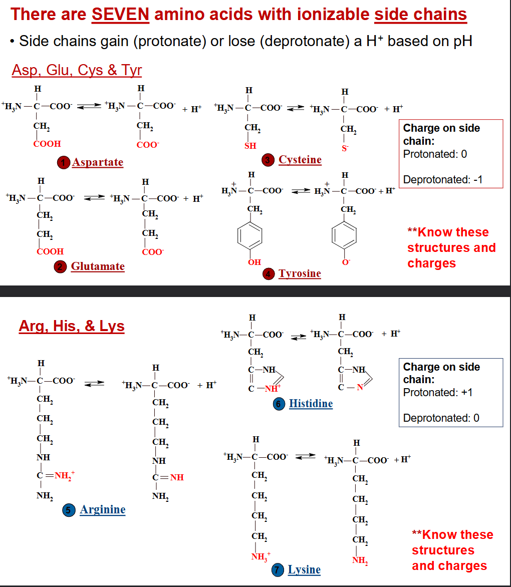
What does the value of pKa tell you in regards to deprotonation?
The value of pKa tells you where in the pH scale a group undergoes deprotonation
A molecule can have several ionizable groups
Each group has its own pKa value
Value of a pKa depends on its chemical context
An amino acid will have a slightly different pKa when it is part of a peptide chain
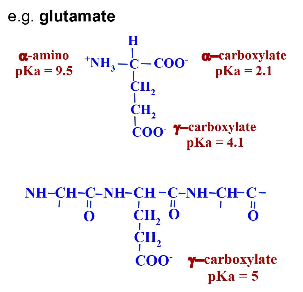
Example/Explanation of Deprotonation’s correlation with pH with Glutamic Acid
Starting with glutamic acid at very low pH, all three functional groups
are fully protonated.As we raise the pH, [H+] becomes less available, so deprotonation is more likely to occur.
Each group undergoes a transition as pH shifts from 0 to 14, starting ~1 unit below its pKa and almost complete by ~1 unit above its pKa.
When pH = pKa, 50% of the group is in the protonated form and 50% is in the deprotonated form
Note that once a group deprotonates, it remains that way for the rest of the pH range
REMEMBER: The ionization state of a group at a given pH depends on its pKa value
![<ul><li><p><span>Starting with glutamic acid at very low pH, all three functional groups<br>are fully protonated.</span></p></li><li><p><span>As we raise the pH, [H+] becomes less available, so deprotonation is more likely to occur.</span></p></li><li><p><span>Each group undergoes a transition as pH shifts from 0 to 14, starting ~1 unit below its pKa and almost complete by ~1 unit above its pKa.</span></p></li><li><p><span>When pH = pKa, 50% of the group is in the protonated form and 50% is in the deprotonated form</span></p></li><li><p><span>Note that once a group deprotonates, it remains that way for the rest of the pH range</span></p></li><li><p><span>REMEMBER: <strong>The ionization state of a group at a given pH depends on its pKa value</strong></span></p></li></ul><p></p>](https://assets.knowt.com/user-attachments/0e66a3d8-4dca-4c43-a96a-3216caab0dc0.png)
Important: How to assess the state of ionization of a functional group
If pH is one unit or more below the pKa, the group is fully protonated
If pH is one unit or more higher than pKa, the group is fully deprotonated
If pH is equal to pKa, the group is 50% deprotonated and 50% protonated
If pH is less than one unit away from pKa, a calculation may be needed to determine the exact state

Does being protonated mean a functional group is always positively charged? How does the ionizing atom determine charge?
Charge of an ionized group depends on the functional group
The relationship of pH and pKa tells you whether a group is protonated or deprotonated, NOT whether it is positive or negative → Make sure to look at those R groups!
Adding H+ always shifts the charge by +1 (more positive), but the net charge depends on the atom involved
Groups that ionize on O or S atoms are neutral when protonated, and negative when deprotonated (e.g., side chains of Asp, Glu)
Groups that ionize on N are positive when protonated, and neutral when deprotonated (e.g., side chains of Arg, Lys, His)
There is no group that goes from positive to negative when it becomes deprotonated!!
REVIEW SLIDES ON HOW TO DO CALCULATIONS 😃 😃😃
What are the two processes of amino acid analysis? Also what three things can it be?
Amino acid analysis helps to determine protein structure
Separation of a mixture into components
Detection of the components of interest
Can be qualitative (tells you what is present)
Can be quantitative (tells you how much is present)
Can be preparative (separated components can be recovered for further experiments)
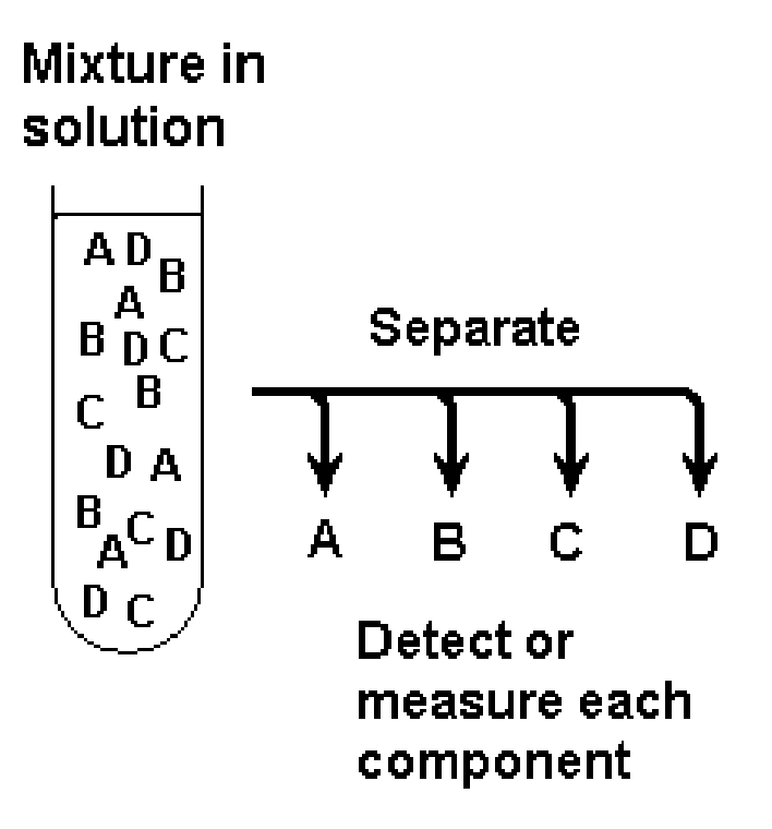
What method is important for separating different parts of a mixture? What are the two phases involved?
Partition Chromatography
Particles of solid are chosen with a specific property, e.g. silica gel has HO-Si-OH groups that can hydrogen-bond to polar amino acids
Stationary phase
Liquid solvent or buffer flows past the particles and is non-polar
Mobile phase
Amino acids exchange (partition) between phases
Polar amino acids P spend more of their time hydrogen bonded to silica and move slowly
Non-polar amino acids N spend more time in solvent, and move almost as fast as solvent
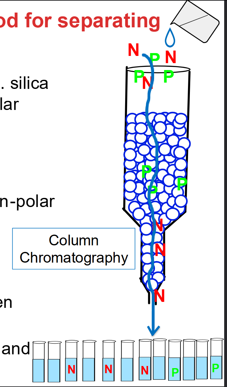
The Stationary and Mobile Phase
Stationary Phase (The "Stick-Around" Phase):
Solid beads packed into a vertical glass column.
Here, silica gel is used, which is covered in exposed hydroxyl groups (HO-SI-OH). Because of these (-OH) groups, the stationary phase is highly polar and readily forms hydrogen bonds.
Mobile Phase (The "Moving" Phase):
A liquid solvent or buffer poured into the top that percolates downward through the packed beads.
In this setup, the solvent chosen is non-polar.
Polar Amino Acids: They bind and unbind repeatedly, spending most of their time stuck to the stationary phase. As a result, they move down the column slowly and come out (elute) last
Non-Polar Amino Acids: They prefer dissolving in the non-polar mobile phase. They travel along with the flowing liquid and exit the bottom first.


Based on this graph and test tubes, how are the Amino Acids Identified?
Concentration of amino acids is measured in each test tube and the results are graphed
To elute more polar amino acids, progressively polar mobile phase is run through the column
Compounds can be identified by their characteristic elution volume
N and A,B,C: Non-Polar (interact weakly with the polar silica stationary phase, so they wash through rapidly with minimal elution volume)
P and D,E: Polar: Amino acids stick tightly to the polar stationary phase via hydrogen bonding and require larger elution volumes
What is another method to identify amino acid properties?
Thin layer chromatography
Silica gel is spread in a thin layer on a plastic sheet
Samples are applied near the lower edge
The lower edge is placed in solvent
As solvent soaks up the sheet, different components of sample move with the solvent at different rates
The highest point reached by solvent is the solvent front
Each amino acid can be identified by its characteristic relative mobility RF
Very polar amino acids have low RF, non-polar amino acids have high RF

How are amino acids detected?
Amino acids are colourless, and samples may be 10-6 to 10-10 moles
They can be detected by adding ninhydrin which reacts with primary and secondary amines
Gives intense purple colour (10-8 moles detectable), or yellow colour for proline
Spray ninhydrin onto TLC plates, or add to amino acid solution, and heat
Colour intensity is proportional to quantity of amino acid, and can be measure
Alternative is fluorescamine, giving yellow fluorescence under UV light (10-10 moles detectable)
Ion Exchange Chromatography
Separates on the basis of charge
Uses charged resins as stationary phase
Cation exchange Chromatography- resins contain negative groups, which bind positive molecules (cations)
Anion exchange Chromatography - resins contain positive groups, which bind negative molecules (anions)
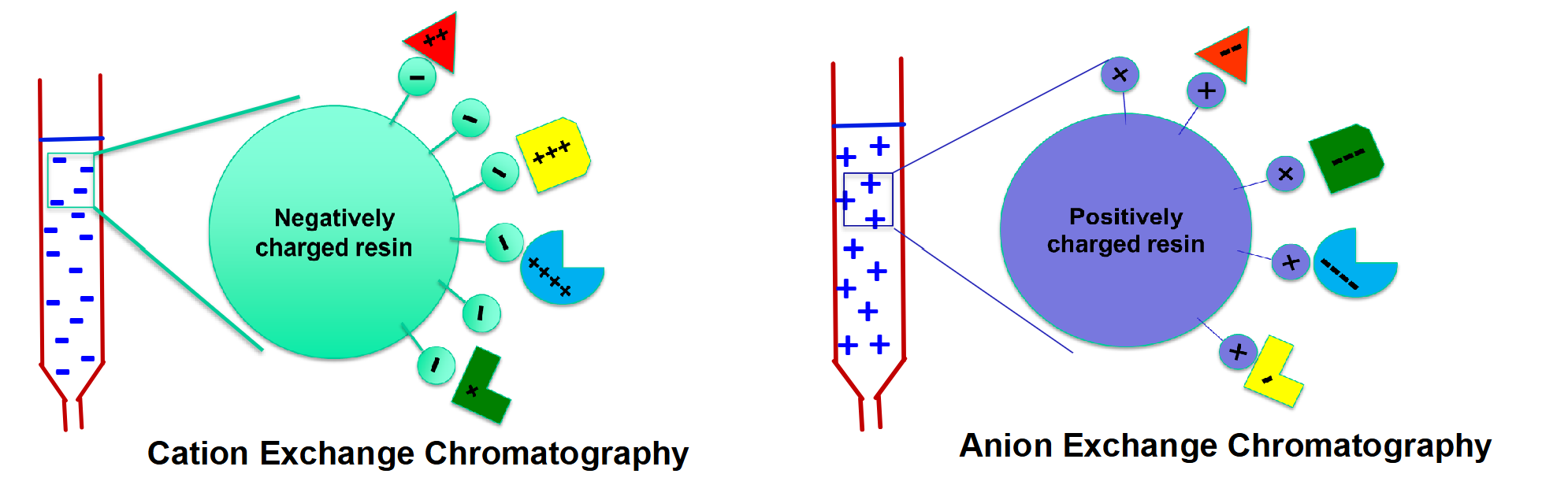
How do you break that ionic attraction to wash them out (elute them?)
Salt Competition (e.g., increasing (NaCl):
Floods the column with counterions (Na+ and Cl-) that physically outcompete the amino acids for the charged binding sites on the resin.
Molecules with lower net charges are displaced first at low salt, while highly charged molecules require higher salt concentrations to release.
Altering pH (Charge Neutralization / Reversal):
Shifting buffer pH changes the protonation state of the amino acid's ionizable groups based on their pKa
This eliminates (or reverses) the molecule's net charge—e.g., raising pH deprotonates positive groups, causing them to lose electrostatic attraction and elute from a cation resin
How does low pH (e.g., pH 2.5) allow amino acids to bind to a cation exchange resin?
Resin charge: Cation exchange resin is negatively charged.
Amino acid charge at pH 2.5:
a-amino group is fully protonated as NH3+ (+1).
a-carboxylate group (pKa approx 2.5) is 50% COO- (-1) and 50% -COOH (0).
Net core charge is overall positive (+0.5), enabling electrostatic binding to the negative beads.
Binding strength: Determined by the magnitude of the amino acid's total net positive charge (including side chains).

What are the two methods used to elute bound amino acids from a cation exchange column?
Na+ Salt Competition (left):
High Na+ counterions compete for and displace amino acids from negative resin binding sites.
Weakly bound (lower positive charge) amino acids displace first at low Na+ tightly bound ones elute as Na+ increases.
Increasing {pH} (right):
Raising pH (e.g., to 6.5) causes amino acids to lose protons (H+).
Neutralizes or reverses the net positive charge, eliminating attraction to the negative resin so they wash out.

Summary of Ion Exchange Slide

Where are these proteins extracted from?
Proteins are derived from natural sources such as microbial cultures, plants, or animal tissues such as liver
The cells are broken open to release the proteins into a solution – crude extract
Extracts may contain thousands of different proteins
Separation by ion exchange is based on charge differences among proteins (Net Charge)
Depends on the relative number of Asp + Glu (negative) versus His + Lys + Arg (positive) in each protein, and on pH
~65% of all proteins are negatively charged at pH 7

Charge Differences Between Proteins and Amino Acids


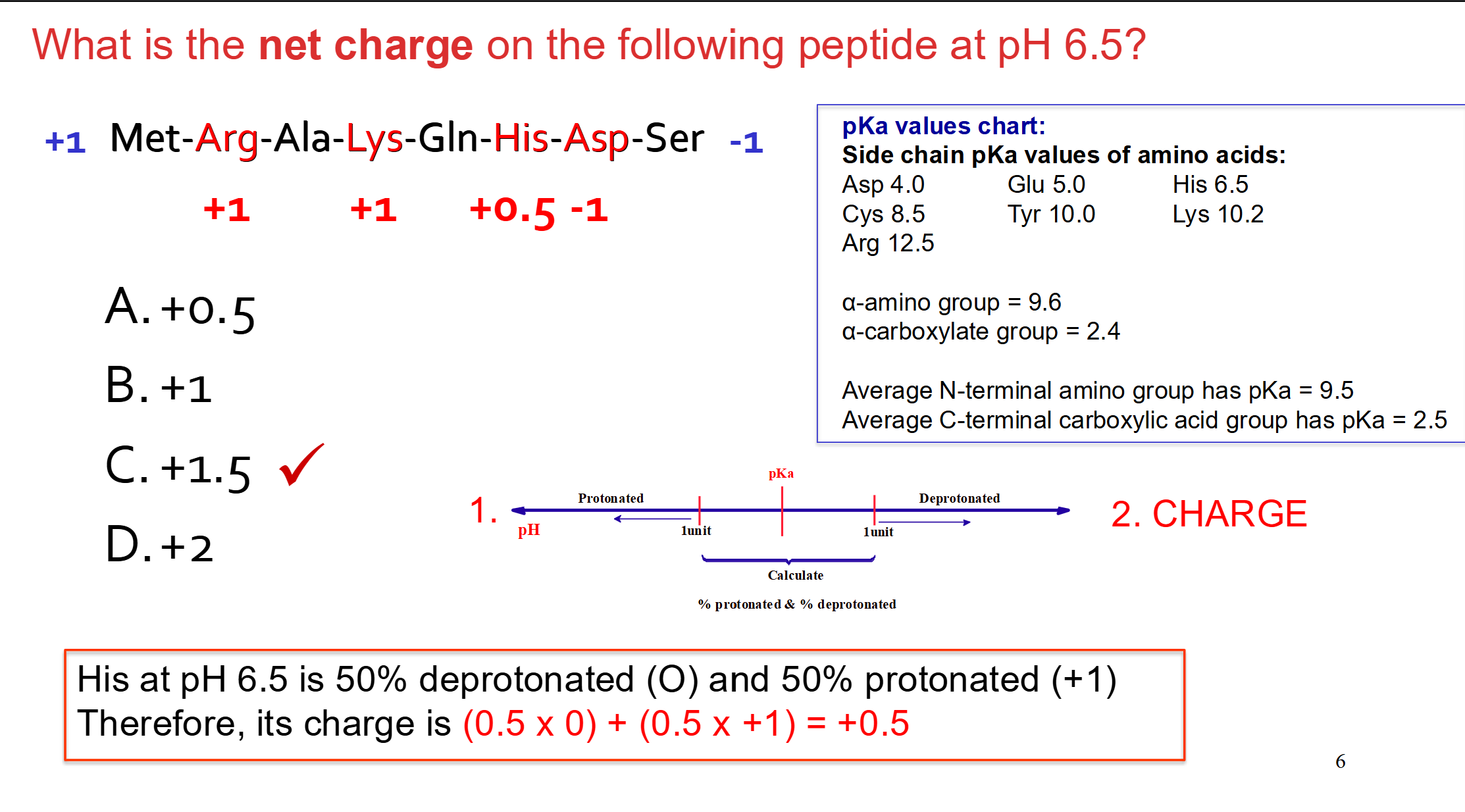
Example Question
Affinity Chromatography
Beads in the column – has covalently attached ligand
Any molecule or ion that binds specifically and reversibly to a target protein or macromolecule to form a complex.
Proteins with affinity towards ligand - bind to it
The ligand doesn't just slow the target protein down—it usually stops it completely (remains anchored in place at the top of the column.)
All other unwanted proteins lack affinity for the ligand and flow straight through the column without sticking (the "flow-through").
The target protein must be detached from the ligand so it can be collected. This is typically done by:
Salt (weakens binding between ligand and proteins) or
Ligand (compete with attached ligands)

Affinity Chromatography - Use of Tagged Proteins
Tag = A peptide or protein that binds a ligand that is fused to the gene encoding target protein

Ligand vs Tag Protein Table

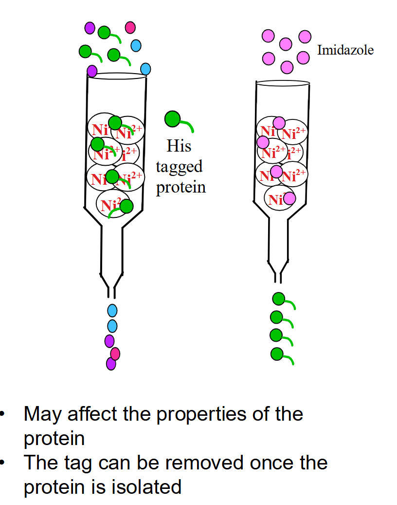
Immobilized Metal Affinity Chromatography (IMAC)
Principle: Target proteins are genetically engineered with a 6–8 His-tag at the N- or C-terminus, which binds tightly to chelating resin charged with {Ni}^{2+} or {Co}^{2+} ions.
Binding/Washing: Column is made up of chelating resin containing
Ni2+. Only the His-tagged protein coordinates with the immobilized metal ions, while non-tagged background proteins wash straight through.Elution: Released by adding imidazole to the buffer. Free imidazole mimics the histidine side chain and out-competes the His-tag for {Ni}^{2+} binding sites.
Pros/Cons: High degree of purification in one step; however, the tag can affect native protein activity and may need to be enzymatically cleaved post-isolation.
Separating proteins on the basis of size
Gel filtration or molecular exclusion chromatography allows separation of proteins on the basis of size
Beads of polymeric gel - a loose network of polymer with many water-filled pores.
Protein molecules can enter the pores if they fit
Larger proteins are excluded from the pores
Proteins of intermediate size may enter some of the pores
Proteins separate by size; larger proteins elute first, smaller proteins elute later

Gel filtration can be used to measure the molar mass of proteins -Sample is compared to proteins of known size
Measure elution volume of proteins of known mass
Elution volume Ve is the volume of buffer needed to move a protein from top to bottom of column
Elution volume is a linear function of log molar mass (negative slope)
Then measure elution volume of unknown protein and project back to the log mass axis
Find antilog to determine the molar mass of unknown
Alternately, find Mr by using coordinates to derive equation for straight line y=mx+b

Proteins can be separated and characterized by electrophoresis
Electrophoresis - movement of charged molecules in an electric field
Does not contribute to purification as structure is commonly affected by electrophoresis
Can visualize and characterize purified proteins
Can be used to estimate:
Number of different proteins in a mixture
Degree of purity of a mixture
Isoelectric point
Approximate molecular weight
Rate of movement depends on size, shape and charge
