KN 230 Exam IV (Chapters 10, 11, 14)
 Call Kai
 Learn
 Practice Test
 Spaced Repetition
 Match
 Flashcards
 Knowt Play
1/417

There's no tags or description
Looks like no tags are added yet.
418 Terms
Functions of skeletal muscle
Move the body
Maintain posture
Protect and support
Regulate elimination of materials
Produce heat
Sphincter
Muscle that encircles a duct, tube, or orifice such that its contraction constricts the lumen or orifice and restricts movement through the tube
Orifice
Aperture or opening
Characteristics of skeletal muscle
Excitability
Conductivity
Contractility
Extensibility
Elasticity
Excitability
The ability of a cell to respond to a stimulus
Conductivity
The ability to propagate an action potential along the plasma membrane
Contractility
When contractile proteins within skeletal muscle cells slide past one another
What enables muscle cells to cause body movement and to perform the other functions of muscles
Extensibility
The lengthening of a muscle cell
Elasticity
The ability of a muscle cell to return to its original length following either shortening or lengthening of the muscle
Dependent upon the release of tension in the springlike connectin protein associated with contractile proteins
Skeletal muscle fibers
The term used to refer to skeletal muscle cells
Also called muscle fibers or myofibers

Skeletal muscle
Location: Generally attached to the skeleton (or to subcutaneous tissue); voluntary movement of skeleton, skin, or other select body components
Associated connective tissue: Epimysium, perimysium, endomysium
Cell appearance and shape: Long, cylindrical fibers with multiple peripheral nuclei; striated; T-tubules; large diameter (10-500 μm); long length (100 μm- 30 cm)
Calcium source: Well-developed sarcoplasmic reticulum
Contractile unit and Ca2+ binding: Sarcomere; Ca2+ binds to troponin
Stimulation: Nervous control is voluntary (regulated by somatic nervous system at neuromuscular junction); excitatory
Rate of response and primary energy source: Slow oxidative (SO) [Slow; aerobic production of ATP]; Fast oxidative (FO) [Rapid and powerful; aerobic production of ATP]; Fast glycolytic (FG) [Rapid and powerful; glycolytic production of ATP]
Tissue repair ability: Limited

Fascicle
Bundle of muscle fibers or axons
Connective tissue layers of muscles
Layers that provide protection and support, a means of attachment of the muscle to the skeleton or other structures within the body, and sites for distribution of blood vessels and nerves
Collectively extend past the muscle fibers to form either a tendon or an aponeurosis
Epimysium
Perimysium
Endomysium
Epimysium
A layer of dense irregular connective tissue that surrounds the whole skeletal muscle
Ensheathes the entire skeletal muscle to protect and support it like a tough leather sleeve
Perimysium
A layer of dense irregular connective tissue around each fascicle
Provides protection and support to each bundle of muscle fibers
Endomysium
A layer of areolar connective tissue that surrounds each muscle fiber
More delicate coverings that function to electrically insulate the muscle fibers
Tendon
A thick, cordlike structure composed of dense regular connective tissue that connects muscle to bone
Aponeurosis
A thin, flattened sheet of dense regular connective tissue
Deep fascia
An additional, expansive sheet of dense irregular connective tissue that is external to the epimysium, but often blends with it
Separates individual muscles
Binds together muscles with similar functions
Contains nerves, blood vessels, and lymph vessels
Fills spaces between muscles
Also called visceral fascia or muscular fascia
Superficial fascia
The layer that the deep fascia is internal to or deep to
Composed of areolar connective tissue and adipose connective tissue that separates muscle from skin
Also called the subcutaneous layer
Somatic motor neurons
Neurons that extend from the brain and spinal cord to skeletal muscle fibers
Each has an axon that branches extensively at its terminal end
Axon
The process of a neuron that propagates nerve signals away from the cell body
Extends through all three connective tissue layers to almost make contact with an individual muscle fiber
Voluntary muscle
A muscle whose fibers can be consciously controlled by the nervous system
Ex: Skeletal muscle
Sarcoplasm
The cytoplasm of a muscle cell
Myoblast
An undifferentiated muscle cell with the potential of becoming a muscle fiber
Groups of the embryonic muscle cells fuse to form single skeletal muscle fibers during development
Each of their nuclei contributes to the eventual total number of nuclei in the muscle fiber (multinucleated)

Multinucleated
Having numerous nuclei
Satellite cells
Adult stem cells
Myoblasts that do not fuse with muscle fibers during development and instead remain in adult skeletal muscle tissue
Some may be stimulated to differentiate and then fuse with a damaged skeletal muscle fiber to assist to a limited extent in its repair and regeneration
Sarcolemma
The plasma membrane of a skeletal muscle fiber
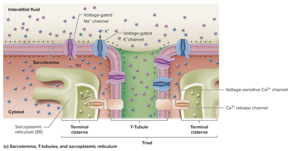
T-tubules
Deep invaginations of the sarcolemma that extend into the skeletal muscle fiber as a network of narrow, membranous tubules to the sarcoplasmic reticulum (ER of the muscle)
Also called transverse tubules
Myofibril
Bundles of myofilaments that are enclosed in portions of the membranous sarcoplasmic reticulum within skeletal and cardiac muscle cells
Composes approximately 80% of the volume of a skeletal muscle fiber
Each extends the entire length of the skeletal muscle fiber and is about 1 to 2 micrometers in diameter
Sarcoplasmic reticulum (SR)
An internal membrane complex found in muscle cells that is similar to the membranous smooth endoplasmic reticulum of other cells
Stores calcium ions required for contraction
Segments of it fit around each myofibril like a sleeve of membrane netting
Terminal cisternae
Blind sacs at either end of individual sections of the sarcoplasmic reticulum
Immediately adjacent to each T-tubule
Triad
A structure formed by two terminal cisternae and a centrally located T-tubule
Voltage-sensitive Ca2+ channels
Dihydropyridine receptors that are responsive to electrical signals
Ca2+ release channels
Ryanodine receptors at the terminal cisternae membrane of the sarcoplasmic reticulum
Myofilaments
Contractile protein filaments that are bundled within myofibrils in both skeletal and cardiac muscle cells
Not as long as a myrofibril and takes many successive units to extend the entire length of the myofibril
Two types: Thick filaments and thin filaments
Thick filaments
Type of myofilament that is assembled from bundles of 200 to 500 myosin protein molecules
Also called thick myofilaments

Thin filaments
Type of myofilament that is primarily composed of two strands of actin protein twisted around each other to form a helical shape
Approximately half of the diameter of thick filaments (about 5 to 6 nanometers)
Also called thin myofilaments

Troponin-tropomyosin complex
The formation of tropomyosin and troponin
Tropomyosin
A short, thin, twisted filament that is a stringlike protein
Regulatory protein associated with thin filaments
Consecutive molecules cover small regions of the actin strands, including the myosin binding sites in a noncontracting muscle
Troponin
A globular, or ball-like, protein attached to tropomyosin
Regulatory protein associated with thin filaments
Contains the binding site for Ca2+
Sarcomere
The functional unit of skeletal muscle and cardiac muscle
Each is composed of overlapping thick filaments and thin filaments

Z discs
Delineate each sarcomere at both ends
Composed of specialized proteins that are positioned perpendicular to the myofilaments and serve as anchors for the thin filaments
I band
Region of the sarcomere that contains only thin filaments and is bisected by the Z disc
Appears light when viewed with a microscope
The bands disappear when the thin filaments are pulled parallel along the thick filaments at maximal muscle shortening
A band
The central region of a sarcomere that contains the entire thick filament
Thin filaments partially overlap the thick filament on each end of the band
Appears dark when viewed with a microscope
Does not change in length during muscle contraction
H zone
The most central portion of the A band in a resting sarcomere
Does not have thin filament overlap (only thick filaments are present)
Disappears when the thin filaments are pulled past thick filaments during maximal muscle shortening
Also called the H band
M line
A thin transverse protein meshwork structure in the center of the H zone
Serves as an attachment site for the thick filaments are keeps the thick filaments aligned during contraction and relaxation events
Striations
The repeating light and dark bands of the overlapping myofilaments that form unique striped patterns within a skeletal muscle fiber
Due to both the size and density differences between thin filaments and thick filaments
Anisotropic
A band whose area appears dark when viewed under polarized light
Isotropic
A band whose area appears light when viewed under polarized light
Connectin
A cable-like protein that extends from the Z discs to the M line through the core of each thick filament
Stabilized the position of the thick filament and maintains thick filament alignment within a sarcomere
Portions of its molecules are coiled and springlike so that during sarcomere shortening, they are compressed to produce passive tension
Contributes to skeletal muscle fiber elasticity
Also called titin
Dystrophin
A protein that is part of a protein complex that anchors myofibrils that are adjacent to the sarcolemma to proteins within the sarcolemma
Extend to the connective tissue of the endomysium that encloses the muscle fiber
Links internal myofilament proteins of a muscle fiber to proteins within the plasma membrane, which are linked to extracellular proteins
Nebulin
An actin-binding protein of the sarcomere that is anchored in the Z-disc and extends the length of the thin filament
Helps determine thin filament length during sarcomere assembly, has structural and mechanical properties, and plays a role during skeletal muscle contraction in cross-bridge cycling
Glycogen
Polysaccharide formed from glucose monomers
Stored in granules called glycosomes in muscle fibers for use as an immediately fuel molecule
Myoglobin
A molecule that binds oxygen when the muscle is at rest and releases it for use during muscular contraction
A reddish, globular protein that is somewhat similar to hemoglobin
Creatine phosphate
A type of molecule that is contained in skeletal muscle fibers
Provides muscle fibers with a very rapid means of supplying ATP
Has a high-energy chemical bond between creatine and Pi
Present in tissues with both large and fluctuating energy needs
Motor unit
A single motor neuron and the skeletal muscle fibers it controls
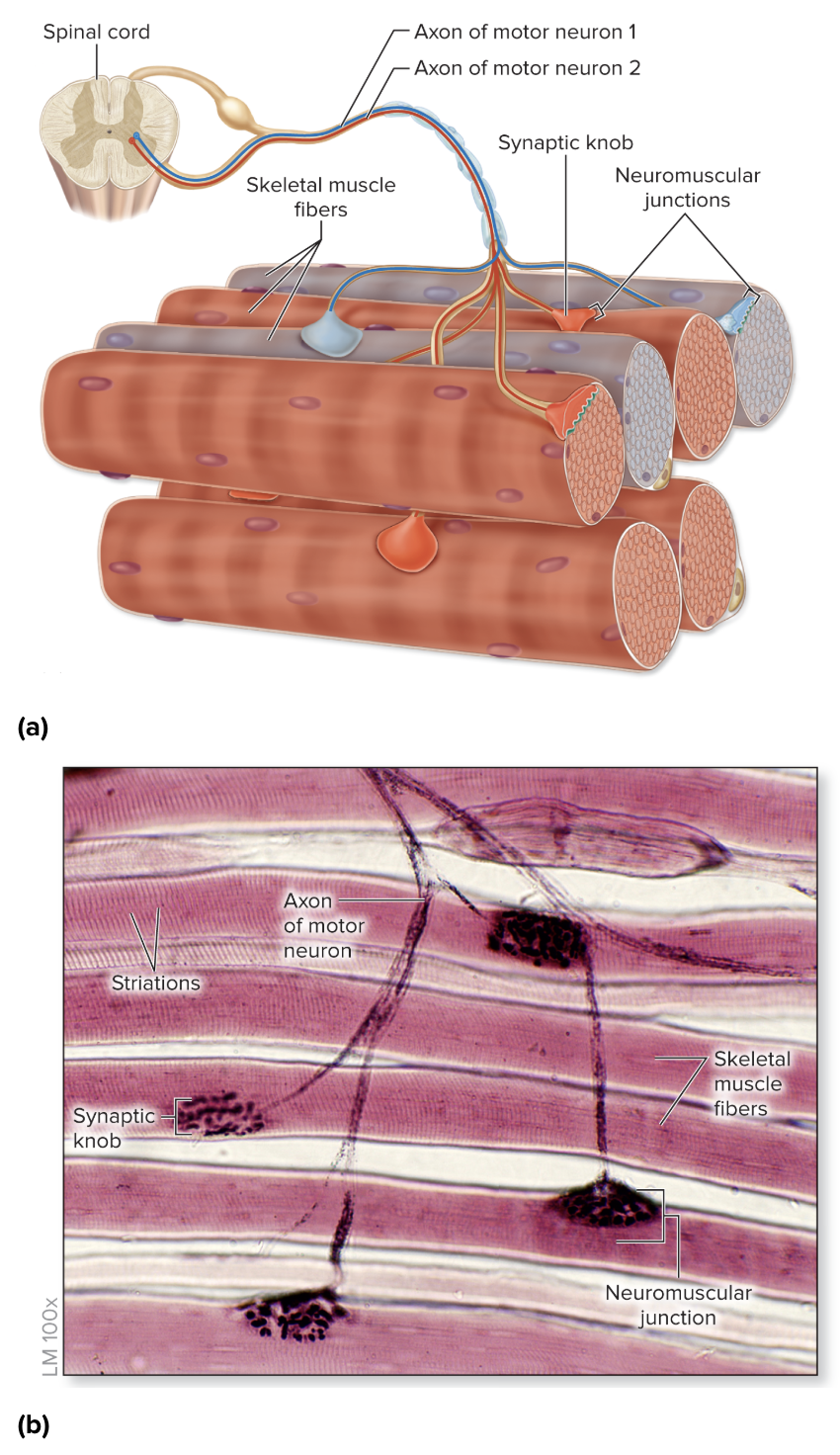
Neuromuscular junction
The specific location, usually in the mid-region of the skeletal muscle fiber, where it is innervated by a motor neuron
Has the following parts: Synaptic knob, motor end plate, and synaptic cleft

Synaptic knob
An expanded tip of an axon of a motor neuron
Is enlarged and flat to cover a relatively large surface area of the sarcolemma
Houses numerous synaptic vesicles filled with molecules of the neurotransmitter acetylcholine
Ca2+ pumps and voltage-gated Ca2+ channels are embedded within its plasma membrane
Synaptic vesicle
A package of membrane enclosing neurotransmitter molecules in the synaptic knob
Acetylcholine (ACh)
Neurotransmitter produced by the central and peripheral nervous systems
Stimulates skeletal muscle
Motor end plate
A specialized region of the sarcolemma of a skeletal muscle fiber
Has numerous folds and indentations (junction folds) to increase the membrane surface area covered by the synaptic knob
Has a vast numbers of ACh receptors
ACh receptors
Plasma membrane protein channels at the motor end plate that bind to ACh to allow Na+ entry into the muscle fiber and K+ to exit
Synaptic cleft
An extremely narrow (30 nanometers), fluid-filled space separating the synaptic knob and the motor end plate
Acetylcholinesterase (AChE)
An enzyme that resides within the synaptic cleft and quickly breaks down ACh molecules following their release into the synaptic cleft
Resting membrane potential (RMP)
The electrical difference at the plasma membrane when a cell is at rest
About -90 mV in skeletal muscle fibers
Muscle fiber excitation
The first physiologic event of skeletal muscle contraction
An event that occurs at the neuromuscular junction and results in the release of ACh and its subsequent binding to ACh receptors
Steps
Calcium entry at synaptic knob
Release of ACh from synaptic knob
Binding of ACh at motor end plate

Nerve signal
An electrical signal that is propagated down an axon
Sent along a motor neuron of the somatic nervous system
Triggers the opening of voltage-gated Ca2+ channels within the synaptic knob plasma membrane
Excitation-contraction coupling
The second physiologic event of muscle contraction
An event that involves the sarcolemma, T-tubules, and sarcoplasmic reticulum of a skeletal muscle fiber
Links the events of skeletal muscle stimulation (excitation) to the events of contraction
Steps
Development of an end-plate potential at the motor end plate
Initiation and propagation of an electrical signal along the sarcolemma and T-tubules
Release of calcium from the sarcoplasmic reticulum

End-plate potential (EPP)
The minimum voltage change (or threshold) in the motor end plate that can trigger opening of voltage-gated channels in the sarcolemma to initiate an action potential
Action potential
Self-propagating change in membrane potential occurring in excitable cells (e.g., neurons, muscle cells)

Depolarization
Change in membrane potential or voltage to a more positive value
Repolarization
Change in membrane potential from a depolarized value back to the resting value
Absolute refractory period
Time period when an excitable cell cannot be restimulated to respond
Calcium binding to troponin
What initiates crossbridge cycling
Crossbridge cycling
A four step process that is repeated
Crossbridge formation (attaching of myosin head to actin)
Power stroke (pulling thin filament by movement of myosin head)
Release of myosin head from actin
Resetting of myosin head

Sliding filament theory
A description of the repetitive movement of thin filaments sliding past thick filaments
Glycolysis
A metabolic pathway that involves the breakdown of glucose into two pyruvate molecules, producing a net of 2 ATP molecules
Occurs within the cytosol
Can function in the presence of oxygen, but oxygen is not required
Aerobic cellular respiration
The breakdown of nutrients (e.g., pyruvate, fatty acids) to produce ATP, water, and carbon dioxide
Occurs within mitochondria and requires oxygen
Involves three stages: Intermediate step, citric acid cycle, electron transport system
Lactate formation
Occurs from pyruvate under conditions of low oxygen availability (ex: during intense exercise when skeletal muscle’s oxygen demands for aerobic cellular respiration cannot be met)
Pyruvate molecules are converted to lactate molecules, an enzymatic reaction catalyzed by lactate dehydrogenase
Lactate can either enter a mitochondrion within a skeletal muscle fiber, where it is converted back to pyruvate and oxidized into carbon dioxide through aerobic cellular respiration, or leave the skeletal muscle fiber and enter the blood
Lactic acid cycle
The cycling of lactate to the liver, where it is converted to glucose, and the subsequent blood transport of glucose from the liver to the muscle
Also called the Cori cycle
Oxygen debt
The amount of additional oxygen that is consumed following exercise to restore pre-exercise conditions
This additional oxygen is required primarily by skeletal muscle fibers and liver cells
Classification of skeletal muscle fibers
The type of contraction generated
The primary means used for supplying ATP
Power
Related to the diameter of a muscle fiber
Large muscle fibers have a larger number of myofibrils in parallel, allowing them to produce a more powerful contraction
Speed
Based on whether the skeletal muscle fiber expresses the relatively slow or fast genetic variant of myosin ATPase, the enzyme that splits ATP
Fast-twitch fibers
Skeletal muscle fibers with a fast variant of myosin ATPase
Typically produce a strong contraction, initiate a contraction more quickly following stimulation, and produce a contraction of shorter duration
Exhibits more power and speed in comparison to slow-twitch fibers
Slow-twitch fibers
Skeletal muscle fibers with the slow variant of myosin ATPase
Exhibits less power and speed in comparison to fast-twitch fibers
Oxidative fibers
Skeletal muscle fibers that specialize in providing ATP through aerobic cellular respiration and have several features that support these processes (extensive capillary network, large numbers of mitochondria, large supply of the red pigment myoglobin)
Sometimes called red fibers due to the presence of both mitochondria and myoglobin giving these skeletal muscle fibers a red appearance
Fatigue-resistant
Oxidative fibers that continue contracting for extended periods of time without tiring due to the higher levels of ATP generated providing energy
Glycolytic fibers
Skeletal muscle fibers that specialize in providing ATP more rapidly through glycolysis
Generally have fewer structures needed for aerobic cellular respiration (less extensive capillary networks, fewer mitochondria, and smaller aounts of myoglobin)
Sometimes called white fibers due to the relatively small amount of mitochondria and myoglobin giving these skeletal muscle fibers a white appearance
Fatigable
Glycolytic fibers that generally tire easily after a short time of sustained muscular activity
Subtypes of skeletal muscle fibers
Slow oxidative (SO) fibers
Fast oxidative (FO) fibers
Fast glycolytic (FG) fibers
Slow oxidative (SO) fibers
Type I fibers
Primary fiber function: Endurance (e.g., maintaining posture, marathon running)
Muscles with a large abundance of fiber type: Trunk and lower limbs
Power of contraction: Relatively less powerful
Fiber diameter: Smallest
Relative number of myofibrils per muscle fiber: Smallest
Speed (contraction velocity): Slow
Myosin ATPase: Slow
Speed of propagation and Ca2+ regulation: Slow
Capacity to make ATP: High
Concentration of capillaries: Extensive
Number of mitochondria: Many
Amount of myoglobin: Large
Color fibers: Red
Resistance to fatigue: Highest
Fast oxidative (FO) fibers
Type IIa fibers
Primary fiber function: Medium duration, moderate movement (e.g., walking, biking)
Muscles with a large abundance of fiber type: Lower limb muscles
Power of contraction: Relatively more powerful
Fiber diameter: Intermediate
Relative number of myofibrils per muscle fiber: Intermediate
Speed (contraction velocity): Fast
Myosin ATPase: Fast
Speed of propagation and Ca2+ regulation: Fast
Capacity to make ATP: Moderate
Concentration of capillaries: Moderately extensive
Number of mitochondria: Many
Amount of myoglobin: Medium
Color fibers: Lighter red
Resistance to fatigue: High
Fast glycolytic (FG) fibers
Type IIx fibers
Primary fiber function: Short duration, intense movement (e.g., sprinting, lifting weights)
Muscles with a large abundance of fiber type: Upper limb muscles
Power of contraction: Relatively more powerful
Fiber diameter: Largest
Relative number of myofibrils per muscle fiber: Largest
Speed (contraction velocity): Fast
Myosin ATPase: Fast
Speed of propagation and Ca2+ regulation: Fast
Capacity to make ATP: Limited
Concentration of capillaries: Sparse
Number of mitochondria: Few
Amount of myoglobin: Small
Color fibers: White (pale)
Resistance to fatigue: Low
Muscle tension
The force generated when a skeletal muscle is stimulated to contract
Myogram
A graphic recording of changes in muscle tension when it is stimulated
Muscle twitch
A single, brief contraction period and then a relaxation period of a skeletal muscle in response to a single stimulation
Three periods: A latent period, a contraction period, and a relaxation period

Threshold
The minimum voltage needed to stimulate the skeletal muscle to generate a twitch
Latent period
The phase of a muscle twitch that occurs after the stimulus is applied and before the contraction of the skeletal muscle fiber begins
No change in fiber length during this period due to the time necessary for all of the events in excitation-contraction coupling, Ca2+ release from the sarcoplasmic reticulum into the cytosol, and the beginning of tension generation within the skeletal muscle fiber
Also called the lag period