Cards APP Fellow | Cath Lab focused
 Call Kai
 Learn
 Practice Test
 Spaced Repetition
 Match
 Flashcards
 Knowt Play
1/11

Earn XP
Description and Tags
Aortic valvular disease, LHC, RHC
12 Terms
Describe parameters of AV hemodynamics that would indicate severe AS requiring TAVR/SAVR procedure
V-max ≥ 4 m/s
AVA (aortic valve area) ≤ 1 cm²
∆P ≥ 40 mmHg (means the LV is pushing 40 mmHg HARDER than the aorta to overcome the stenosis)
What sign on PE may suggest critical stenosis?
Lack of a 2nd heart sound
Describe C and D stagings of aortic stenosis?
C1 = asx severe AS
C2 = asx severe AS with LV dysfunction (LVEF < 50%)
D1 = symptomatic severe high gradient
D2 = symptomatic low flow/gradient w/ rEF (LVEF < 50%, with LVH, diasytolic dysfunction)
Describe the appropriate follow up DAPT after a PCI/CABG?
ASA:
loading dose: 325 mg
maintenance: 75-100 mg (81mg MC formulation)
Ticagrelor (Brilinta)
loading dose: 180 mg
maintenance: 90 mg
Recommended to reduce MACE
Prasugrel
loading dose: 60 mg
maintenance: 5-10 mg (depends on if > or < 60kg)
DO NOT GIVE in patients w/ history of CVA/TIA (worsens outcomes)
Match the access sites to what they carry
R femoral artery = deploys the valve
L radial artery = pigtail; allows pressure readings at aorta, ventricle before and after valve placement
femoral vein = allows for ventricular pacing DURING the installment of the valve
Describe normal readings for the following sites for a RHC:
RA
RV
PA
PCWP
RA: 2-5 mmHg
RV: 15-30 mmHg (s) or ≤ 8 (d)
PA: 8-20 (usually = RV pressure)
PCWP: ≤ 15 mmHg
Describe RHC findings suggestive of cardiogenic shock
increased SVR, CO
*NORMAL SVR is 800-1200, normal CO is 4-8
What site of a coronary blockage is a LIMA best equipped to be used for a CABG?
the LAD
Describe how to attain the incomplete ‘Fick’
Using thermodilution by the means of obtaining the average of 3 values within 10% of each other; which is done by flushing 10cc of saline into the catheter
Describe the formula for cardiac index
CI = CO / BSA
Describe the formula for SVR
[(MAP - RAP) / CO] x 80
answer comes out to dyns
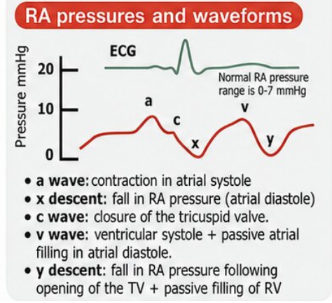
Describe the morphology of the plethforms as it relates to a RHC and the a, c, v waves as well as the x, y descents
a wave- aligns with atrial systole and thus correlates with the p-wave on EKG
c wave - aligns with tricuspid valve closing
x descent - atrial diastole
v wave - aligns with ventricular systole and thus correlates with the T-wave on EKG
y descent - aligns with passive RV filling and tricuspid valve opening