12: Cranial Nerves
 Call Kai
 Learn
Learn Practice Test
 Spaced Repetition
 Match
Match Flashcards
 Knowt Play
1/134

There's no tags or description
Looks like no tags are added yet.
Name | Mastery | Learn | Test | Matching | Spaced | Call with Kai | Chat |
|---|
No analytics yet
Send a link to your students to track their progress
135 Terms
cranial nerve 1
olfactory
cranial nerve 2
optic nerve
cranial nerve 3
oculomotor nerve
cranial nerve 4
trochlear nerve
cranial nerve 5
trigeminal nerve
cranial nerve 6
abducens nerve
cranial nerve 7
facial nerve
cranial nerve 8
vestibulo-cochlear
cranial nerve 9
glossopharyngeal nerve
cranial nerve 10
vagus nerve
cranial nerve 11
accessory nerve
cranial nerve 12
hypoglossal
functional components of olfactory nerve
SVA → olfaction, taste, conveys input from olfactory receptors about odorants in the nasal cavity
supporting cells for olfactory nerve
similar to glia, provide nutrition to receptor cells and produce mucus
basal cells for olfactory nerve
replenish olfactory receptor cells every 1-2 cells
olfactory nerve fibers
connect olfactory receptor cells with Mitral cells
second order olfactory neurons in the Glomeruli, which or through cribiform plate of the bone
information progression in olfactory nerve
odorant molecules dissolve in the mucus and stimulate olfactory receptors located in cilia of the olfactory receptor cells (olfactory sensory neurons). the olfactory receptor cells’ axons make up the olfactory nerve. signals from olfactory receptor neurons are relayed to 2nd order olfactory neyrons (mital cells) in the olfactory bulb and makes synapses with olfactory glomeruli.
common issues with the olfactory nerve
commonly lesioned by viral infections, stem cell disruptions, and chronic inflammation in the epithelium

label with: olfactory bulb, 2nd order olfactory neurons, olfactory tract, olfactory glomeruli, cribiform plate, olfactory receptor cells


label with: olfactory nerve fibers, olfactory receptor cells, olfactory receptors (in the cilia), odor molecules, basal cells


label with: olfactory bulb (cell bodies of secondary sensory neurons - glomeruli), olfactory tract, uncus, amygdala, entorhinal area

olfactory bulb and olfactory tract
bulb: cell bodies of secondary sensory neurons (glomeruli)
tract: connects olfactory blub to primary olfactory areas. no thalamic relay nuclei for olfactory sensation. connected to limbic system, mediates emotional response to smell
primary olfactory areas that receive and process olfactory input to generate sense of smell
uncus/olfactory cortex, amygdala, entorhinal area
functional components of optic nerve
SSA → vision, hearing, balance (head only)
progression of info in optic nerve
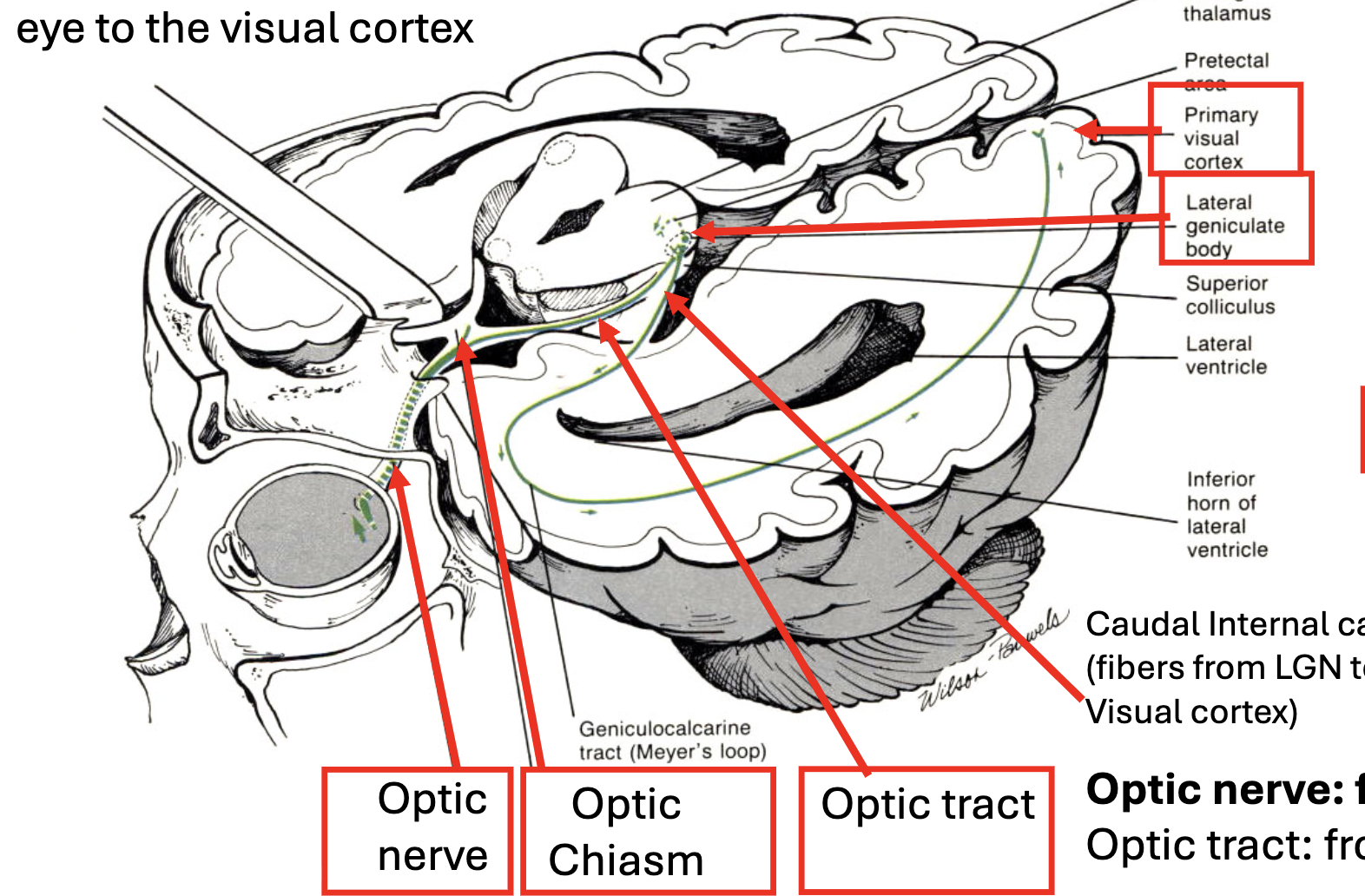
optic nerve formed by axons from retina ganglion cells (RGCs) projecting out of eye toward lateral geniculate nucleus (LGN) in thalamus. from LGN, a second neuron projects to primary visual cortex through the internal capsule
eye → optic nerve → optic chiasm → optic tract → LGN
transection of optic nerve results in
loss of vision in the eye on the side of the lesion → loss of monocular field on same side as lesion
transection of optic tract results in
loss of visual hemifield contralateral to side of the lesion
transection AT optic chiasm results in
crossing fibers from both nasal retinas are damaged, results in loss of monocular fields of view (loss of peripheral vision)
eye movements rely on which cranial nerves
III (oculomotor), IV (trochlear), VI (abducens)
explain partial cross of fibers at optic chiasm
optic nerves carry information from both visual hemifields (ex: outer L, inner R). optic tracts carry all information from one hemifield (ex, inner and outer L). at the optic chiasm, fibers from the nasal retina cross to the contralateral side. this results in left hemifield information appearing in the right visual cortex and vice versa.

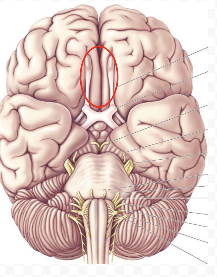
where is optic nerve


where is olfactory nerve

label binocular, left, right fields of vision; optic chiasm, optic nerves, optic tracts
tracts would extend under the nerves, with only red on the left (right hemifield) and blue on the right (left hemifield)


label with: optic chiasm, primary visual cortex, lateral geniculate body, caudal IC (fibers from LGN → visual cortex), optic tract, optic nerve
what components make up optic nerve nasal and outer sides?
outer is from ipsilateral tract, inner/nasal is from contralateral tract
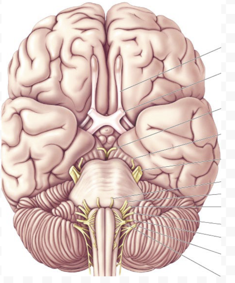
where is oculomotor nerve

oculomotor functional components
GSE and GVE
GSE component of oculomotor nerve
fibers from III provide innervation to 4 of 6 extraocular muscles and muscle that raises upper eyelid; nerve innervates muscles in ipsilateral eye
which muscles does the oculomotor nerve innervate
extraocular (all ipsilateral)
inferior rectus
inferior oblique
medial rectus
superior rectus
levator palpebrae superior (raises upper eyelid)
how does superior rectus muscle move eye
moves up, and assists with inward rotation/intorsion towards nose
how does medial rectus muscle move eye
inwards/towards nose, called adduct
how does inferior rectus muscle move eye
moves down, assists with moving inwards
how does inferior oblique muscle move eye
rotates top of eye outward (extorsion) while helping elevate and move eye outward

label superior rectus, superior oblique, medial rectus, lateral rectus, inferior rectus, and inferior oblique muscles

what are the 4 directions of eye movement
upwards = elevation
downwards = depression
outwards (away from nose) = adduction
inwards (towards nose) = abduction
GVE component of oculomotor nerve
fibers innervating edinger-westphal nuc provide parasympathetic innervation of pupillary sphincter (which is the iris muscle for constriction of the pupil) and ciliary muscles (for accomodation of the lens - focus)
which cranial nerve nuclei are associated with oculomotor nerve
oculomotor (GSE)
edinger-westphal (GVE)
exploration of oculomotor nerve allows evaluation of…
status of rostral midbrain, specifically ventral and medial region

label oculomotor nucleus, oculomotor nerve

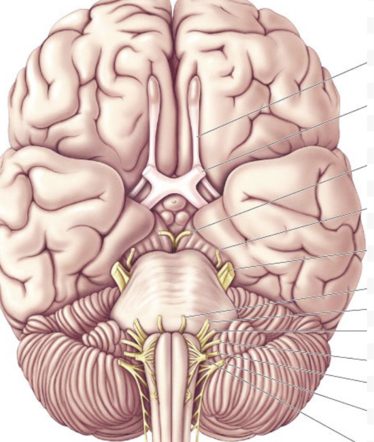
where is the trochlear nerve

functional components of trochlear nerve
GSE → carries fibers from trochlear nuclei, which innervate superior oblique muscle
how does superior oblique muscle move eye, assoc w which cranial nerve
IV (trochlear); rotates eye outwards and downwards → laterally, depression
trochlear nerve palsy
incorrect function of superior oblique muscle; eyes tilt up and in, which is the opposite of typical movement of down and out
only nerve that exists brainstem through dorsal surface is
IV, trochlear nerve
shortest cranial nerve
I, olfactory
exploration of the trochlear nerve allows evaluation of
status of caudal midbrain, specifically dorsal and medial region

label trochlear nerve, nucleus

which cranial nerve nuclei are associated with trochlear nerve
trochlear, GSE

where is abducens nerve

functional components of abducens nerve
GSE: controls lateral rectus muscle of the eye
how does lateral rectus muscle move the eye, what nerve is it assoc w
VI, abducens; moves eye outwards/away from nose/adduct
is abducens innervation ipsi or contralateral
ipsilateral only
abducens palsy
pull from medial rectus isn’t balanced by the lateral rectus
ex: in the right eye → left eye is normal and can look outwards, right eye is affected and can’t look outwards

exploration of the abducens nerve allows evaluation of
status of the pons, specifically ventral and medial regions
which cranial nerve nuclei are associated with abducens nerve
abducens, GSE

label lateral rectus and abducens nucleus

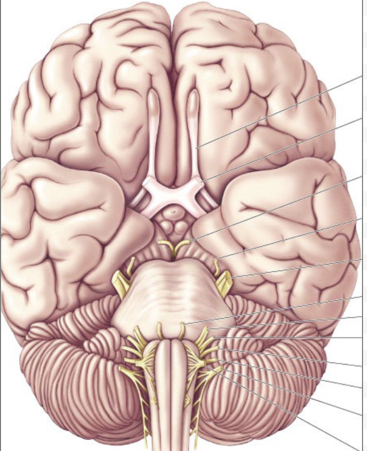
where is trigeminal nerve

functional components of trigeminal nerve
GSA
SVE
GSA in trigeminal nerve
sensitivity from most of the face/head → touch, pressure, temp, pain sensation from skin of face (exteroception), musculoskeletal sensitivity (proprioception)
SVE in trigeminal nerve
chewing muscles and some of the larynx muscles
most neurons located in the motor nucleus of the trigeminal
motor fibers go into nerve together with sensory fibers in the V3/mandibular branch (only branch with motor component)
3 nerves that trigeminal nerve innervates/splits into
opthalmic nerve (VI branch)
maxillary nerve (V2 branch)
mandibular nerve (V3 branch)
trigeminal VI branch - opthalmic nerve
skin of forehead, upper eyelids, lateral aspects of nose
input to spinal nucleus of trigeminal nucleus
trigeminal V2 branch - maxillary nerve
lower eyelids and cheeks
input to principal sensory nucleus of V
trigeminal V3 branch - mandibular nerve
lateral scalp, skin anterior to ear, lower cheeks, lips
input to mesenphalic nucleus of trigeminal nucleus
what trigeminal branch(es) has motor component
V3/mandibular only
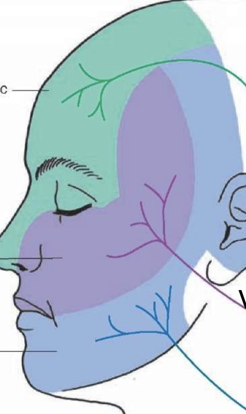
which regions are innervated by which trigeminal branch

which cranial nerve nuclei are associated with trigeminal nerve
trigeminal sensory nucleus (mesencephalic, principal, and spinal regions), GSA
trigeminal motor nucleus, SVE

where is facial nerve

functional components of facial nerve
SVE
GVE
SVA
GSA
SVE in facial nerve
banchial motor nerve
innervation of muscles of facial expression, stylohyoid, stapedius → fibers from facial motor nucleus
GVE in facial nerve
visceral motor nerve
innervation of lacrimal, submandibular salivary, submandibular salivary glands
preganglionic neurons in superior salivary nucleus
postganglionic neurons in pterygopalatine ganglia and submandibular ganglia
these are parasympathetic innervations of the salivary glands!!
SVA in facial nerve
special sensory nerve
receives taste to anterior 2/3 of tongue (solitary nucleus)
GSA in facial nerve
general sensory
receives touch, pressure, temperature, pain sensitive from external ear area (spinal trigeminal nucleus)

which are branchial, visceral, special sensory, and general sensory branches?
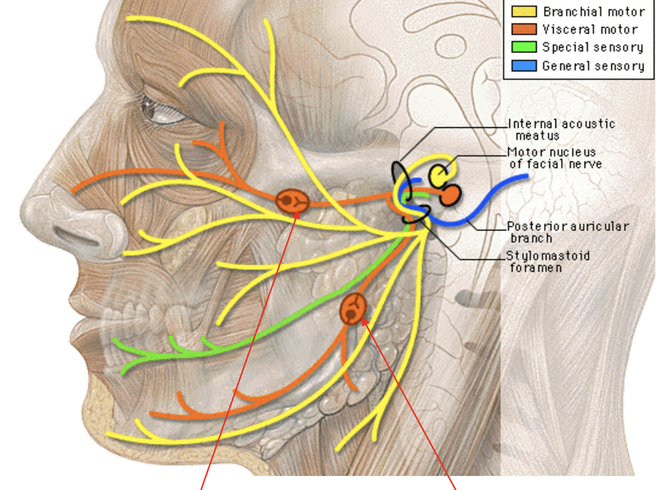
which cranial nerve nuclei are associated with facial nerve
superior salivatory
facial motor
solitary nucleus
spinal/trigeminal
facial expression muscles rare controlled by…
bilateral descending projections from the cortex
upper facial muscles
contralateral descending projections from the cortex
lower facial muscles
lesion in cortical fibers controlling facial motor nucleus causes…
contralateral paralysis in lower half of the face
lesion in facial nerve causes…
paralysis of the ipsilateral half of the face (ipsilateral to lesioned nerve)
Bell’s palsy
facial nerve lower motor neuron lesion (LMNL)

where is vestibulo-cochlear nerve

functional components of vestibulo-ocular nerve
SSA
components of SSA in vestibulo-ocular nerve
auditory component
vestibular component
auditory component of VIII
cochlear nerve → primary sensory neurons located in the spiral (cochlear) ganglion), brings auditory signals to the cochlear nuclei
vestibular component of VIII
vestibular nerve → primary sensory neurons located in scarpa (vestibular) ganglion, brings auditory signals to vestibular nuclei

label VIII vestibular component, VIII auditory component, organ of corti (spiral), spiral ganglion

vestibulo-ocular connections and reflex function
mechanism to maintain focused gaze (in same spot) while head or body is moving/in an unstable environment
how does VOR (vestibulo-ocular reflex) work
elicits compensatory eye movements in opposite direction of head movements to stabilize gaze on a fixed point, stabilizes images in retina
cranial nerves involved in VOR
sensory info from vestibular system → nucleus controlling eye movement to adjust eye position: III, IV, VI (oculomotor, trochlear, abducens)
coordinates sensory input from vestibular nuclei with the activity of extraocular muscles to correct eye position
how to mimic compensatory eye movement using water?
COWS: cold opposite warm same
cold water into an ear mimics head turn to opposite side; eyes slowly move SAME way to compensate, then Nystagmus (fast correction movement to original side) (mimics contralateral head turn)
warm water into an ear mimics head turn to same side; expect compensatory movement to OPPOSITE side (mimics ipsilateral head turn)
two general steps of COWS movement
1) slow phase correctional movement
2) Nystagmus → eyes snap back to center/toward stimulated ear