Developmental Biology Exam 3
 Call Kai
 Learn
 Practice Test
 Spaced Repetition
 Match
 Flashcards
 Knowt Play
1/80

There's no tags or description
Looks like no tags are added yet.
81 Terms
What are homeotic/hox genes?
Homeotic: master regulatory genes that control the body plan and segment identity of developing embryos across plants and animals
Hox: control the anterior-to-posterior body plan and segment identity in animal embryos
What are the homeotic genes called in flies? What two clusters of homeotic genes make Drosophila segments different from each other?
HOM-C:
Antp-C (antennapedia complex)
Bx-C (bithorax complex)

What are the segments of wild-type Drosophila?
T1-T3 and A1-A8

What three hox genes are active in the bithorax complex? What is the arrangement of their expression domains?
Abdominal-B (abd-B)
abdominal-A (abd-A)
Ultrabithorax (ubx)
The lines at the bottom show where they are expressed
Adding complexity going from anterior to posterior

What do loss-of-function hox genes do?
They cause posterior segments to transform into more anterior segments


What happens if you delete ubx (ultrabithorax)?
T3 and A1 both turn into T2

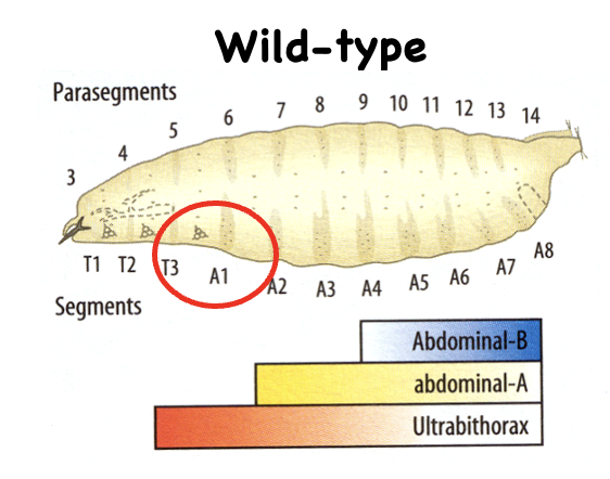
What happens if you delete the entire Bx-C (bithorax complex genes)?
T3-A8 all turn into T2

What is notable about the order of homeotic genes along the chromosome?
The order in which they are found along the chromosome is the same order in which they are expressed along the body
This is conserved in humans

While the bithorax complex (Bx-C) has only three protein coding genes, it has many…
Parasegment-specific enhancers

What are the enhancers for ubx?
abx/bx
bxd/pbx

What are the enhancers for abd-A?
iab-2
iab-3
iab-4

What are the enhancers for Abd-B?
iab-5
iab-6
iab-7
iab-8,9

What is the result of a mutation in an ehancer of the bithorax complex?
The mutation is in an intron, so it changes the expression pattern (where the gene is expressed)
Doesn’t change the protein itself
How are segments of the bithorax complex made different from one another?
Segments become different because Bx-C genes are turned on in more complex patterns in each successive posterior segment (the pattern of gene expression becomes more complex the closer you go to the posterior)

What does this show?
Each panel shows the DNA with enhancer regions labeled along it, plus the gene underneath (Ubx, then abdA once it appears)
The curved arrows show enhancers reaching over to activate the gene’s promoter
The colored circle in each panel marks the new enhancer being added at that step
Each grid is a set of cells across parasegments PS4 to PS8 (labeled below as segments T2, T3, A1, A2, A3)
The colored circles show which enhancer is active in which cells, using the same colors as the circled enhancers on the left:
Notice the staircase: PS5 has one color, PS6 has two, PS7 has three, PS8 has four. The “>>>> PS9>13” on the right means the pattern keeps going with more enhancers (the iab-4 through iab-8,9 regions) for the more posterior segments
Each parasegment is defined by a unique combination of active enhancers, and the combination gets more complex towards the posterior

What would happen if you lost pbx (labeled enhancer D)
T3 would become more similar to T2, resulting in the haltere starting to make a wing


What would happen if you lost A, B, and D enhancers?
T3 would become identical to T2, and therefore 4 legs and 4 wings would develop
This is what Ed Lewis did


What does this show? What trans-acting factors act through these enhancers?
Using a reporter gene to see where the Bx-C enhancers are expressed
Engrailed marks the posterior edge of every parasegment
Enhancers are cis-acting DNA elements, and they only work because trans-acting factors (diffusible proteins, i.e., transcription factors) bind them
The answer is the upstream segmentation gene products: gap genes and pair-rule genes, whose protein gradients differ along the anterior-posterior axis and tell each enhancer where to turn on
What do gain-of-function (misexpression) hox genes do? Name an example
Causes anterior segments to adopt identities of more posterior segments

The gap and pair-rule gene products decay around the time of cellularization, but the homeotic gene products are needed throughout development. What keeps them active after gap and pairrule genes are off?
Epigenetics; the polycomb (PcG) and Trithorax (TrG) groups of chromatic modifiers act as repressors and activators to maintain Hom-C expression
In the anterior, ubx is off
PcG proteins form large complexes that bind the gene.
They deposit the histone mark H3K27me3 (trimethylation of lysine 27 on histone H3)
This compacts the chromatin, and the gene is off, shown by the blocked arrow with an X.
PcG keeps Hox genes silent where they shouldn’t be expressed, such as Ubx in the head and anterior segments
In the posterior, ubx is on
TrG proteins keep genes in an active state
They are associated with H3K27ac (acetylation of the same lysine), an open chromatin mark, so the gene is on
TrG keeps Hox genes active where they were turned on, such as Ubx in PS5 and posterior.
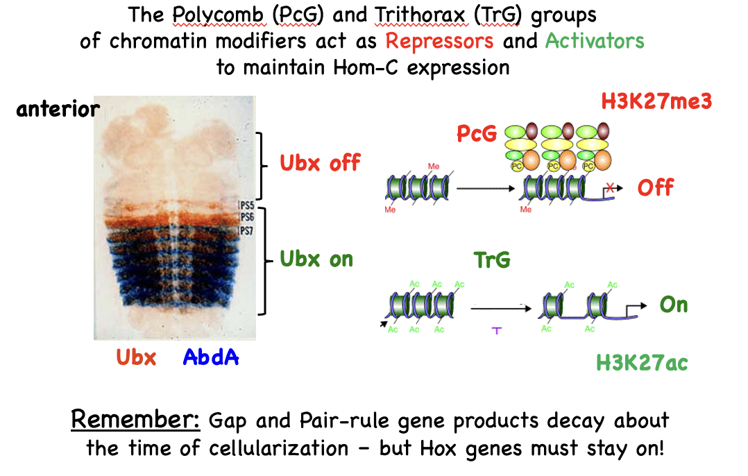

What does this show?
This is the physical basis of the “Bithorax code”: a segment’s identity depends on how many domains are open and which enhancers can therefore reach their genes
Shows that PcG repression isn’t all-or-nothing across the complex. It’s controlled domain by domain, with boundaries between them.
Enhancers say which parasegment a gene turns on in.
PcG/TrG keep the open or closed state stable.
This slide shows what that looks like in 3D: open domains fold separately and can be activated, closed domains are compact and silent.
What do homeotic genes encode? What is their pattern of expression?
They encode transcription factors with restricted expression on the A/P axis
Expressed along the body axis in the same order as found along the chromosome

What do hox genes (type of homeotic gene) encode?
Homeodomain transcription factors
Homeodomain: 60aa region that forms a 3-helix structure that binds DNA
All Hox genes contain a homeodomain, but not all homeodomain-containing proteins are homeotic genes
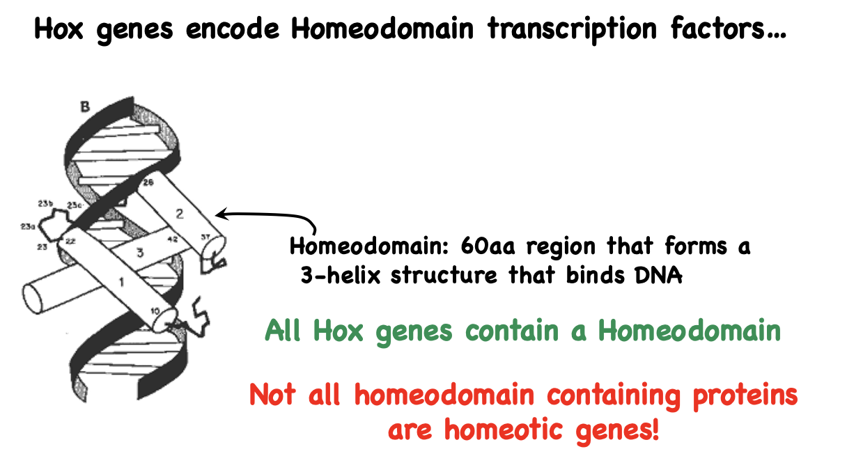
How do Hox family members regulate many related genes? What does this do?
Hox proteins bind to other genes to activate or repress expression to give segment identity

Why is there redundancy in homeotic genes? What does this mean for lose it experiments?
From duplication followed by specialization
Lose-it experiments must knock out genes in multiple Hox clusters to overcome the redundancy
Is there conservation of homeotic genes between flies and mice?
Yes
Each Hoxx is a cluster

How do chick and mouse vertebrae differ in development?
They both have the same number of vertebra, but different expression boundaries of Hox genes
The Hox genes are the same
Shows conservation in protein sequence, order of chromosomes, and expression

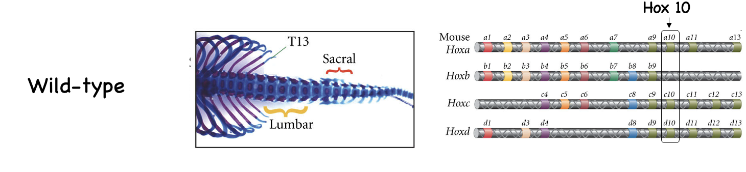
What would you expect from a knockout of Hox10 across all Hox regions?

Are hox genes conserved in C. elegans?
Yes; Hox genes are an ancient invention that was duplicated and functionally diversified during evolution
C. elegans is not obviously segmented, but still shows regionalized expression of Hox genes in different tissues

What is the function of homeotic genes? What do mutations cause?
To make segments different from one another
Mutations cause spatial transformations in cell fate
What do homeotic genes encode?
Transcription factors (contain homeodomain)
How many hox gene complexes do mice and humans have
4
What components of Hox-c gene regulation are also conserved in mammals
Epigenetic mechanisms to lock on (TrG) or lock off (PcG) homeotic gene expression
What makes Xenopus laevis a good model organism?
Vertebrate
Good for injection and transplantation studies
Large eggs
Eggs develop externally
Can obtain lots of eggs, synchronous fertilization
Eggs develop rapidly
Can do pseudo loss-of-function studies (knock-down) using dominant negatives, morpholinos
What are the limits of Xenopus as a model organism?
CRISPR-Cas9 is tricky because it is a tetraploid
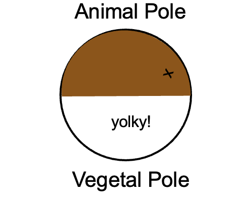
Yolky, not clear embryos
1-2 years to sexual maturity
Is the symmetry of frog eggs broken before or after fertilization? What does an unfertilized egg look like?
Symmetry is broken after fertilization
Unfertilized oocyte has animal-vegetal polarity
Animal pole is pigmented
What breaks symmetry in frog eggs and how does this process work?
shows that motor proteins will become important in establishing A/P polarity
Side opposite from SEP becomes dorsal

What is cortical rotation?
Cortical cytoplasm rotation starts when the cell roughly 70% through the first cell cycle
Sperm entry at the SEP (sperm entry point) triggers a rotation of the egg’s outer layer, the cortical cytoplasm, relative to the inner cytoplasm. The cortex rotates toward the side opposite the SEP, and this starts when the cell is about 70% through the first cell cycle.
The microtubules (red lines and blue bars) drawn inside are part of what drives the movement
The animal cortex has dark pigment granules. When the cortex shifts, the pigmented layer moves away from the side opposite the SEP, exposing the lighter underlying cytoplasm. That lighter, “gray” band of exposed cytoplasm is the gray crescent. It forms on the side opposite sperm entry
What happens if an egg does not have a gray crescent?

What is key about the gray crescent region?
Sperm entry and cortical rotation break the symmetry of the egg
Gray crescent indicates symmetry has been broken
Gray crescent/cortical rotation is required for correct A/P and D/V axis determination
Gray crescent marks site where gastrulation initiated and future dorsal side of embryo

What type of cleavage does Xenopus laevis have? What kind of egg?
Unequal radially symmetric holoblastic cleavage
Reductive divisions
Mesolecithal egg: The yolk is distributed unevenly and is heavily concentrated in the lower hemisphere, known as the vegetal pole
Has a blastocoel

What does gastrulation look like in frogs? what is the goal?
Goal: rearrange cells to make 3 germ layers
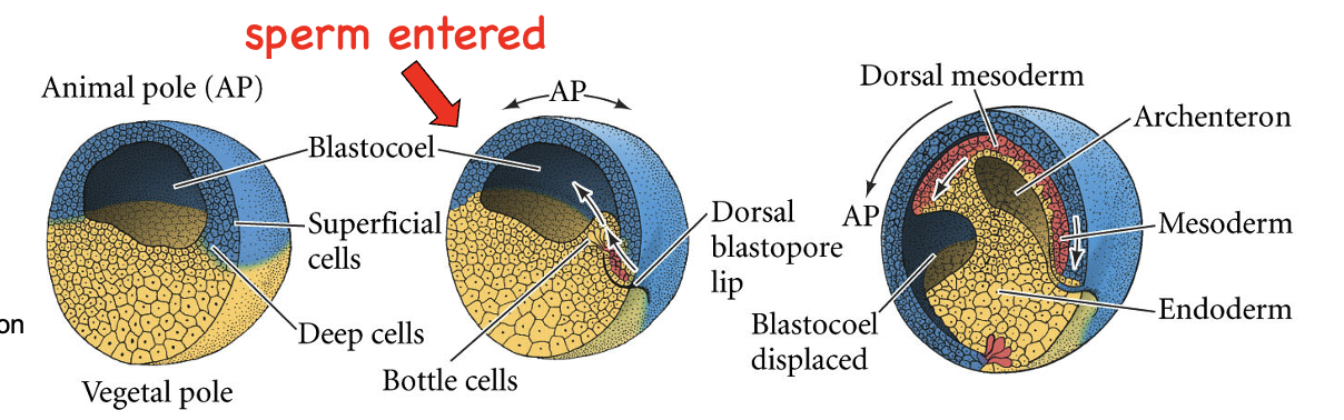
Left: the embryo is a ball with a fluid-filled cavity, the blastocoel, shifted toward the animal pole (top). The wall has superficial cells on the outside and deep cells underneath, and the yellow yolky cells of the vegetal pole make up the bottom.
Middle: gastrulation begins. The red arrow marks where the sperm entered. Gastrulation starts on the opposite side, which is where the gray crescent formed. Here, a group of bottle cells constrict their outer ends and sink inward, creating a groove called the dorsal blastopore lip. The white arrows show cells rolling over this lip and moving inside the embryo.
Right: gastrulation well underway. Cells have moved inward and the embryo has been rearranged:
The archenteron (the future gut cavity) has formed and is growing.
The blastocoel is displaced, squeezed toward the animal side as the archenteron expands.
Three germ layers are now in place: endoderm (yellow, lining the archenteron), mesoderm (red, a middle layer, with the dorsal mesoderm along the back), and ectoderm (blue, the outer layer).
Arrows show the dorsal mesoderm migrating forward along the inside of the roof of the blastocoel.
When is the MBT for frogs?
12th cycle
How can you test what components make up drive early cell divisions in frogs?
Right: the problem. If you want to know what a maternal product does, you need to remove it and see what breaks (“lose it” approaches). But Xenopus is a hard system for that:
No forward genetics: the frog has a long generation time, so you can’t easily run mutant screens.
RNAi doesn’t work well in frogs without extra help.
CRISPR/Cas9 is difficult because Xenopus laevis is allotetraploid, so you’d have to knock out all 4 copies of a gene to see an effect.
The solution, knockdown. Instead of deleting the gene, you block its product:
Morpholino oligonucleotides (MOs): These are small, stable, synthetic antisense molecules. As the diagram shows, the MO is designed to base-pair with the mRNA at the start codon (AUG) region near the 5' end. That physically blocks the ribosome from starting translation, so the mRNA is still there but no protein is made. You inject the MO into the egg or early embryo and look for defects. Or →
Injecting mRNA encoding dominant negative proteins: A dominant negative is a mutant version of a protein that interferes with the normal one (for example, by occupying its binding partners or receptor without signaling). Injecting its mRNA lets you inhibit the pathway even though the normal protein is still present.

Where are VegT and Vg1 localized in an unfertilized Xenopus egg?

What is VegT? What does it do? What would be the phenotype of an egg in which VegT is depleted using morpholino oligos?
VegT is a transcription factor that activates a few early genes (genes that are transcribed before the MBT), including
Sox17 → endoderm
Nodals (Xnrs is the specific one) → mesoderm
A VegT-deficient egg would not develop endoderm or mesoderm
What is Vg1? What does it do? What would be the phenotype of an egg in which Vg1 is depleted using morpholino oligos?
Vg1 is a signaling molecule involved in the formation of the mesoderm
A Vg1-deficient egg would develop little or no mesoderm
What does a late blastula-stage Xenopus egg look like? How are the three germ layers distributed?

How does the unfertilized Xenopus egg display polarity?
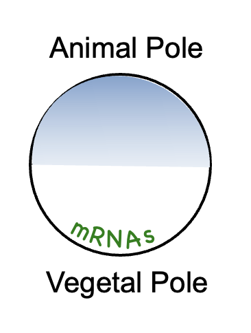
The oocyte appears radially symmetric, but is yolk-dense in the vegetal pole
Many key mRNAs are vegetally stored, these mRNAs help specify germ layers

How does the blastula stage embryo of Xenopus display polarity?
Embryo still appears radially symmetric, but important molecules are no longer radially symmetric
Explain the Spemann-Mangold experiment
Took a piece of tissue from the dorsal lip of the blastopore (DLP) of one newt embryo and transplanted it into the ventral side of a second newt embryo
The transplanted tissue induced the surrounding host cells to change their developmental fate, resulting in the growth of a secondary, conjoined embryo (twinned larvae) with its own neural tube, notochord, and somites
This was the first demonstration of induction

In the Spemann-Mangold experiment, what three things did the dorsal lip of the blastopore (DLP) cells do?
Retain their fate: make notochord and other mesoderm structures; self-differentiate into dorsal mesoderm
Induce ventral tissue at a new location to change fate: “dorsalizes” surrounding tissue, making neural tube and dorsal mesoderm: induction
“Organizes” host/donor tissue to make secondary embryo: initiated new site of gastrulation, new embryo with clear A/P and D/V axis
Do organizers like DLP exist in other organisms?
Yes, they exist in fish, birds, and humans
How does the organizer form?
From the convergence of two signals:
B-catenin: be dorsal
Vg1 and nodals: be mesoderm
Which experiment found how the mesoderm fate is specified?
Nieuwkoop took animal cap cells (that would normally become the ectoderm) and cultured them with vegetal cells (eventual endoderm) and no marginal/mesoderm cells)
Found that the vegetal cells produce a signal that induces the formation of the mesoderm
Animal cap cells are competent, and able to turn into mesoderm cells

Is the vegetal signal that induces mesoderm formation homogeneous across all blastomeres? What experiment shows this?
No
If you repeat the Nieuwkoop experiment with the D1 and D4 blastomeres:
D1, which is more dorsal, produces more dorsal mesoderm cells
D4, which is more ventral, produces more ventral mesoderm
By the 32 cell stage, the dorsal-most (D1) vegetal blastomeres are “special”, called the Nieuwkoop center

What is the Nieuwkoop center?
The dorsal-most vegetal cells
They signal to induce the Spemann (DLP) Organizer in the overlying marginal zone cells


Explain the purpose of this experiment
This experiment tests how the dorsal mesoderm-inducing signals reach the dorsal side
The dorsal-ventral axis is specified at fertilization
Cortical rotation and the cortical microtubule arrays occur when the 1st cell cycle is roughly 70% complete
The experiment destroys the cortical microtubule arrays, but then injects vegetal cortical cytoplasm into the egg on the dorsal end to see if this saves the UV-treated embryo
If it does, this shows:
The dorsal determinant is localized in the vegetal cortex from the start
Cortical rotation relocates it to the dorsal side
The result is rescue
What is the distribution of β-catenin mRNA and protein in Xenopus initially? Where does it end up?
Maternal β-catenin mRNA is not localized, and the protein is initially made everywhere
Eventually, it becomes enriched dorsally during movements associated with fertilization after cortical rotation
At the blastula stage, it is greatly enriched in dorsal nuclei
What happens when you deplete B-catenin using morpholino oligos in Xenopus? What happens if you UV treat the embryo?
MO: no dorsal structures form
UV treatment destroys cortical microtubule arrays, resulting in no nuclear dorsal β-catenin localization
What happens if β-catenin is moved to ventral nuclei?
New gastrulation site and body axis forms
What parts of the wnt pathway are moved during cortical roation?
Disheveled
GSK-3 binding protein (CBP; helps inhibit GSK-3)
wnt11 mRNA

How are GBP, DSH, and wnt11b mRNA moved in cortical rotation? What does the localization of wnt11b mRNA on the dorsal side do?
Kinesin carries GBP and DSH
wnt11b mRNA is physically moved by cortical rotation
Wnt11 mRNA likely provides a localized source of Wnt signal on the Dorsal side

After GBP, DSH, and wnt11b mRNA moved in cortical rotation, what effect does this have?
DSH/GBP inhibits GSK, allowing B-cat to enter dorsal nuclei
B-cat and TCF3 activate genes for dorsal fate

What two signals converge in the Nieuwkoop center? Are they maternal or zygotic? How does this all work?
Stage 8:
The whole vegetal region (blue) contains VegT and Vg1. This is the “Be mesoderm” signal, present everywhere in the vegetal half, ventral and dorsal.
β-catenin (orange dots) is concentrated only on the dorsal side, the result of cortical rotation. This is the “Be dorsal mesoderm” signal.
Stage 9:
VegT/Vg1 drive Xnr expression, shown by the green arrows. The arrows get bigger toward the dorsal side because β-catenin boosts Xnr expression there. So you get a gradient of Xnr, highest dorsally.
Stage 10:
By gastrula onset, the signals have set up the pattern in the marginal zone:
The Organizer (blue) sits dorsally.
A gradient of BMP4 and Xwnt8 (red) runs ventrally, highest on the ventral side and fading toward the Organizer. The organizer secretes BMP inhibitors, which is why BMP activity is low there.
The dashed lines link the Xnr signaling region at stage 9 to the mesoderm it patterns at stage 10.
The note at the bottom left adds that VegT represses organizer genes in the vegetal region itself. That keeps the Organizer from forming in the endoderm and restricts it to the overlying marginal zone.
Main idea: VegT/Vg1 alone give low Xnr and ventral mesoderm. Adding dorsal β-catenin gives high Xnr, and high Xnr plus β-catenin specifies the Organizer. Two maternal signals together, not either one alone, make the dorsal organizer.

What family of secreted signaling molecules do Vg1 and Xnrs belong to? What type of signaling molecule are they? What other molecule that we studied is also in this family
Members of the TGF-β superfamily
Often function as morphogens
BMP is also part of this family

What are the two pathways that TGF-β superfamily members signal through?
Binding of the ligand causes the two receptors to dimerize →
Smads are phosphorylated →
Phosphorylated Smad forms a complex with another Smad →
Goes to the nucleus to either active or repress expression
Know the smad number differences (R-smads are different)

What two pathways activate the organizer genes? Where does this occur in the embryo? (Just name the two pathways)
β-catenin/Tcf3 pathway
Xnrs pathway
Since both are needed to activate organizer genes, organizer genes are only going to be activated on the dorsal side

The Nodals (Xnrs) pathway is already described in another flashcard. Name and describe the other pathway that is needed to activate organizer genes

What are the different organizer genes and what do they do? There are two types
Transcription factors
Goosecoid, Xlim1
Specify dorsal mesoderm
Growth factor antagonists (inhibitors)
Noggin, Chordin, Follistatin, Frzb, Cerberus
Required for neural ectoderm fate
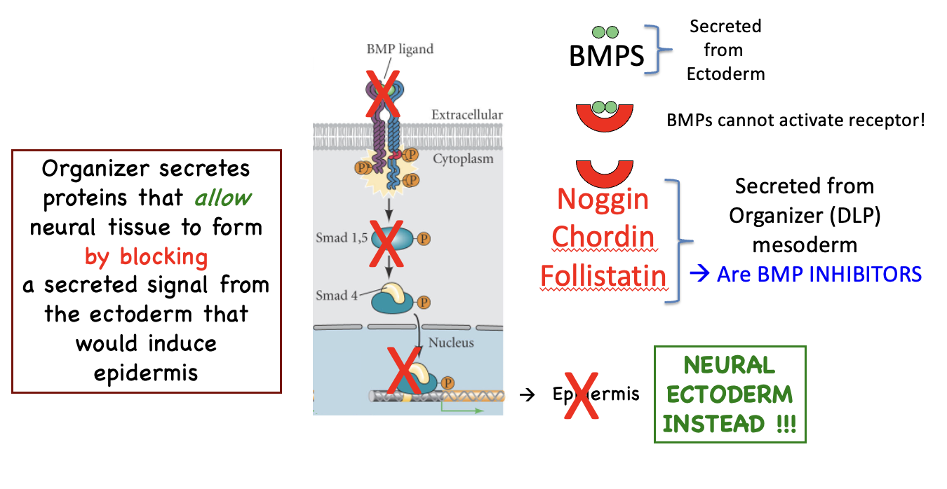
Which organizer gene products are used to block BMP? What effect does this have?
Noggin, Chordin, Follistatin (growth factor antagonists)
BMP is secreted from the ectoderm as an epidermal inducer
Blocking it allows neural ectoderm to form (and build the central nervous system)
What does the BMP signal cause and how does it work?
Signals ectoderm to become epidermis
Signal come from ectoderm (autocrine signaling)
Note that BMP must dimerize for the signal to occur

Show how the organizer gene products that block BMP work in the BMP pathway
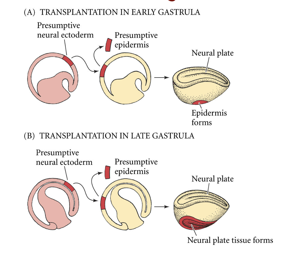
What does this show?
When presumptive neural ectoderm is transplanted, the time of gastrulation at which the transplant occurs affects whether or not the neural plate tissue forms
If the transplant happens:
Early in gastrulation: becomes epidermis
Later in gastrulation: becomes neural plate tissue
This is because a secreted organizer molecule (BMP) blocks a signal that specifies epidermis
Later in gastrulation, the organizer becomes closer to the presumptive neural ectoderm, allowing the signal to be transmitted between
What would happen if you got rid of noggin entirely through UV irradiation? What if you added a high concentration? What concentration of noggin could fix it? What type of signaling molecule is noggin?
No noggin: too much BMP signaling, only epidermis, no dorsal structures formed
Too much noggin: no BMP signaling, only neural ectoderm, embryo becomes dorsalized
Middle concentration would result in normal phenotype
Noggin is a morphogen

The organizer patterns the D/V axis in all three germ layers and is required for proper gastrulation. What does this look like?
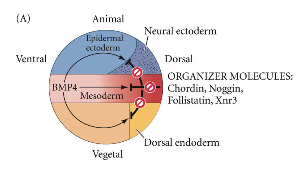
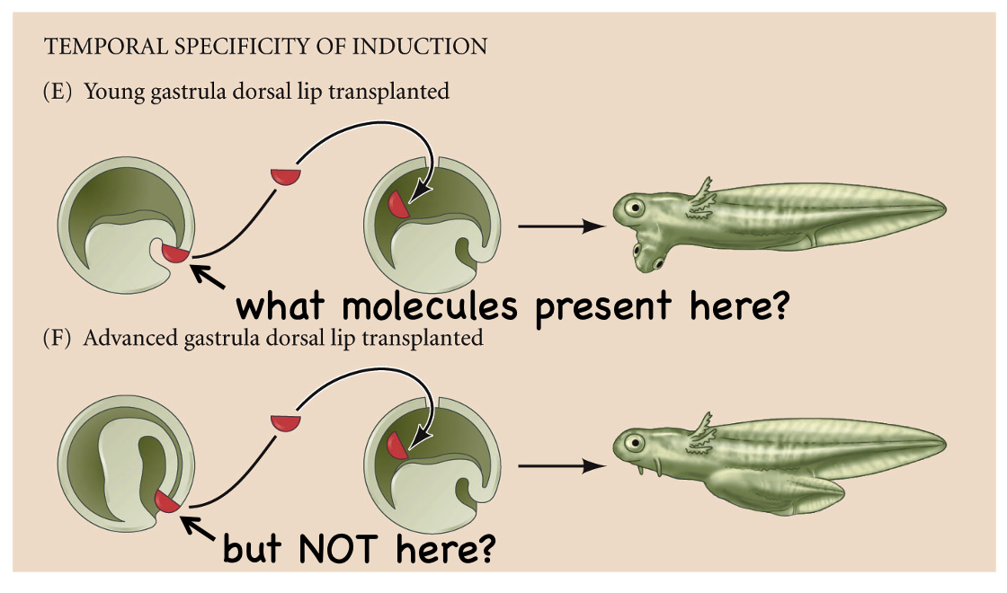
What does this show?
This slide asks which molecules do the work along the anterior-posterior (A-P) axis by doing 2 transplants:
Young gastrula dorsal lip (E): the host develops a second head (note the extra eye and mouth region).
Advanced gastrula dorsal lip (F): the host develops only trunk and tail structures, with no extra head.
So the organizer changes over time. Early dorsal lip tissue (which becomes head mesoderm and endoderm) carries something that induces head, and later dorsal lip tissue (which becomes notochord) does not
Which molecule induces anterior head structures and how?
Cerberus: expressed in the early dorsal lip, and injecting its mRNA into the vegetal (D4) region of an early embryo produces ectopic head structures, such as the extra eye shown at the arrow.
Cerberus is a secreted, multi-purpose inhibitor that binds and blocks three different signals:
BMPs. Xnrs (Xenopus nodal-related proteins), XWnt8
It doesn’t turn on head genes directly. It removes the posteriorizing signals, and head forms by default where they’re absent
Cerberus turns on head genes by removing BMPs, etc

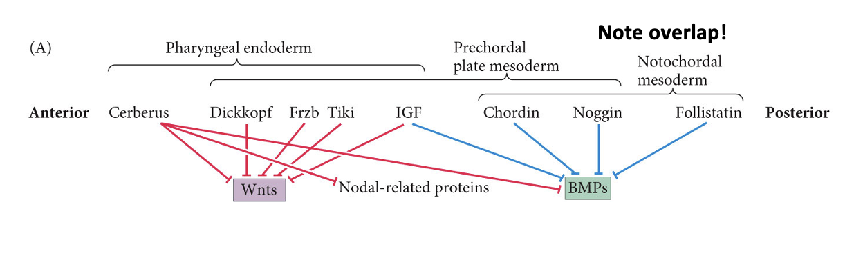
How is regionalization set along the A/P axis?
Differential expression of antagonists
The organizer secretes a set of antagonists, each expressed in a different, overlapping region of the axis
The regions aren’t sharply separated. The key principle is that the anterior end blocks the most signals (Wnt + BMP + Nodal), while the posterior end of the organizer blocks only BMP. The more pathways inhibited, the more anterior the identity
How is ectoderm patterned?
Head and brain: BMP and Wnt are both inhibited (by Chordin/Noggin/etc. and Dickkopf/Cerberus/etc.).
Trunk and spinal cord: only BMP is inhibited; Wnt remains active, which posteriorizes the neural tissue.
Epidermis: neither is inhibited, so both signals act and the ectoderm becomes skin.
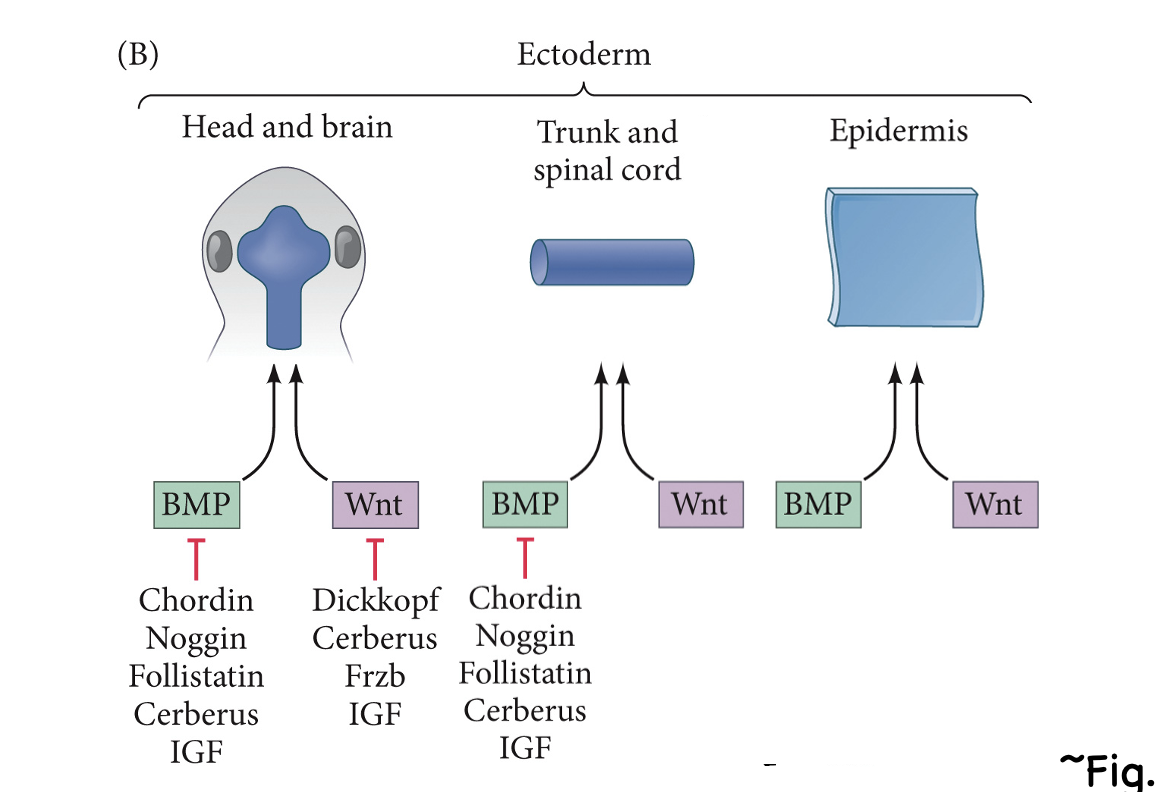

Summary. Use this to study
Yes