Abdominal- Test 1
 Call Kai
 Learn
Learn Practice Test
 Spaced Repetition
 Match
Match Flashcards
 Knowt Play
1/54

There's no tags or description
Looks like no tags are added yet.
Name | Mastery | Learn | Test | Matching | Spaced | Call with Kai | Chat |
|---|
No analytics yet
Send a link to your students to track their progress
55 Terms
Which vessel courses within the main lobar fissure
Main portal vein
Lt portal vein
Middle hepatic vein
Right hepatic vein
Middle hepatic vein
Abdominal aorta bifurcates into right and left common iliac arteries at the level of
4th. thoracic vertebra
5th. thoracic vertebra
4th. lumbar vertebra
5th. lumbar vertebra
4th. lumbar vertebra
You are performing a sonogram on a slender female and notice a long, thin extension of the inferior aspect of the right lobe of the liver. This most likely represents
Caudate lobe
Quadrate lobe
Accessory liver
Reidel's lobe
Reidel's lobe
Which of the following divides the left lobe of the liver into lateral and medial segments?
Ligamentum venosum
Left hepatic vein
Left portal vein
Main lobar fissure
Left hepatic vein
What ligament separates the left lobe of liver from caudate lobe?
Ligamentum venosum
Ligamentum teres
Hepatoduodenal ligament
Coronary ligament
Ligamentum venosum
The main portal vein divides into
Middle, left, and right branches
Left and right branches
Anterior and posterior branches
Medial and lateral branches
Left and right branches
You have detected a mass anterior and to the ligamentum venosum. This mass is located in what lobe of the liver
Left lobe
Caudate lobe
Right lobe
Quadrate lobe
Left lobe
The thin capsule surrounding the liver is known as
Reidel's capsule
Glisson's capsule
Teres capsule
Wirsung's capsule
Glisson's capsule
Which of the following course interlobar and intersegmental within the liver
Bile ducts
Portal veins
Hepatic arteries
Hepatic veins
Hepatic veins
Which of the following correctly describes the probe placement and imaging plane you would use to demonstrate the three hepatic veins and IVC on one view
Subcostal oblique approach with the probe angled slightly to the patient's right
Intercostal approach with the probe oriented in a coronal plane
Subcostal approach with the probe just to the right of midline
Sagittal subcostal approach with the probe just to the right of the midline
Subcostal oblique approach with the probe angled slightly to the patient's right
You are imaging a patient with a high liver. Subcostal images do not clearly demonstrate the liver tissue. What should you do
Scan the patient in deep inspiration
Scan the patient in expiration
Place the patient in a Trendelenburg position and rescan
Scan with the patient in quiet respiration
Scan the patient in deep inspiration
A patient is referred for ultrasound evaluation of a questionable mass in the dome of the liver seen on a CAT scan. Which method below would improve visualization in this area of the liver
Perform a subcostal scan with the probe angled superior and the patient in deep inspiration
Perform a intercostal scan with the probe in a coronal plane and the patient in expiration
Perform a subcostal scan with the patient performing a Valsalva maneuver
Roll the patient into a right lateral decubitus position and scan from a subcostal approach with the patient in expiration
Perform a subcostal scan with the probe angled superior and the patient in deep inspiration
Which of the following is not a branch of celiac artery?
Hepatic artery
Left gastric artery
Superior mesenteric artery
Splenic artery
Superior mesenteric artery
The majority of blood supply to the liver is provided from the
Hepatic veins
Portal vein
Hepatic artery
SMV
Portal vein
What three structures comprise the portal triad (seen at porta hepatis)
Portal vein, bile duct, hepatic artery
Portal vein, bile duct, hepatic vein
Bile duct, hepatic vein, hepatic artery
Hepatic vein, hepatic artery, portal vein
Portal vein, bile duct, hepatic artery
The ligamentum venosum forms the anterior border of what lobe of the liver
Left lobe
Right lobe
Caudate lobe
Quadrate lobe
Caudate lobe

The yellow line is pointing to the
The middle hepatic vein
The common hepatic artery
The portal vein
A thick walled hepatic cyst
The portal vein

The red line is pointing to the
Falciform ligament
Ligamentum of teres
Ligamentum venosum
Main lobar fissure
Main lobar fissure

The yellow line is pointing to the
Caudate lobe
Quadrate lobe
Right hepatic lobe
Posterior segment right lower lobe
Caudate lobe

The red line is pointing to the
Falciform ligament
Ligamentum of teres
Ligamentum venosum
Main lobar fissure
Ligamentum venosum

The medial segment of the left lobe corresponds to which number
1
2
3
4
3
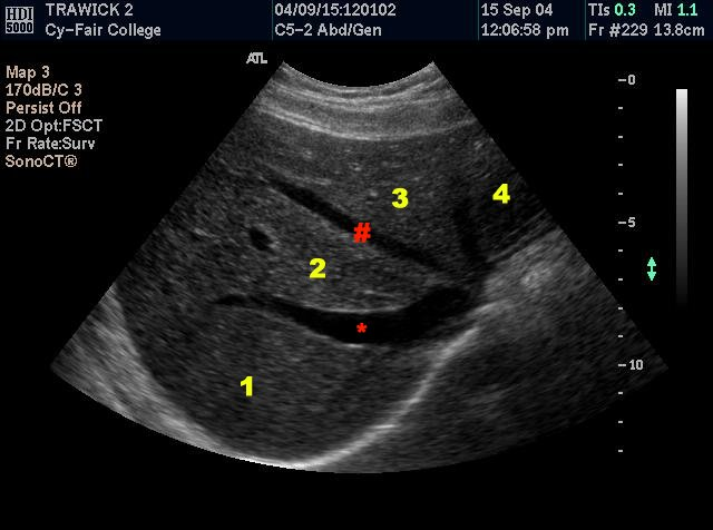
The number 4 corresponds specifically to which of the following
Anterior segment left lobe
Lateral segment left lobe
The left lobe
Left branch of the left hepatic artery
Lateral segment left lobe

The red line points to the
Falciform ligament
Ligamentum of teres
Ligamentum venosum
Main lobar fissure
Ligamentum venosum

The image is taken
Anterior to the SMA
Proximal to the SMA
Inferior to the SMA
Distal to the SMA
Proximal to the SMA

This image is taken
Inferior to the renal veins
Inferior to the aortic bifurcation
Cranial to the right portal vein
Caudal to the SMA
Cranial to the right portal vein
Which of the three structures compose the portal triad?
Hepatic artery, portal vein, and bile duct
Hepatic vein, hepatic artery, and bile duct
Lt portal vein, Lt hepatic artery, and ligamentum teres
Hepatic vein, hepatic artery, and portal vein
Hepatic artery, portal vein, and bile duct
Which of the following arteries are branches of the celiac trunk?
Splenic, Lt gastric, and common hepatic
Splenic, Rt gastric, and proper hepatic
Superior mesenteric, Rt gastric, and proper hepatic
Inferior mesenteric, Rt gastric, and common hepatic
Splenic, Lt gastric, and common hepatic
What is the relationship of the Lt renal vein to the aorta, and the SMA?
Posterior to both the Ao and the SMA
Anterior to both the Ao and the SMA
Anterior to the Ao and posterior to the SMA
Superior to the Ao and inferior to the SMA
Anterior to the Ao and posterior to the SMA
The IVC empties blood into which of the following
Rt atrium
Lt atrium
Rt ventricle
Lt ventricle
Rt atrium
Which of the following does not drain directly into the IVC?
Renal vein
Rt hepatic vein
Common iliac vein
Splenic vein
Splenic vein
You are imaging the common hepatic artery and detect its division into two branches. What are these two branches?
Proper hepatic artery and Rt gastric artery
Rt gastric artery and gastroduodenal artery
Lt gastric artery and proper hepatic artery
Proper hepatic artery and gastroduodenal artery
Proper hepatic artery and gastroduodenal artery
Which of the following describes the anatomic course of abdominal aorta?
Enters peritoneal cavity anterior to IVC
Lies posterior to the left lobe of the liver
Anterior and slightly to the right of the vertebral column
Lies anterior to left renal vein
Lies posterior to the left lobe of the liver
Which of the following vessels courses posterior to the IVC
Rt hepatic artery
Lt renal vein
Rt renal artery
Lt renal artery
Rt renal artery
You have been asked to identify the lobes of the liver in a patient with a hepatic mass. Which vessels course intersegmental in the liver
Portal veins
Hepatic veins
Hepatic arteries
Both the portal veins and hepatic veins
Hepatic veins
You are performing an abdominal ultrasound on a patient with a mass in the pancreatic head. In order to be sure you are imaging the entire pancreas, you must identify the vessel that lies immediately posterior to the pancreas. What is this vessel
Abdominal aorta
IVC
Splenic vein
Portal vein
Splenic vein
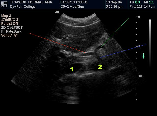
The red line is pointing to
The main portal vein
The hepatic artery
The splenic vein
The SMA
The hepatic artery
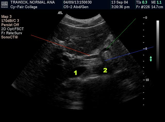
The green line is pointing to
The main portal vein
The hepatic artery
The splenic artery
The SMA
The splenic artery
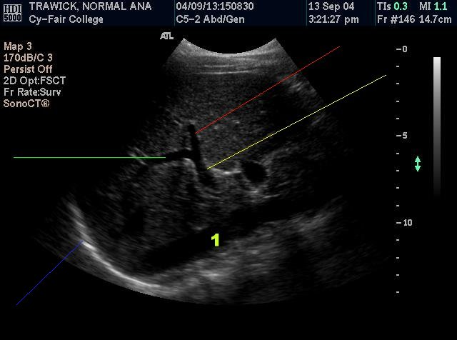
The green line is pointing to the
Rt hepatic artery
Rt hepatic vein
Rt. Portal vein
Main portal vein
Rt. Portal vein

The yellow line is pointing to the
Rt hepatic artery
Rt hepatic vein
Rt. Portal vein
Main portal vein
Main portal vein
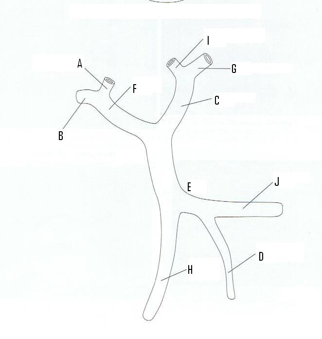
The Splenic vein is identified by letter
D
E
C
J
J

The Rt. portal vein is identified by the letter
E
A
C
F
F

The SMV is identified by the letter
H
D
J
E
H

The purple line points to
The Aorta
The Left renal vein
Superior mesenteric artery
The IVC
Superior mesenteric artery

The yellow line points to
The SMV
The SMA
The Rt renal vein
The portal vein
The SMA

The green line points to
The celiac axis
The SMA
The Rt hepatic artery
The Splenic artery
The celiac axis

The blue line points to the
SMA
IVC
Proper hepatic artery
Portal splenic confluence
IVC
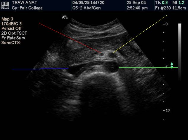
The green line points to the
Aorta
SMA
Proper hepatic artery
Portal splenic confluence
Aorta

The green line points to the
Right hepatic vein
Rt portal vein
Left hepatic vein
Medial Lt portal vein
Left hepatic vein

The green line points to the
IVC
Aorta
Portal splenic confluence
Rt Renal artery
Rt Renal artery
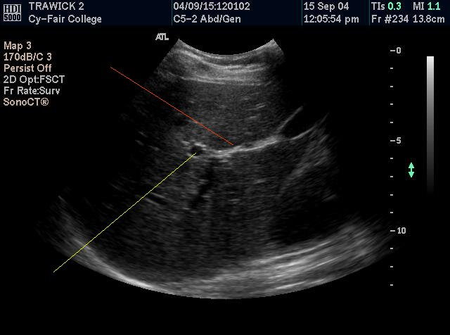
The yellow line points to the
IVC
Aorta
Portal vein
Rt Renal artery
Portal vein

The red line points to the
Rt renal artery
Hepatic artery
Lt renal artery
Aorta
Rt renal artery

The purple line points to the
Rt renal artery
Left portal vein
Lt renal artery
Aorta
Left portal vein

The image on the left is a sagittal image of the upper abdomen. The image on the right is a transverse image of the upper abdomen. Which of the lines on the image on the right correspond with the sagittal image on the left.
Red
Purple
Yellow
Green
Green
You are performing a follow up sonogram on a patient in which a 5mm cyst was previously identified at the anterior border of the left lobe of the liver. Although you are using a 3.5 MHz curved linear array probe, you do not see the cyst. Which of the following would be most helpful in improving visibility of this cyst?
Increase the overall gain
Increase the dynamic range
Rescan the left lobe with a lower frequency transducer
Rescan the left lobe with a higher frequency transducer
Rescan the left lobe with a higher frequency transducer
You are performing an ultrasound exam of the liver on a small patient with a 5MHz curved linear array. Although you have increased the overall gain to its maximum setting, the posterior border of the liver and diaphragm are not visualized. What should you do?
Call the service representative to repair your equipment
Decrease the transmit power on the ultrasound unit
Rescan the liver with a lower frequency transducer
Move the focal zone into the near field
Rescan the liver with a lower frequency transducer