CNS: higher function, protections and spinal cord

0.0(0)
Studied by 0 people
call kaiCall Kai
learnLearn
examPractice Test
spaced repetitionSpaced Repetition
heart puzzleMatch
flashcardsFlashcards
GameKnowt Play
Card Sorting

1/38

encourage image

There's no tags or description

Looks like no tags are added yet.

39 Terms

1
New cards

Pyramidal cells

  • output cells of the neocortex excitatory (glutamatergic)

  • integrating information and sending it to a different location


<ul><li><p>output cells of the neocortex excitatory (glutamatergic)</p></li><li><p>integrating information and sending it to a different location </p></li></ul><p></p>
2
New cards

There are also many local interneurons

  • mostly inhibitory-basket cells and chandeliers cells (GABAergic-peptidergic)

  • some excitatory- stellate cells(layer 4, glutamatergic)


<ul><li><p>mostly inhibitory-basket cells and chandeliers cells (GABAergic-peptidergic) </p></li><li><p>some excitatory- stellate cells(layer 4, glutamatergic) </p></li></ul><p></p>
3
New cards

Layer lll of cerebral cortex

  • external pyramidal cell layer

  • small pyramidal cells and local interneurons

  • information flow to other cortical areas


<ul><li><p>external pyramidal cell layer</p></li><li><p>small pyramidal cells and local interneurons</p></li><li><p>information flow to other cortical areas </p></li></ul><p></p>
4
New cards

Layer lV of cerebral cortex

  • internal granule cells

  • granule cells-local interneurons

  • information flow to other cortical areas and Thalamus


<ul><li><p>internal granule cells</p></li><li><p>granule cells-local interneurons </p></li><li><p>information flow to other cortical areas and Thalamus </p></li></ul><p></p>
5
New cards

Layer V of cerebral cortex

  • internal pyramidal cell

  • mostly large pyramidal cells

  • Information flows to other cortical areas and oppostie hemisphere


<ul><li><p>internal pyramidal cell </p></li><li><p>mostly large pyramidal cells </p></li><li><p>Information flows to other cortical areas and oppostie hemisphere </p></li></ul><p></p>
6
New cards

EEG

  • electrical recordings from the surface of the scalp-larger amplitude=more synchronization of many APs from many neurons


<ul><li><p>electrical recordings from the surface of the scalp-larger amplitude=more synchronization of many APs from many neurons</p></li></ul><p></p>
7
New cards

Alpha

  • low amplitude, regular: alert relaxed state


<ul><li><p>low amplitude, regular: alert relaxed state </p></li></ul><p></p>
8
New cards

Beta

  • more rapid and more irregular: alert and concentrating


<ul><li><p>more rapid and more irregular: alert and concentrating </p></li></ul><p></p>
9
New cards

Theta

  • larger and slower: unusual in adults


<ul><li><p>larger and slower: unusual in adults </p></li></ul><p></p>
10
New cards

Delta

  • high amplitude, slower: seen when reticular system is suppressed during sleep or under anesthesia


<ul><li><p>high amplitude, slower: seen when reticular system is suppressed during sleep or under anesthesia </p></li></ul><p></p>
11
New cards

Sleep

  • common patterning of health sleep progression


<ul><li><p>common patterning of health sleep progression</p></li></ul><p></p>
12
New cards

REM

  • skeletal muscles (except ocular and diaphragm) are actively inhibited; most dreaming occurs


<ul><li><p>skeletal muscles (except ocular and diaphragm) are actively inhibited; most dreaming occurs </p></li></ul><p></p>
13
New cards

State N1

  • relaxation begins; EEG shows low-amplitude mixed-frequency and alpha waves; arousal is easy


<ul><li><p>relaxation begins; EEG shows low-amplitude mixed-frequency and alpha waves; arousal is easy</p></li></ul><p></p>
14
New cards

Stage N2

  • irregular EEG with sleep spindles(short high-amplitude bursts); arousal is more difficult


<ul><li><p>irregular EEG with sleep spindles(short high-amplitude bursts); arousal is more difficult </p></li></ul><p></p>
15
New cards

Stage N3

  • EEG has high-amplitude theta and delta waves; vital signs decline; arousal is difficult; bed-wetting, night terrors, and sleepwalking may occur


<ul><li><p>EEG has high-amplitude theta and delta waves; vital signs decline; arousal is difficult; bed-wetting, night terrors, and sleepwalking may occur</p></li></ul><p></p>
16
New cards

Spinal cord

  • extends from foramen magnum (medulla oblongata) to 2nd lumbar vertebra

  • 16-18 inches long

  • cervical and lumbar enlargements-more info being processed and usage\


<ul><li><p>extends from foramen magnum (medulla oblongata) to 2nd lumbar vertebra </p></li><li><p>16-18 inches long</p></li><li><p>cervical and lumbar enlargements-more info being processed and usage\ </p></li></ul><p></p>
17
New cards

Conus medullaris

  • tapered end of spinal cord


<ul><li><p>tapered end of spinal cord</p></li></ul><p></p>
18
New cards

Filum terminale

  • connects meninges to coccyx


<ul><li><p>connects meninges to coccyx </p></li></ul><p></p>
19
New cards

Cauda equina

  • (horse tail): bundle of descending spinal nerves


<ul><li><p>(horse tail): bundle of descending spinal nerves</p></li></ul><p></p>
20
New cards

Dorsal root

  • sensory(afferent) axons


<ul><li><p>sensory(afferent) axons </p></li></ul><p></p>
21
New cards

Dorsal root ganglion

  • contains afferent cell bodies


<ul><li><p>contains afferent cell bodies </p></li></ul><p></p>
22
New cards

ventral root

  • Motor (efferent) axons


<ul><li><p>Motor (efferent) axons</p></li></ul><p></p>
23
New cards

Spinal nerve

  • mixed sensory and motor nerve fibers


<ul><li><p>mixed sensory and motor nerve fibers </p></li></ul><p></p>
24
New cards

Local flow of information

  1. Peripheral stimulation activates sensory neuron

  2. Signal propagates down sensory neuron to dorsal horn

  3. Synapse onto inter neuron or directly to motor neuron

  4. Activation of motor neuron

  5. Activation of muscle


<ol><li><p>Peripheral stimulation activates sensory neuron</p></li><li><p>Signal propagates down sensory neuron to dorsal horn </p></li><li><p>Synapse onto inter neuron or directly to motor neuron </p></li><li><p>Activation of motor neuron</p></li><li><p>Activation of muscle </p></li></ol><p></p>
25
New cards

Ascending tracts of spinal cord

  • conduct sensory impulses to the brain


<ul><li><p>conduct sensory impulses to the brain</p></li></ul><p></p>
26
New cards

Descending tracts

  • conduct motor impulses from the brain to motor neurons reaching muscles and glands


<ul><li><p>conduct motor impulses from the brain to motor neurons reaching muscles and glands</p></li></ul><p></p>
27
New cards

Fasciculus gracilis and cuneatus of ascending tracts

  • touch, pressure vibration and proprioception of upper and lower limbs and chest

  • They cross over information in the medulla oblongata


<ul><li><p>touch, pressure vibration and proprioception of upper and lower limbs and chest</p></li><li><p>They cross over information in the medulla oblongata</p></li></ul><p></p>
28
New cards

Spinothalamic of ascending tract

  • the anterolateral or spinothalamic pathways carry mainly pain and temperature impulses

  • Lateral spinothalamic tract carries pain, temperature

  • Anterior(ventral) tract carries touch and pressure

  • Axon in the spinothalamic tract cross over information in the spinal cord


<ul><li><p>the anterolateral or spinothalamic pathways carry mainly pain and temperature impulses</p></li><li><p>Lateral spinothalamic tract carries pain, temperature</p></li><li><p>Anterior(ventral) tract carries touch and pressure</p></li><li><p>Axon in the spinothalamic tract cross over information in the spinal cord</p></li></ul><p></p>
29
New cards

Corticospinal

  • lateral and anterior

  • Control of skeletal muscles


<ul><li><p>lateral and anterior</p></li><li><p>Control of skeletal muscles</p></li></ul><p></p>
30
New cards

Reticulospinal

  • lateral, anterior, medial

  • Control muscle tone and sweat glands


<ul><li><p>lateral, anterior, medial</p></li><li><p>Control muscle tone and sweat glands</p></li></ul><p></p>
31
New cards

Corticospinal tracts

  • carry motor commands

  • Fibers originate in the cerebral cortex

  • Most information crosses over in the medulla, some in the spinal cord

  • Synapse either directly onto motor neurons or onto an interneuron that synapses onto a motor neuron


<ul><li><p>carry motor commands</p></li><li><p>Fibers originate in the cerebral cortex</p></li><li><p>Most information crosses over in the medulla, some in the spinal cord</p></li><li><p>Synapse either directly onto motor neurons or onto an interneuron that synapses onto a motor neuron</p></li></ul><p></p>
32
New cards

Dura mater: meninges of the brain

  • periosteal layer: fused to periosteum

  • Meningeal layer

  • Dural sinus between layers(some areas)


<ul><li><p>periosteal layer: fused to periosteum </p></li><li><p>Meningeal layer</p></li><li><p>Dural sinus between layers(some areas)</p></li></ul><p></p>
33
New cards

Arachnoid mater

  • attaches to dura mater

  • May be separated by subdural space


<ul><li><p>attaches to dura mater </p></li><li><p>May be separated by subdural space</p></li></ul><p></p>
34
New cards

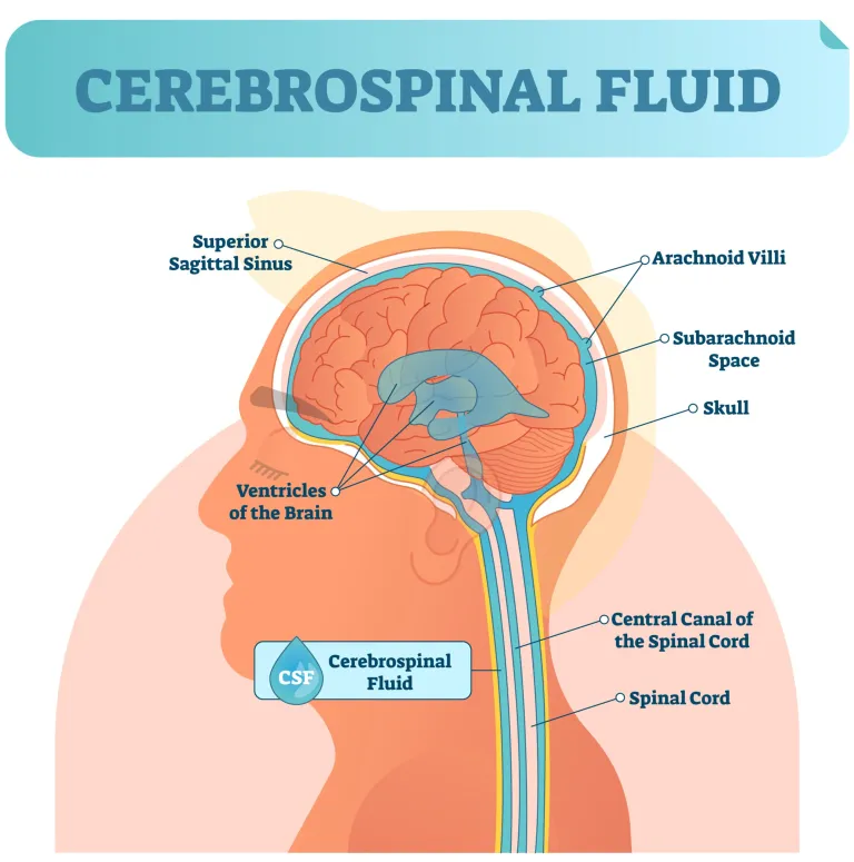
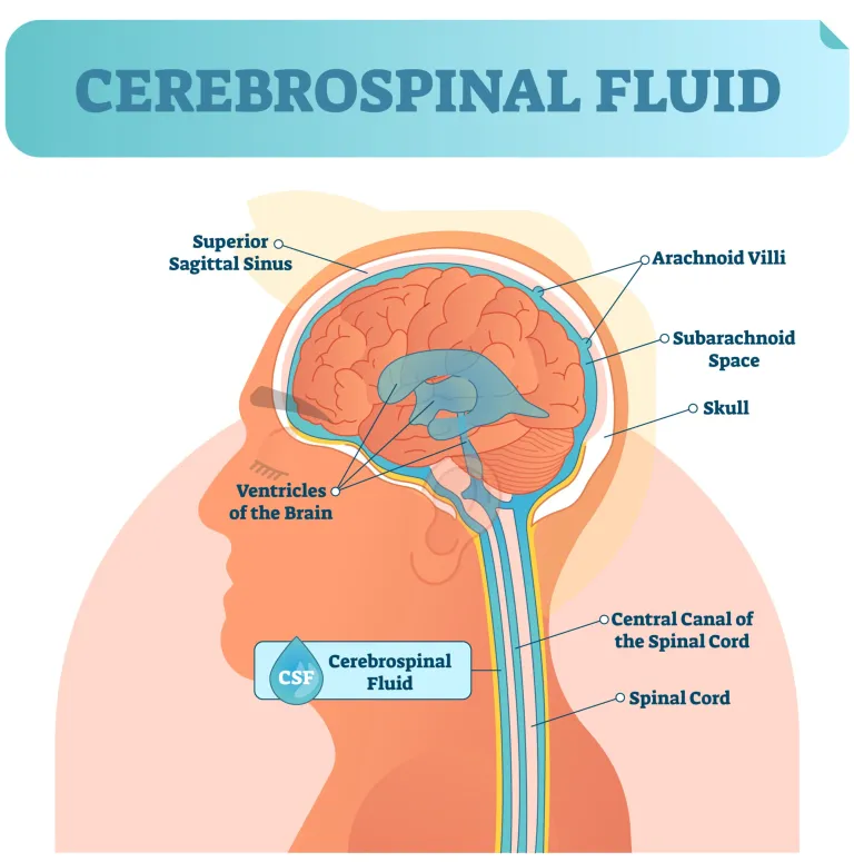
Subarachnoid space

  • filled with CSF, trabeculae


<ul><li><p>filled with CSF, trabeculae </p></li></ul><p></p>
35
New cards

Pia mater

  • attached to the surface of the brain, astrocyte interaction


<ul><li><p>attached to the surface of the brain, astrocyte interaction </p></li></ul><p></p>
36
New cards

Dural folds

  • extensions of the meningeal cranial dura into cranial cavity

  • Theses folds stabilize and support the brain

  • Contain collecting veins(Dural venous sinuses)

  • Three largest Dural folds: flax cerebri, tentorium cerebelli, falx cerebelli


<ul><li><p>extensions of the meningeal cranial dura into cranial cavity </p></li><li><p>Theses folds stabilize and support the brain</p></li><li><p>Contain collecting veins(Dural venous sinuses) </p></li><li><p>Three largest Dural folds: flax cerebri, tentorium cerebelli, falx cerebelli </p></li></ul><p></p>
37
New cards

Cerebrospinal fluid

  • mechanical and metabolic protection


<ul><li><p>mechanical and metabolic protection </p></li></ul><p></p>
38
New cards

Choroid plexus

  • many infoldings-villi

  • Extract water from blood and transport to CSF


<ul><li><p>many infoldings-villi </p></li><li><p>Extract water from blood and transport to CSF</p></li></ul><p></p>
39
New cards

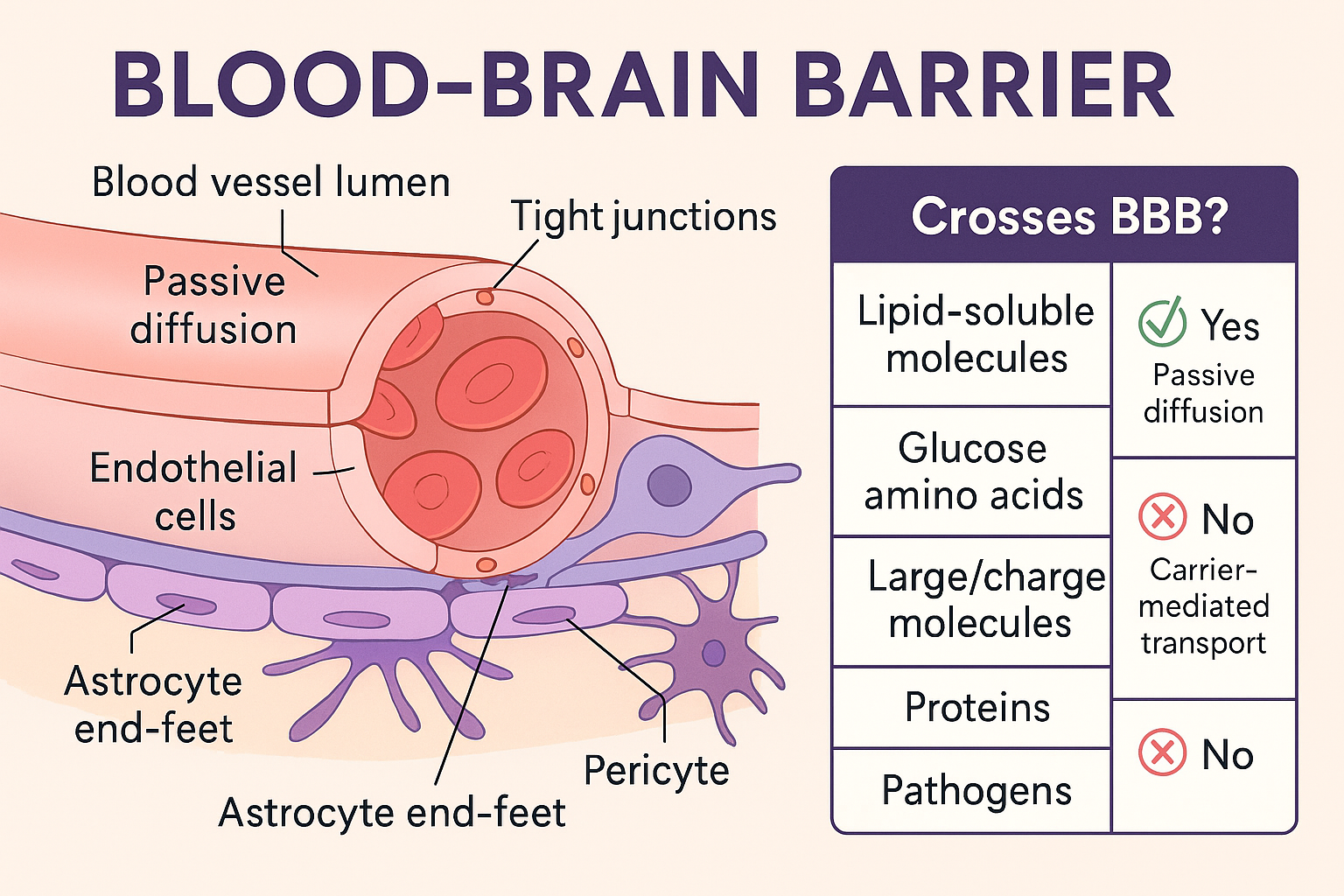
Blood brain barrier- highly distributed structure

  • Primary: tight junctions between capillary endothelial cells. Some role for end feet of astrocytes-molecular uptake, induction

  • BBB not present in choroid plexus, arachnoid villi and regions of the brain responsible for monitoring blood quality or neuro-hormonal secretory areas(hypothalamus, pituitary gland, pineal gland)

<ul><li><p>Primary: tight junctions between capillary endothelial cells. Some role for end feet of astrocytes-molecular uptake, induction</p></li><li><p>BBB not present in choroid plexus, arachnoid villi and regions of the brain responsible for monitoring blood quality or neuro-hormonal secretory areas(hypothalamus, pituitary gland, pineal gland) </p></li></ul><p></p>