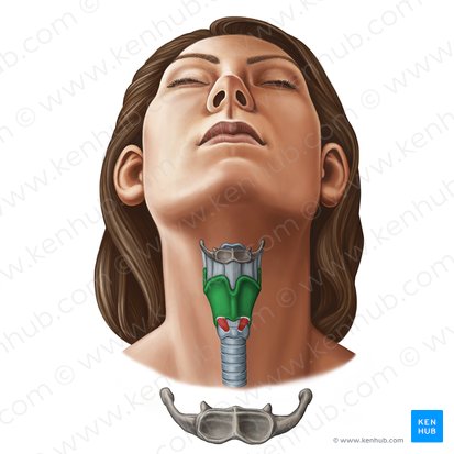
Axial Skeleton (Skull, auditory ossicles, hyoid)
 Call Kai
 Learn
 Practice Test
 Spaced Repetition
 Match
 Flashcards
 Knowt Play
1/41

Earn XP
Description and Tags
The axial skeleton consists of all the bones along the central axis of the skill, auditory ossicles, hyoid bone, vertebral column and thoracic cage
42 Terms
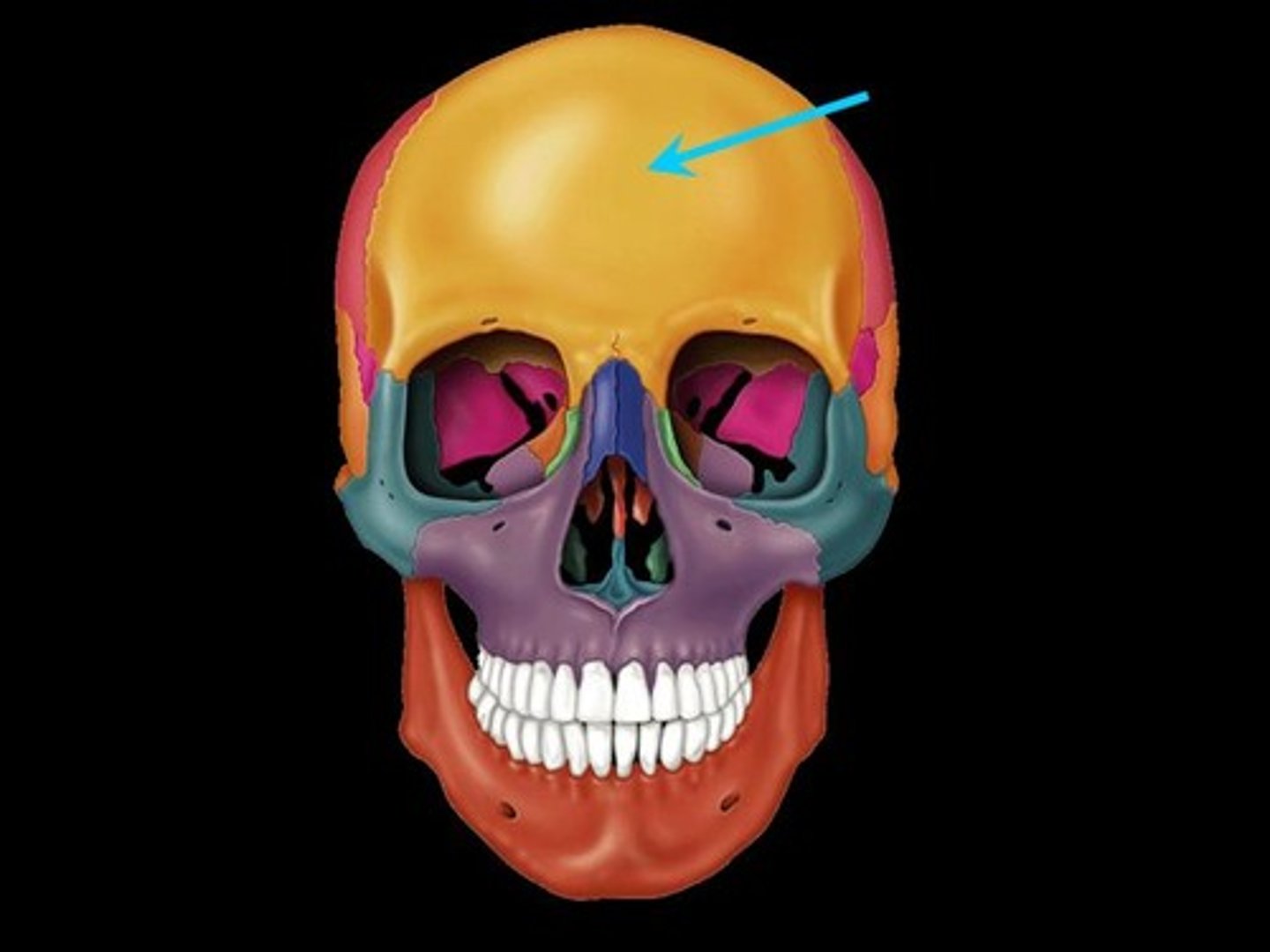
Frontal Bone
shell shaped bone, forming the anterior cranium and superior orbits
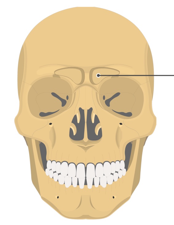
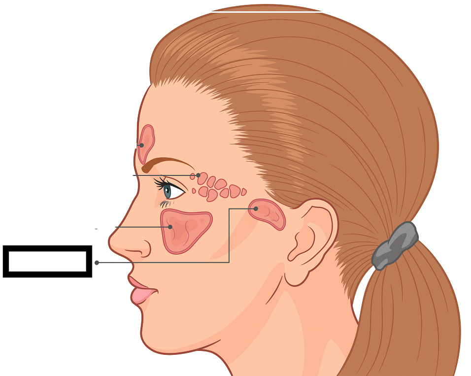
Frontal Sinus
(Frontal Bone)
The sinuses are air filled cavities within skull bones that open into the nasal cavity. The sinus functions to lighten the weight of the skull and help with voice resonance.
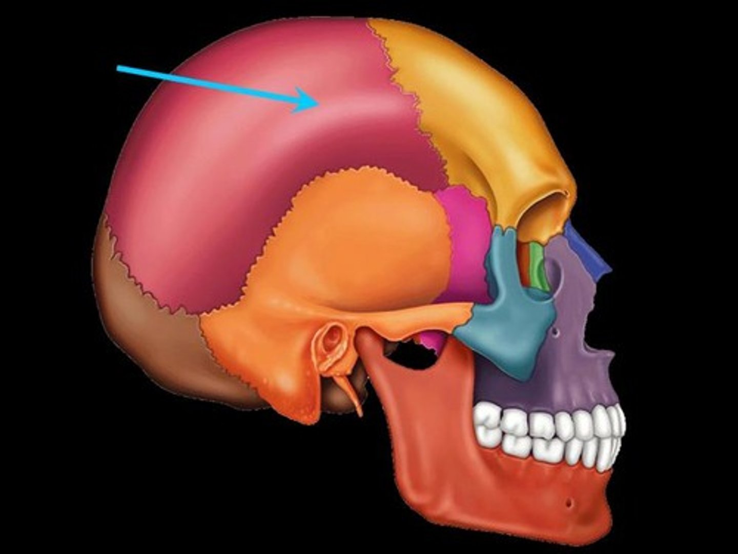
Parietal bones
2 of them
large bones forming most of the superior and lateral aspects of the cranium
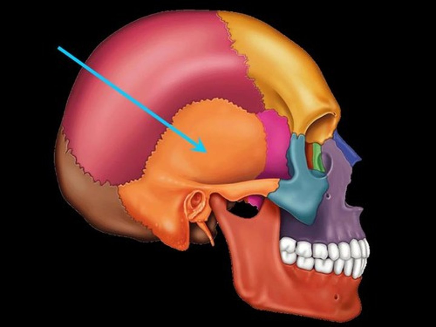
Temporal Bones
2 of them
bones that form the inferolateral aspects of the skull
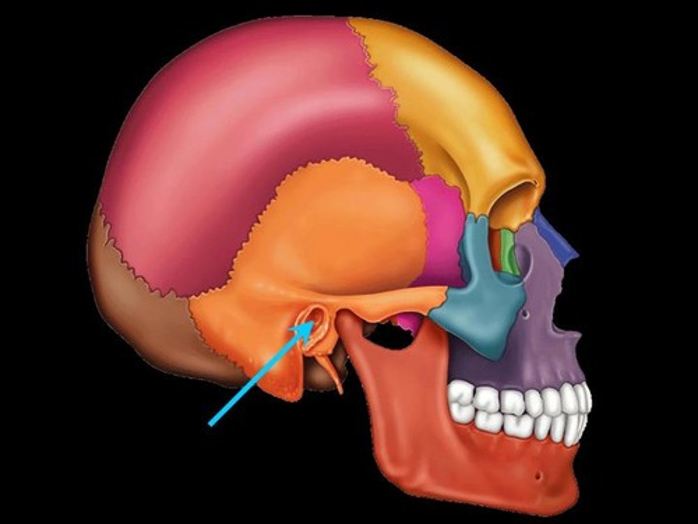
External auditory/acoustic meatus
(Temporal Bone)
Canal leading to the middle ear (malleus, incus and stapes)
responsible for directing sound waves from tympanic membrane to inner ear
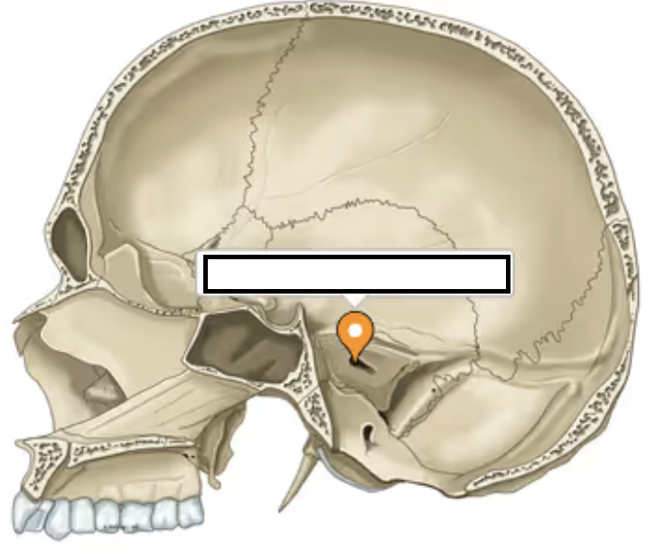
Internal Auditory/Acoustic Meatus
(Temporal Bone)
A passageway for the vestibulocochlear nerve. This nerve extends from inner ear to the brain. The vestinulocochlear nerve carries nerve impulses for hearing and balance.
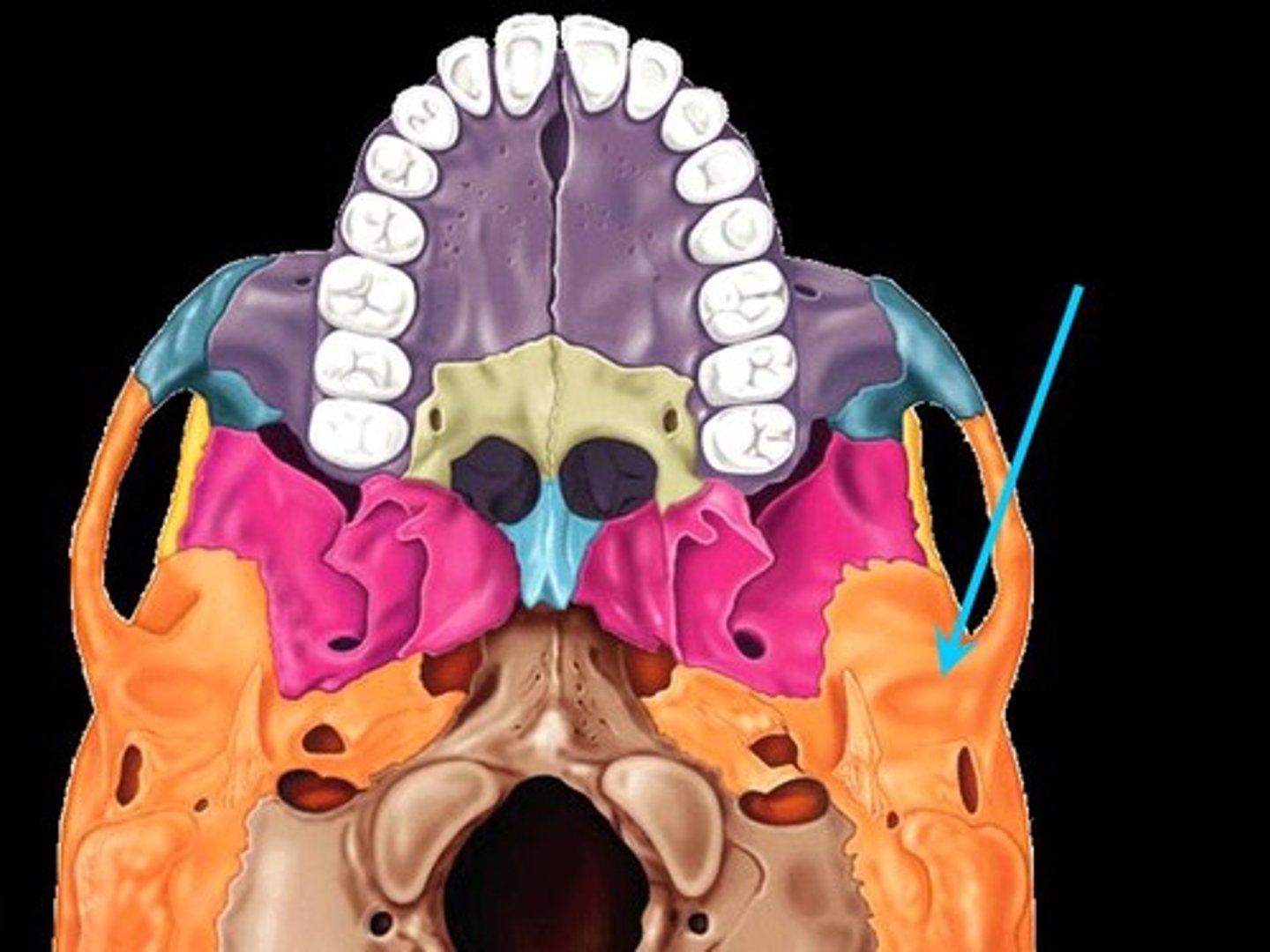
Mandibular fossa
(Temporal Bone)
a depression on the temporal bone where the mandible articulates with the skull. The mandibular fossa of the temporal bone and the condyle of the mandible come together to form a joint called the temporomandibular joint
Mastoid process
(Temporal Bone)
enlargement on the skull posterior to the earlobe. Site of attachment for muscles that move the skill
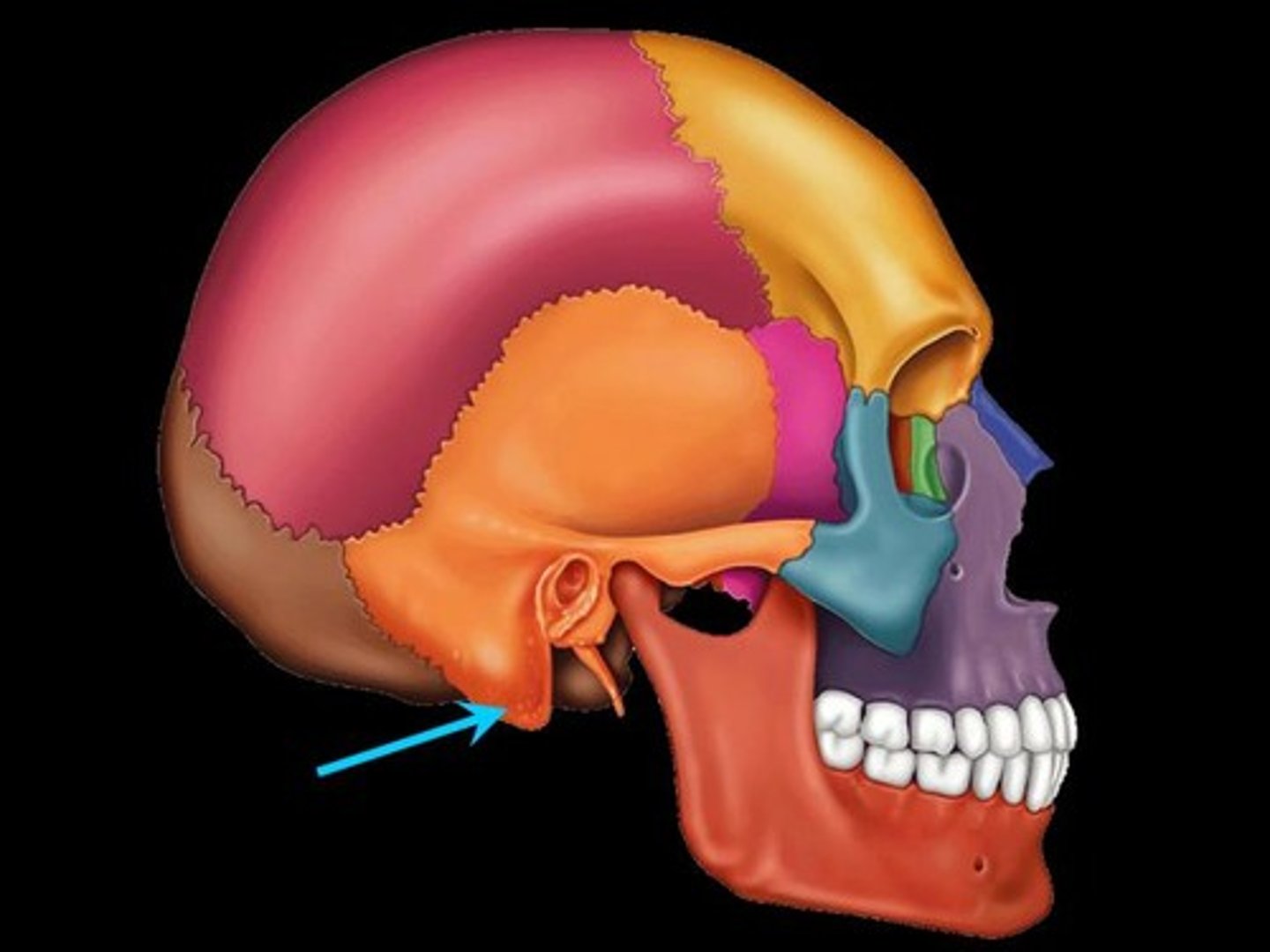
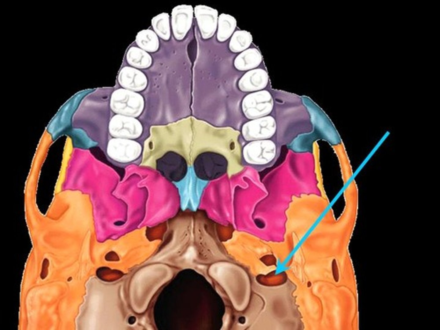
Jugular foramen
(Temporal Bone)
Located where the petrous part of the temporal bone joins the occipital bone.
The jugular vein which drains blood from the brain and the vagus nerve which innervartes the thoracic and abdominal organs both pass throughthis foramen.
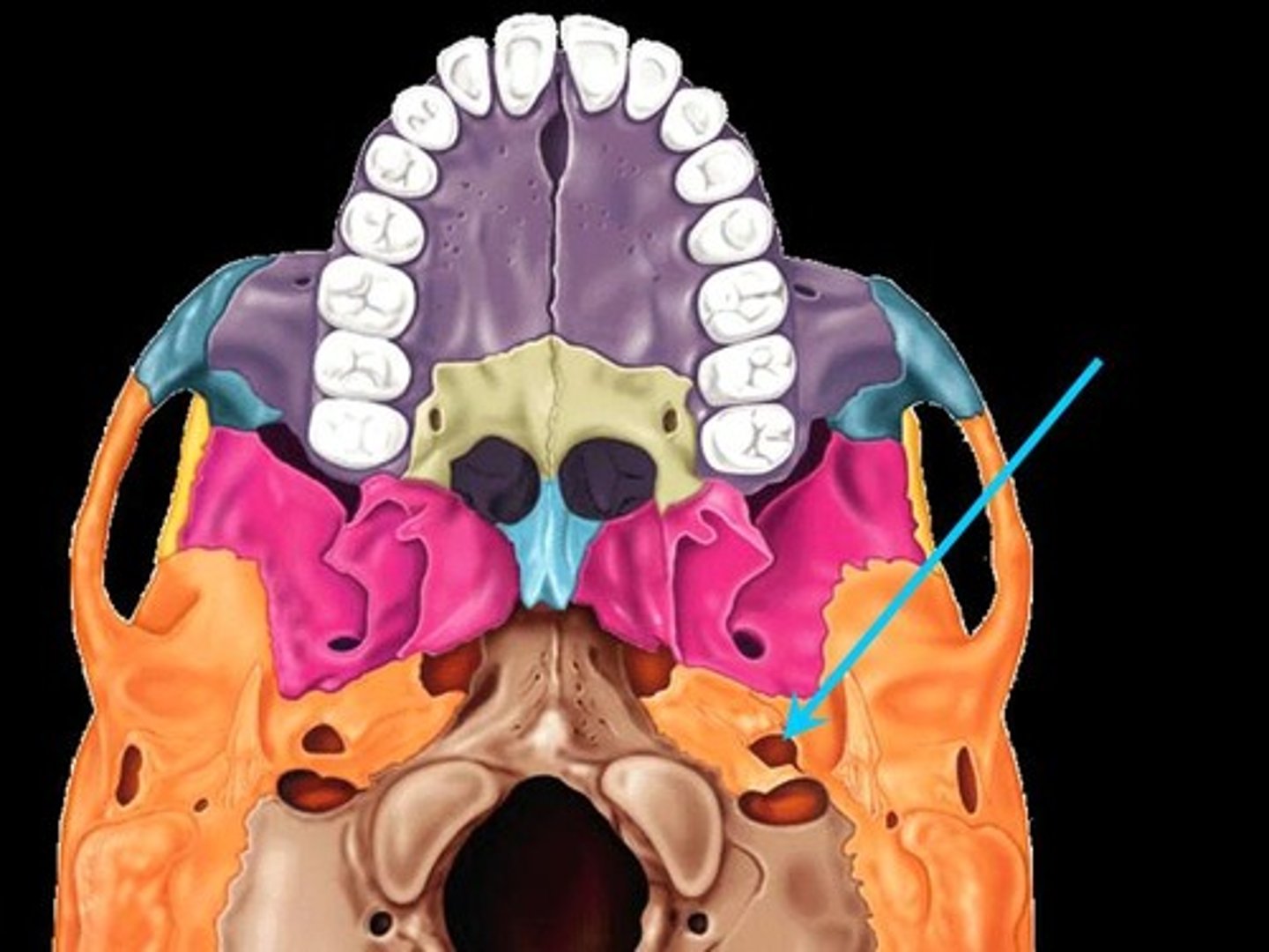
Carotid canal/foramen
(Temporal Bone)
Passageway for a blood vessel called the interal carotid artery which supplies blood to the brain.
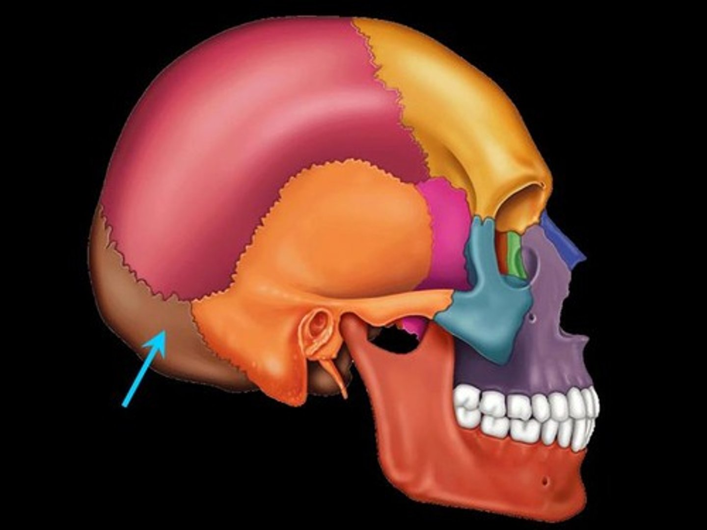
Occipital Bone
Large bone forming most of the posterior wall and base of the skull
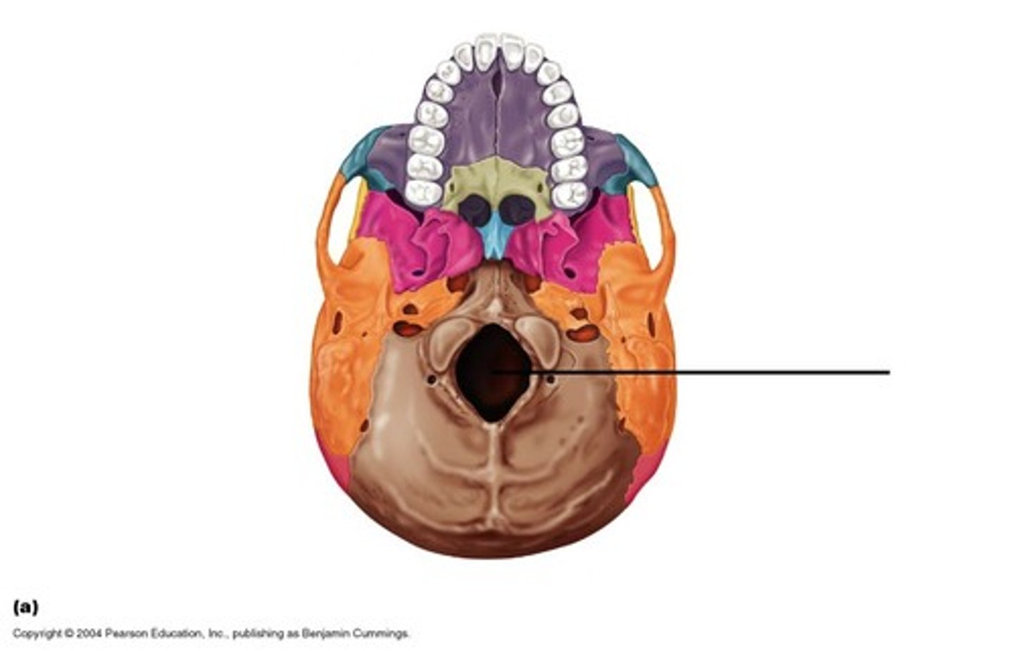
Foramen magnum
(Occipital Bone)
the brain stem extends through the foramen magnum to connect to the spinal cord
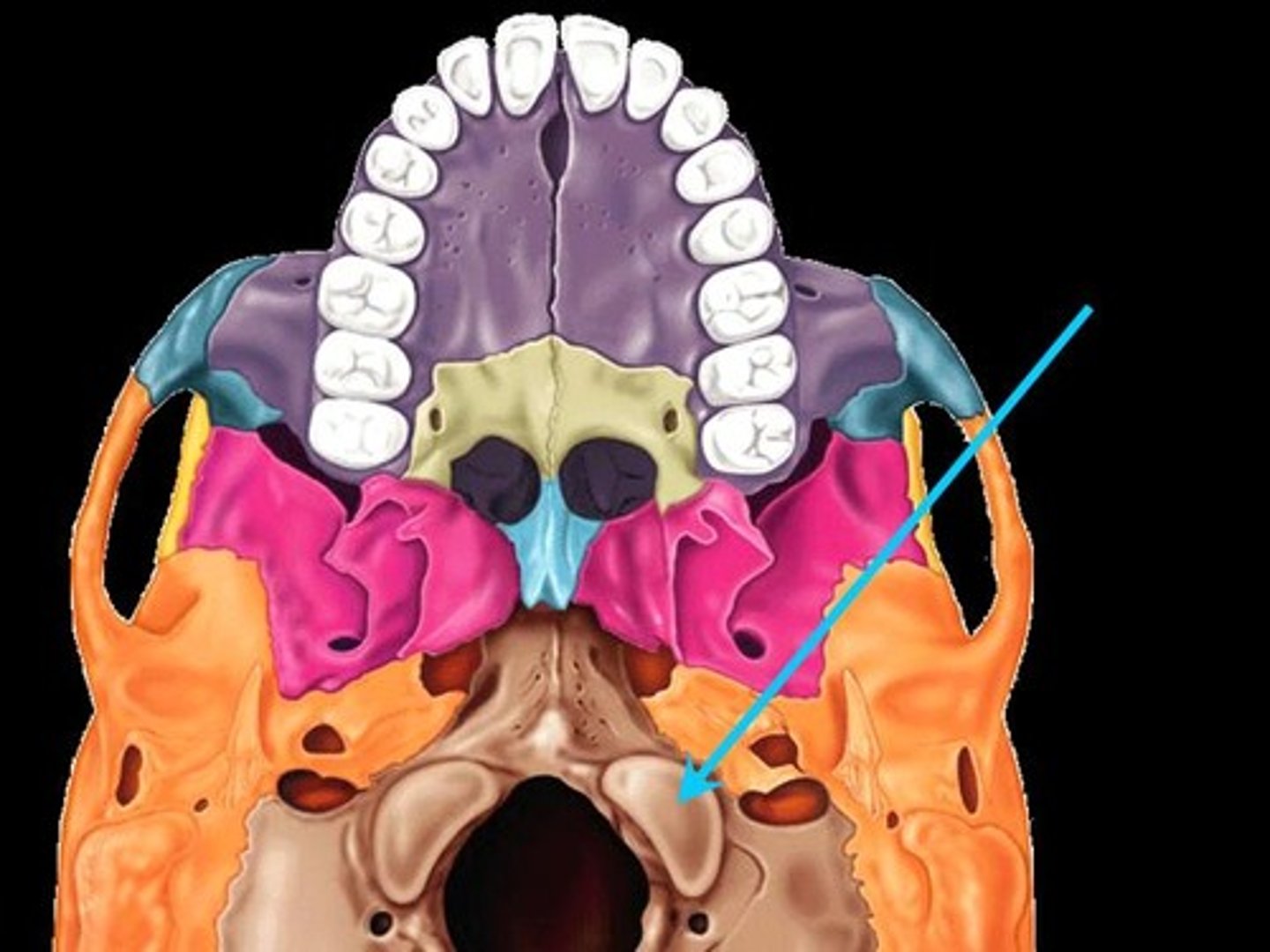
Occipital condyles
(Occipital Bone)
articulate with the atlas vertebra (C1), allowing the head to move up and down in a “yes” movement and a side-to-side tilt movement.
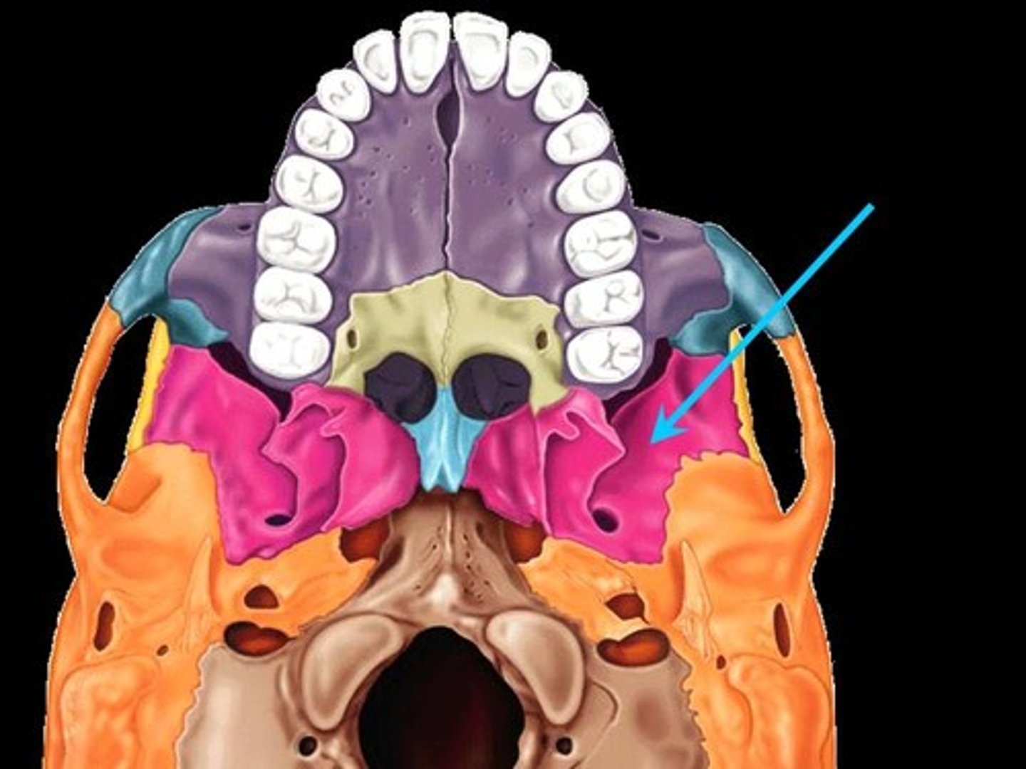
Sphenoid bone
A butterfly shaped bone in the middle of the skull forming the posterior wall of the orbits. It is considered the keystone of the skull because it articulates with all of the cranial bones.
Optic Foramen
(Sphenoid Bone)
Round hole in each posterior orbit. It is a passageway for the optic nerve which carries visual information to the brain.
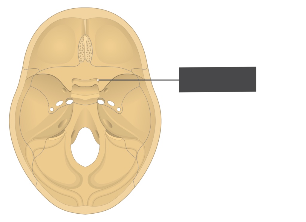
Sella turcica
(Sphenoid Bone)
"Turkish saddle"
depression on the sphenoid bone (inside the brain case) that protects and houses the pituitary gland
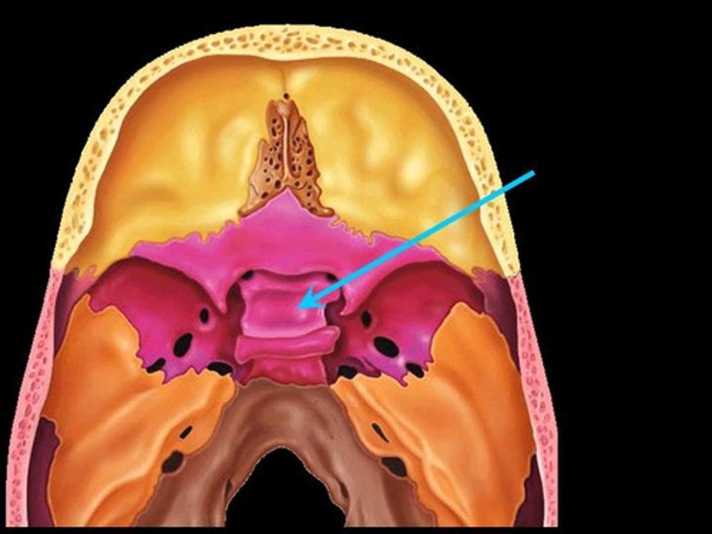
Sphenoidal Sinus
The sinuses are air-filled cavities within skull bones that open into the nasal cavity. The sinuses decrease the weight of the skull and act as resonating chambers during sound production.
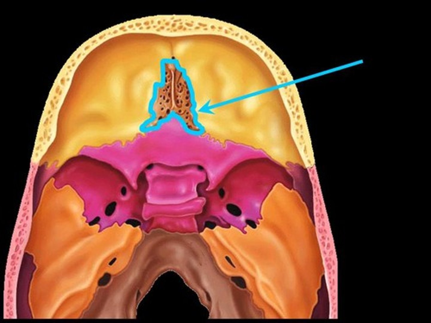
Ethmoid
the deepest bone of the skull, it lies between the nasal bones and the sphenoid
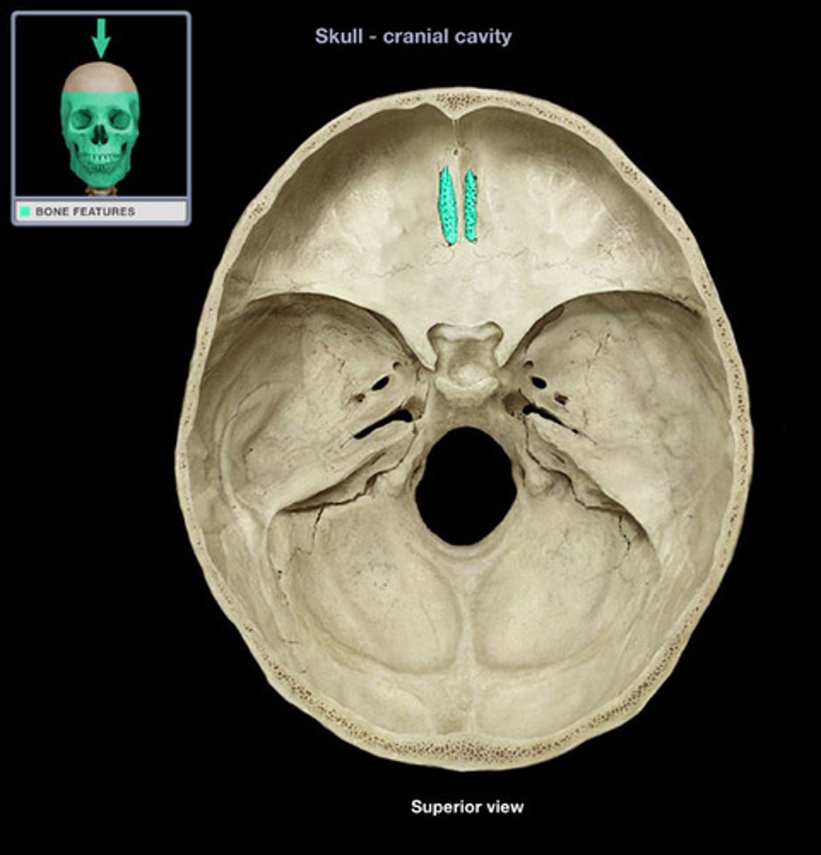
Cribriform plates
(Ethmoid Bone)
solid flat portion of the ethmoid bone on either side of the crista galli (inside the brain case)
Cribriform/Olfacotry foramina
(Ethmoid Bone)
small holes in the cribriform plate that allow olfactory nerves to pass through the skull from the nasal cavity into the olfactory bulbs within the skull
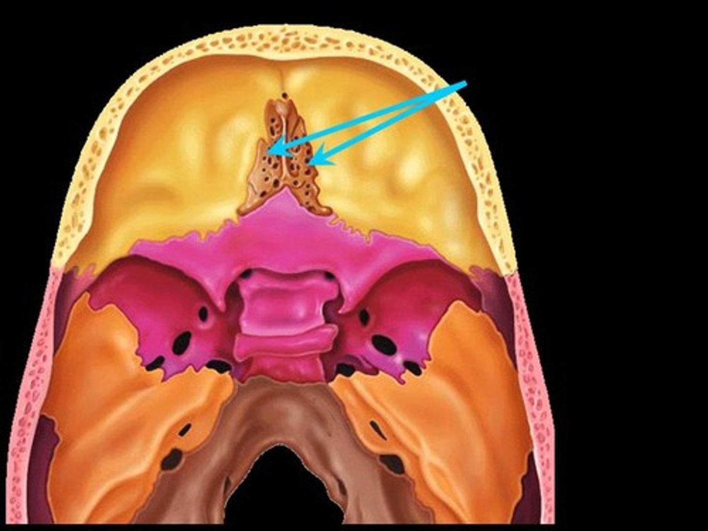
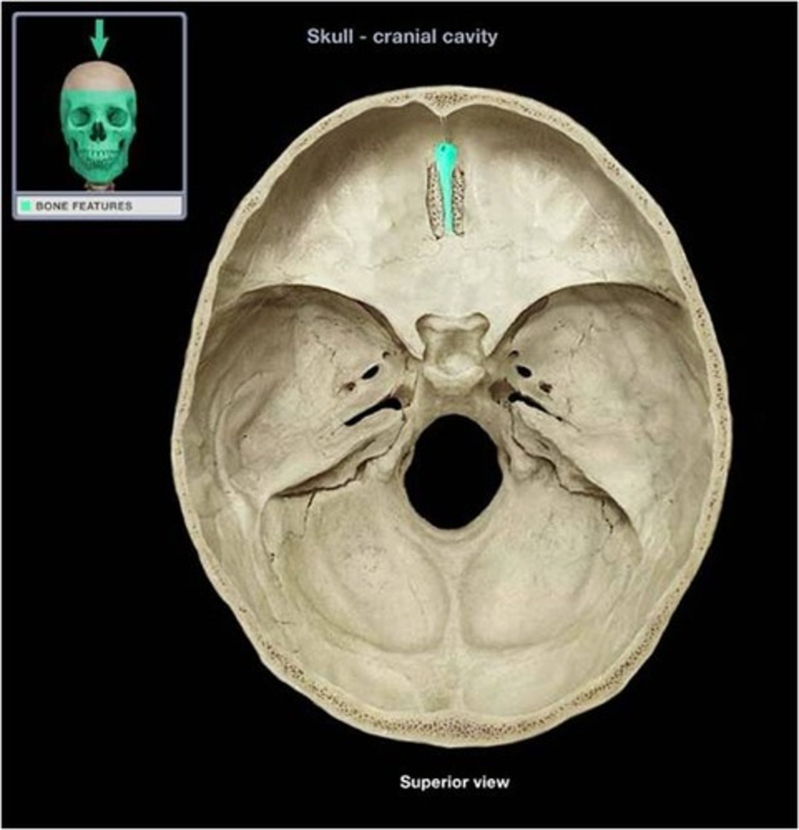
Crista galli (Rooster Crest)
(Ethmoid Bone)
midline projection of the ethmoid bone into the brain case. A membrane holding the brain in place attaches to this structrue. (The fin that sticks out)
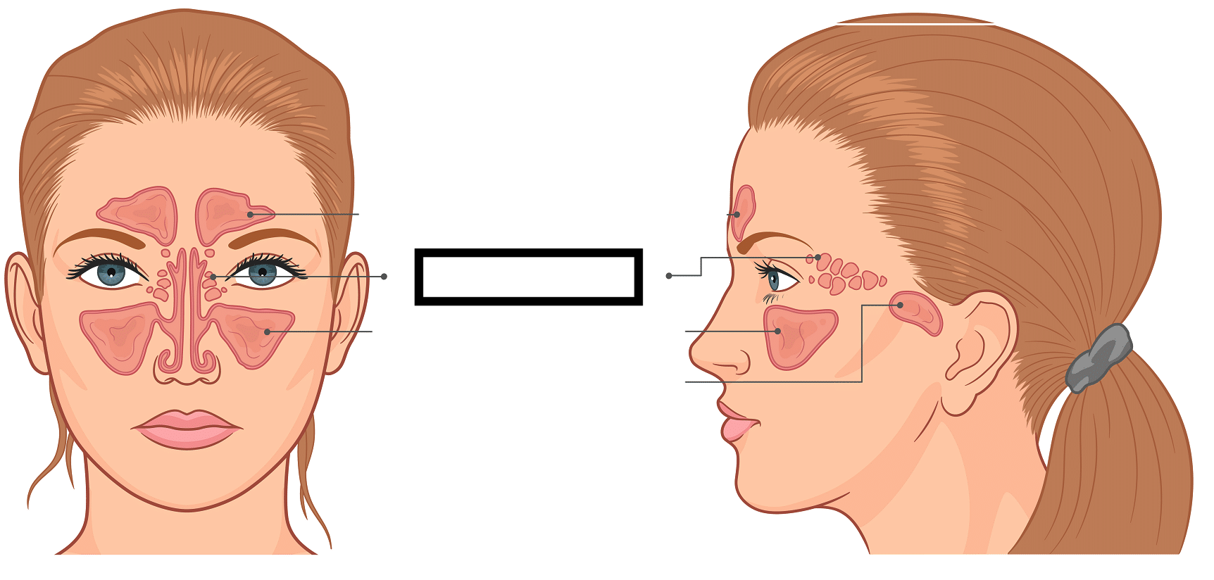
Ethmoidal Sinus
(Ethmoid Bone)
the sinuses are air-filled cavities within skull bones that open into the nasal cavity. The sinuses function to decrease the weight of the skull and act as resonating chambers during sound production.
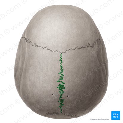
Sagittal Suture
Right and Left side of skull
Fibrous joint between the two parietal bones along the midline of the skull
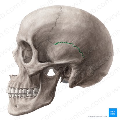
Sqaumosal/Squamous Suture
Fibrous joint between the parietal boe and the temporal bone on either side of the skull
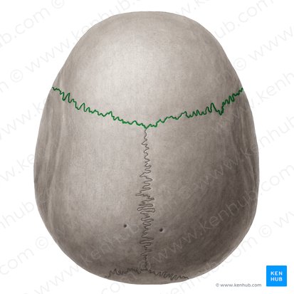
Coronal Suture
Fibrous joint between the parietal pones and the frontal bone
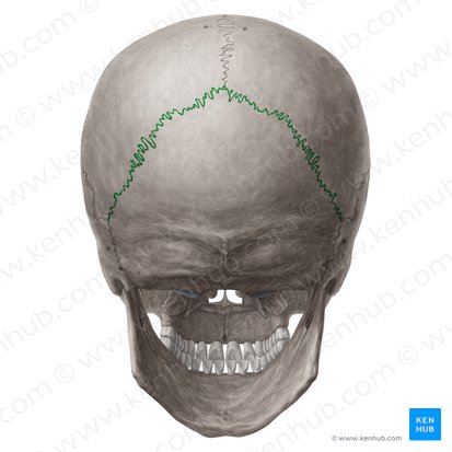
Lambdoidal/Lambdoid Suture
Fibrous joint between the parietal bones and the occipital bone
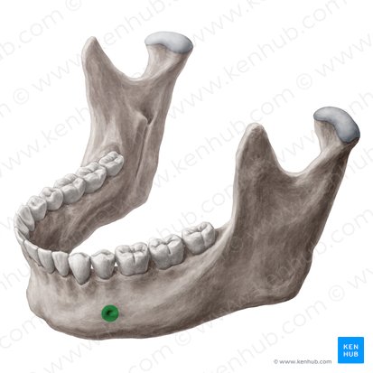
Mandible
U-shaped lower jaw bone, lower teeth are embeddedin it and it is the only movable bone of the skull.
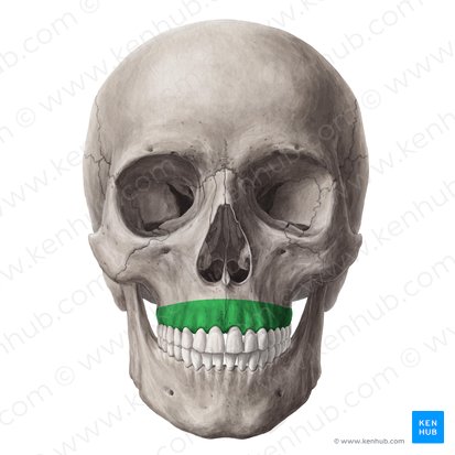
Alveolar Processes
(Mandible or Maxillae Bone)
Ridges on the mandible and maxilla that help to form the sockets that hold the teeth
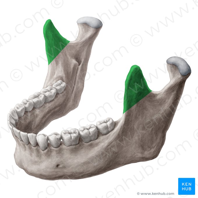
Coronoid Process
(Mandible Bone)
A projection of the mandible to which a muscle moving the mandible upward attaches
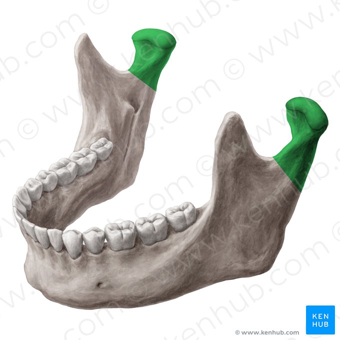
Mandibular Condyle
(Mandible Bone)
Rounded structure on the mandible that fits into mandibular fossa on the temporal bone to form a synovial joint that allows the mandible to move. The mandibular fossa of the temporal bone and the condyle of the mandible create the temporomandibular joint (TMJ)
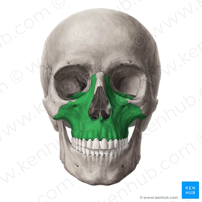
Maxillae
The two bones are fused medially and form the upper jaw. This bone articulates with all the facial bones except for the mandible. Teeth are embedded.
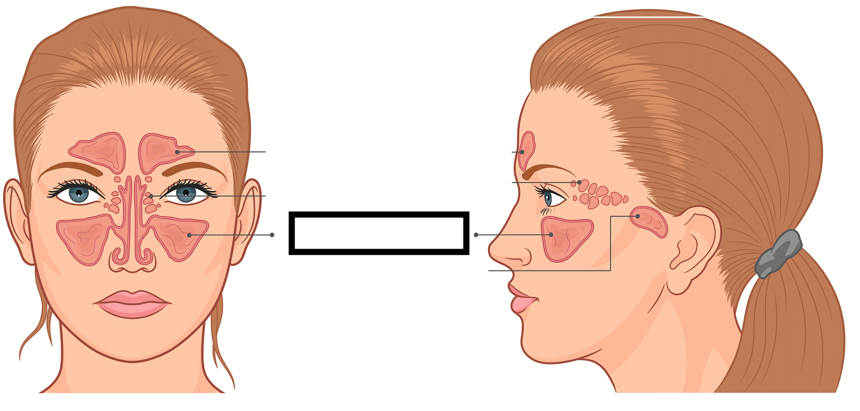
Maxillary Sinus
(Maxillary bone)
the sinuses are air-filled cavities within skull bones that open into the nasal cavity. The sinuses function to decrease the weight of the skull and act as resonating chambers during sound production.
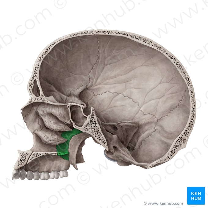
Palatine Bones
A pair of bones forming the posterior portion of the hard palate
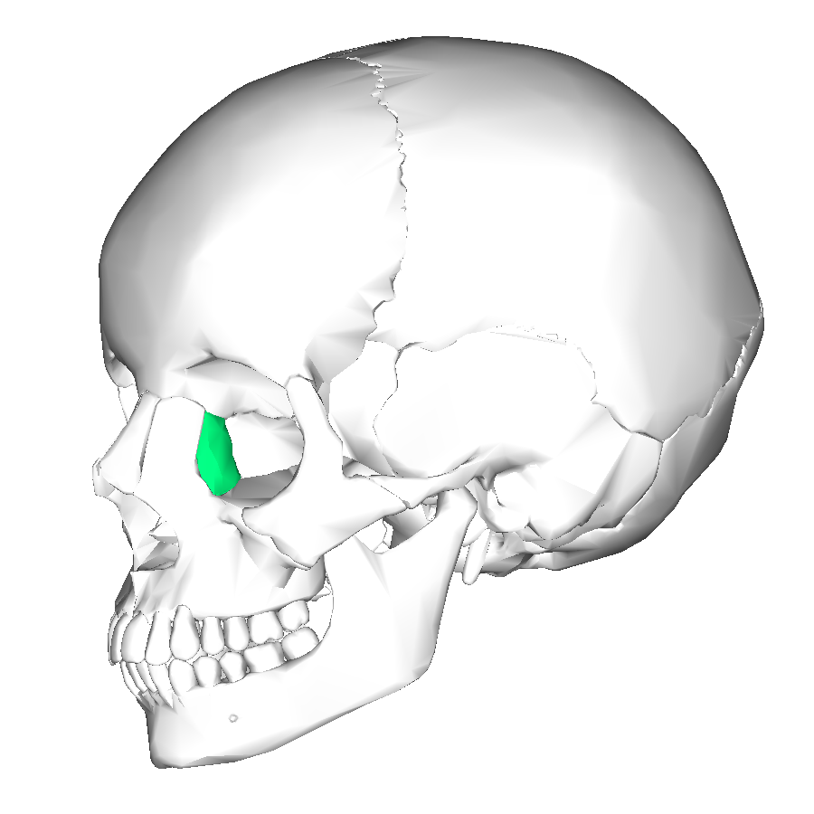
Lacrimal Bones
delicate small bones on the anterior medial wall of the orbits
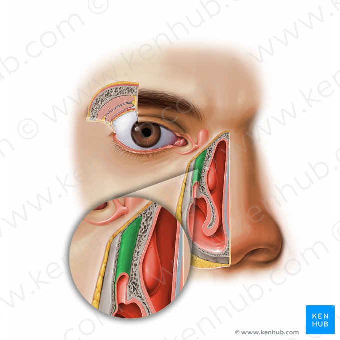
Nasolacrimal Canal
(Lacrimal Bone)
Passagew which drains tears from the surface of the eye to the nasal cavity
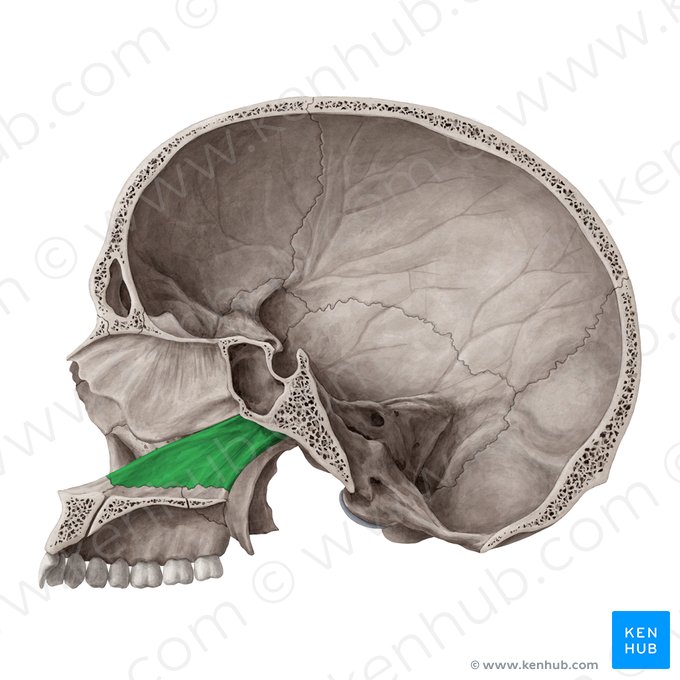
Vomer
A slender bone that forms in the inferior part of the nasal septum
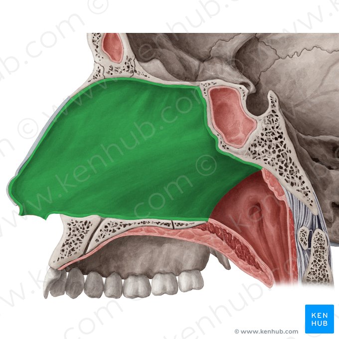
Nasal Septum
The wall in the nasal cavity fromed by cartilage, the vomer, and the medial portion of the ethmoid bone.
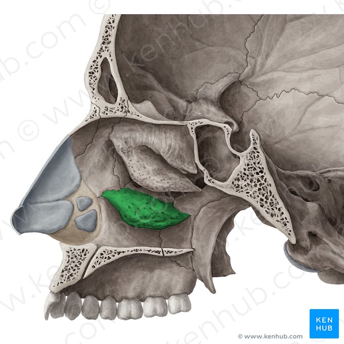
Inferior Nasal Conchae
Thin curved bones on the lateral walls of the nasal cavity. They function to increase the surface area of the nasal cavity
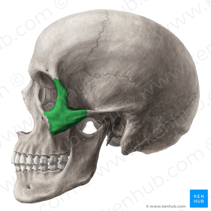
Zygomatic Bones
Temporal Bone
Two. bones form the cheeks ad lateral orbits that give shape to the face and articulate with the maxillae, frontal bone, and temporal bone.
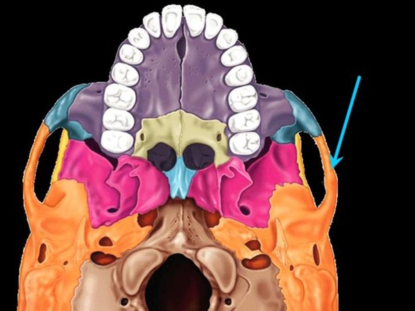
Zygomatic arch
(Zygomatic Bone)
A bridge of bone fromed by projections of the temporal and zygomatic bones. It functions as an attachment site for a muscle movie the mandible and ligaments holding the mandible in place.
Nasal bones
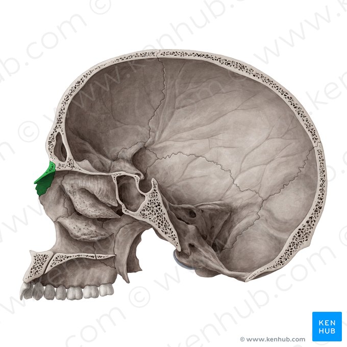
two bones forming the bridge of the nose
Hyoid Bone
this bone is not part of the skull and lies just inferior to the mandible. It functions to anchor throat, larynx and tongue muscles.