Thorax-thoracic muscle wall
 Call Kai
 Learn
Learn Practice Test
 Spaced Repetition
 Match
Match Flashcards
 Knowt Play
1/21

There's no tags or description
Looks like no tags are added yet.
Name | Mastery | Learn | Test | Matching | Spaced | Call with Kai | Chat |
|---|
No analytics yet
Send a link to your students to track their progress
22 Terms
what is the thorax
the span of the thoracic vertebrae

name the thoracic body wall muscle, function
rectus sternalis
—flexes sternum
—think a cat’s arched bacl (disappearing in human evolution)
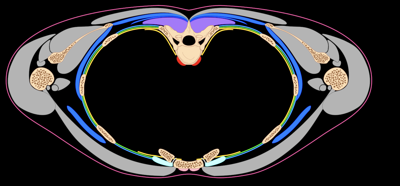
find it, also names the bones in this picture
rectus sternalis (pink) (bottom on anterior portion of sternum)
—vertebrae, clavicle, humerus
what type of thoracic musculature is the rectus sternalis (this is helping us understand the whole picture)
ventral musculature
—the rectus sternalis is anterior)

name, attachment, func
what muscle layer is this
outermost muscle layer
serratus posterior superior muscle
—anchored to spinous process of vertebae
—hinge ribs upwards
serratus posterior inferior muscle
—still anchored to spinous process of vertebrae
—pulls ribs downwards

find it
just the thinnest dark blue line posterior to the vertebrae behind the purple epaxial muscle

name, struc, func
what muscle layer is this
external muscle layer
external intercostal muscles
struc
—inbetween the ribs, oblique angled
—END AT COSTAL CARTILAGES (doesn’t come to the anterior portion of the body)
func
—breathing —→ inhaling —→ expanding lungs (inhalation)
within the external intercostal muscles, in the section between the ribs, what parts are more stable and which parts are more mobile
(top) more stable
(bottom) more mobile

find it
external intercostal muscles (blue) in lateral muscle layers
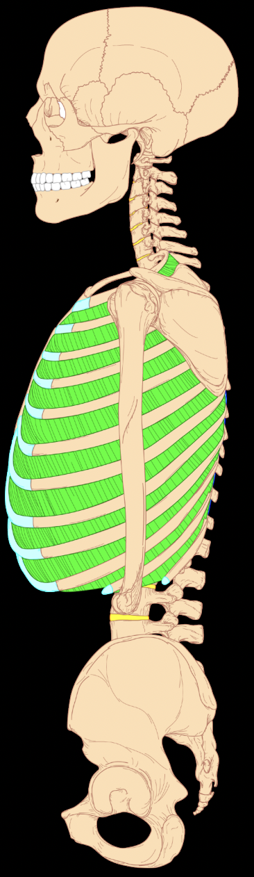
name, struc, func
what muscle layer is this
internal intercostal muscles
middle muscle layer
struc
—go all the way anterior (to the sternum)
—angled in other direction compared to external intercostal muscles
func
—breathing —→ exhalation
which parts of the internal intercostal muscle are more stable and more mobile
what identifies a portion more stable?
more mobile (top)
more stable (bottom)
more stable=closer to the vertebral column

find it
green in lateral body wall muscles

name, struc, func
what muscle layer of the lateral musculature of the thorax is this
transverse thoracis muscles
internal mucle layer (anterior muscle of the internal layer)
struc
—attaches to sternum and ribs
func
—pulls ribs down (hinge downwards)
—exhalation!

find it
(yellow) on anterior side branching off of thorax-smaller line

name, struc, func
innermost intercostal muscles
-lateral muscle of the internal layer
struc
—muscles segments INTERUPTED BY RIBS
—only on lateral side
—same fiber orientation as internal intercostals
func
—depression of ribs
-exhalation
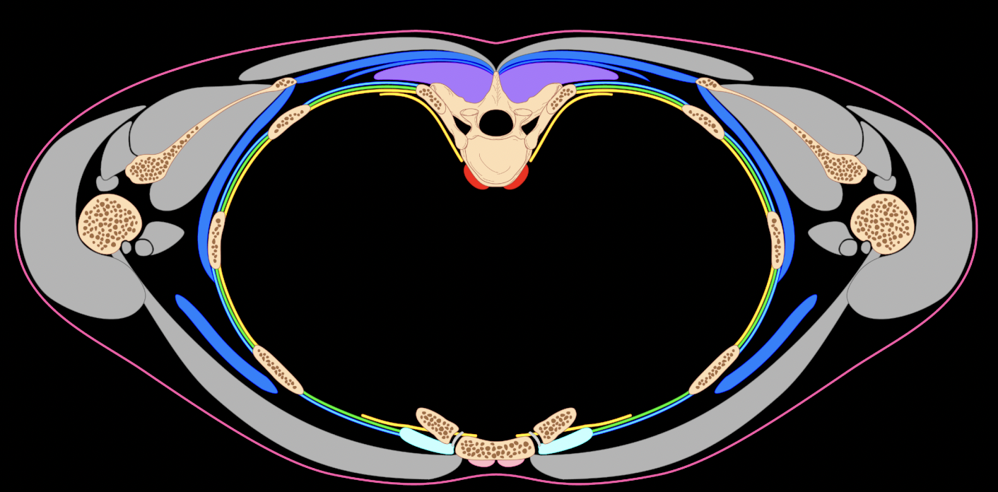
find it
innermost later muscle wall layer (yellow)

name, struc, func
subcostal muscules
—posterior muscle of the internal layer
struc
—attaches rib to rib
func
—depress the ribs
—exhalation

find it
yellow line adjacent to vertebrae

name, struc, func
diaphram
-specialized floor of thorax from internal layer
struc
—skeletal muscle (voluntary)
func
—inhalation (diaphram contracts) (balloon downwards) (balloon upwards is relaxed)

name, struc, func
what type of musculature is this now
longus colli muscle
-subvertebral musculature (upper quarter of thorax)
struc
—since this is in the subvertebral musculature, the upper thoracic region in the anchor pt, and the cervical region is where the function will take place
—it is pulling on vertebral bodies in the cervical region
func
—cervical flexion, pulls head down
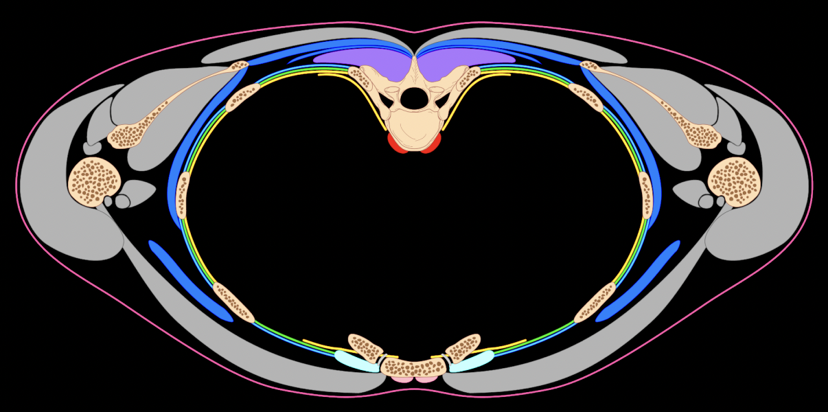
find it
red bumps on posterior portion of body wall, sitting on vertebrae
How was the functional significance of the thoracic muscle wall repurposed?
from lateral angulation/locomotion —→ respiratory movements