SBMS1431 Human Anatomy and Physiology I
 Call Kai
 Learn
 Practice Test
 Spaced Repetition
 Match
 Flashcards
 Knowt Play
1/58

Earn XP
Description and Tags
Cardiac Functions
59 Terms
Cardiac Cycle Phases:
Phase 1 : Atrial systole
Phase 2 : Isovolumetric contraction
Phase 3 : Ventricular ejection
Phase 4 : Isovolumetric relaxation
Phase 5 : Ventricular filing
What happens during Phase 1: Atrial systole?
The atria contract.
This pushes blood into the ventricles to top them off before they contract.
What happens during Phase 2: Isovolumetric contraction?
The ventricles begin to contract.
Both the AV valves and semilunar valves are closed.
"Isovolumetric" means the volume of blood is not changing, so pressure builds up rapidly.
What happens during Phase 3: Ventricular ejection?
Ventricular pressure exceeds artery pressure.
Semilunar valves open, and blood is ejected out of the ventricles into the aorta and pulmonary artery.
What happens during Phase 4: Isovolumetric relaxation?
The ventricles relax.
Semilunar valves close (preventing backflow).
All valves are closed again, and volume stays the same while pressure drops.
What happens during Phase 5: Ventricular filling?
Ventricular pressure drops below atrial pressure.
AV valves open.
Blood passively flows from the atria into the ventricles.
How do the phases divide into overall Diastole and Systole?
Ventricular Diastole (Relaxation): Includes Phase 4 (Isovolumetric relaxation) and Phase 5 (Ventricular filling).
Ventricular Systole (Contraction): Includes Phase 2 (Isovolumetric contraction) and Phase 3 (Ventricular ejection).
What is the formula for Mean Arterial Pressure (MAP)?
MAP = Cardiac Output (CO) × Total Peripheral Resistance (TPR)
Think of it like a hose: Pressure depends on how much water you pump (CO) and how narrow the nozzle is (TPR).
What two factors make up Cardiac Output (CO)?
Stroke Volume: How much blood is pumped per beat.
Heart Rate: How many times the heart beats per minute.
(CO = Stroke Volume × Heart Rate)
What is the main factor that determines Stroke Volume (how much blood the heart pumps per beat)?
End-diastolic ventricular volume (how much blood is in the ventricle right before it contracts).
More blood in = more blood out.
This volume is determined by venous return (how much blood comes back to the heart).
What factors increase venous return (blood going back to the heart)?
Venous pressure (driven by sympathetic nerves to veins).
Blood volume.
Skeletal muscle pump (muscles squeezing veins).
Inspiration movements (breathing acts like a pump).
What controls the SA node (Heart Rate)?
Sympathetic nerves ↑ Heart Rate.
Parasympathetic nerves ↓ Heart Rate.
Plasma Epinephrine ↑ Heart Rate.
What determines Total Peripheral Resistance (TPR)?
Arteriolar radius (the size of the blood vessels). This is the biggest factor.
Blood viscosity (thickness of blood, determined by hematocrit).
What three types of control change the arteriolar radius?
Local controls: Respond to tissue needs (e.g., ↓ Oxygen causes vasodilation).
Neural controls: Sympathetic nerves cause vasoconstriction.
Hormonal controls: Chemicals like Epinephrine and Angiotensin II cause vasoconstriction (narrowing).
What is Stroke Volume (SV)?
The volume of blood pumped from the ventricle per heartbeat.
Essentially, it is the amount of blood ejected into the aorta during systole.
How is Stroke Volume (SV) calculated?
SV = EDV − ESV
EDV (End-Diastolic Volume): Volume of blood just prior to the beat.
ESV (End-Systolic Volume): Volume of blood left at the end of a beat.
Simply put: Blood before beat minus blood left over = blood ejected.
How does Stroke Volume relate to Cardiac Output?
CO = SV × HR
Cardiac Output equals Stroke Volume multiplied by Heart Rate.
Factors affecting Stroke Volume
Preload
Afterload
Contractility
What is Preload?
The volume of blood the ventricle can hold during diastole.
(Think of it as the "stretch" on the heart before it squeezes.)
What is Afterload?
The resistance the heart must overcome to eject blood into the aorta during systole.
It is closely related to aortic pressure (how hard the heart has to push to open the valve).
it is influenced by Arteriolar Elasticity.
What is Contractility?
The ability of the heart muscle to contract and pump blood (how strong the squeeze is).
It is influenced by wall stretch (a more stretched heart contracts harder) and autonomic nerves.
How do Preload, Afterload, and Contractility fit into the Cardiac Output formula?
Preload + Afterload + Contractility all determine Stroke Volume (SV).
Stroke Volume × Heart Rate = Cardiac Output (CO).
(Note: Heart Rate is controlled separately by the Sympathetic and Parasympathetic nerves)
What factors raise Preload, and what factors lower it?
Raised by: Fast filling time, increased venous return.
Result: Increases end-diastolic volume, which increases stroke volume.
Lowered by: Decreased thyroid hormones, decreased calcium, abnormal ions/pH, low temp, hypoxia, drugs (calcium channel blockers).
Result: Decreases end-diastolic volume, which decreases stroke volume.
What factors raise Contractility, and what factors lower it?
Raised by: Sympathetic stimulation, epinephrine/norepinephrine, high intracellular/blood calcium, thyroid hormones, glucagon.
Result: Decreases end-systolic volume, which increases stroke volume.
Lowered by: Parasympathetic stimulation, acetylcholine, hypoxia, hyperkalemia.
Result: Increases end-systolic volume, which decreases stroke volume.
What factors raise Afterload, and what factors lower it?
Raised by: Increased vascular resistance, semilunar valve damage.
Result: Increases end-systolic volume, which decreases stroke volume.
Lowered by: Decreased vascular resistance.
Result: Decreases end-systolic volume, which increases stroke volume.
How do changes in End-Diastolic Volume (EDV) and End-Systolic Volume (ESV) affect Stroke Volume?
Stroke Volume increases when you increase EDV (more blood in) OR decrease ESV (less blood left over).
Stroke Volume decreases when you decrease EDV (less blood in) OR increase ESV (more blood left over).
What are the formulas for Stroke Volume (SV), Ejection Fraction (EF), and Cardiac Output (CO)?
Stroke Volume (SV) = EDV minus ESV
Ejection Fraction (EF) = SV divided by EDV
Cardiac Output (CO) = Heart Rate (HR) multiplied by SV
What does Ejection Fraction (EF) actually reflect?
It reflects how effectively the left ventricle pumps blood into the systemic circulation.
A higher EF means better systolic function (the heart squeezes well).
A lower EF means impaired systolic function (the heart squeezes poorly).
What are the clinical interpretations of different Ejection Fraction (EF) percentages?
55–70%: Normal
50–55%: Borderline
40–49%: Mildly reduced
Less than 40%: Heart failure with reduced EF (HFrEF)
Less than 30%: Severe systolic dysfunction
If EDV is 120 mL and ESV is 50 mL, what are the SV and EF?
Stroke Volume: 120 mL minus 50 mL = 70 mL
Ejection Fraction: 70 mL divided by 120 mL = 58% (Normal)
What is the Frank-Starling Law of the Heart?
It describes the relationship between Stroke Volume (SV) and End-Diastolic Volume (EDV).
Simply put: The heart pumps out more blood when it receives more blood.
What is the physical mechanism behind the Frank-Starling Law?
It is based on the length-tension relationship of cardiac muscle.
At rest, cardiac muscle fibers are shorter than their optimal length. When they are stretched, they can contract with more force.
What is the key factor that stretches the heart muscle fibers?
Venous return (the amount of blood returning to the heart).
When venous return increases (e.g., during exercise or a slower heart rate), the ventricles are stretched more, leading to a stronger contraction.
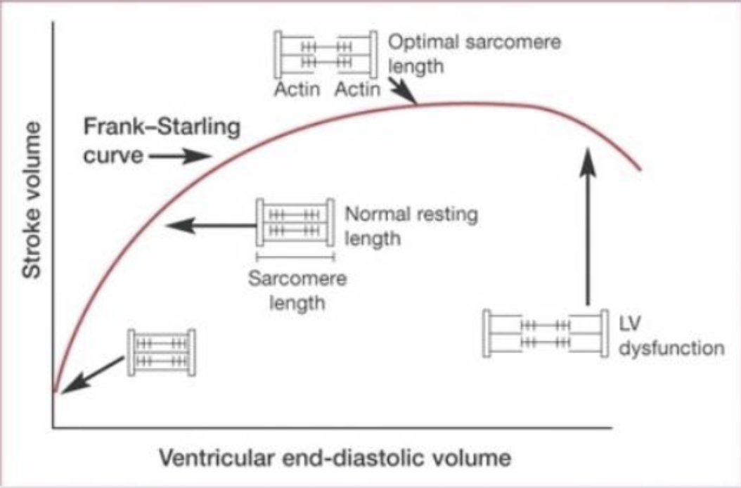
What does the Frank-Starling curve show, and what happens in LV dysfunction?
The curve shows that as Ventricular end-diastolic volume increases, Stroke volume increases, up to a point.
There is an optimal sarcomere length (peak of the curve). If stretched too far, the curve drops.
In LV (Left Ventricular) dysfunction, the curve is shifted down and to the right, meaning the heart has to be stretched much more just to maintain a normal stroke volume.
What is Preload (overall definition), and what causes it?
Preload is the amount of stretch on the heart muscle during diastole (when the heart is filling).
It is caused by the volume of blood filling the left ventricle before it contracts (the white arrows in the diagram show this outward stretch).
What clinical terms are used to measure Preload?
LVEDV (Left Ventricular End-Diastolic Volume): The actual amount of blood stretching the ventricle.
LVEDP (Left Ventricular End-Diastolic Pressure): The pressure inside the ventricle, which is used clinically to estimate the preload.
What is Preload in the heart? (More specific definition)
It is the initial stretching of cardiac muscle cells (cardiomyocytes) before contraction.
Specifically, it is the amount of sarcomere stretch at the end of ventricular filling during diastole.
How is Preload Measured?
We cannot directly measure preload in a living heart.
Instead, we estimate it using end-diastolic ventricular pressure or volume (how full the ventricle is).
What do the x-axis and y-axis represent on this Preload graph, and what are the "Normal resting values"?
X-axis (Stretch): Indicated by ventricular end-diastolic volume (mL).
Y-axis (Force): Indicated by stroke volume (mL).
Normal resting values: About 135 mL of EDV produces a stroke volume of 70 mL.


What relationship does the Preload graph show, and what formula is provided on the right?
Relationship: As stretch (EDV) increases, the force (Stroke Volume) increases—but only up to a point, after which the curve flattens out.
Formula: SV = EDV - ESV (Stroke Volume = End-Diastolic Volume minus End-Systolic Volume).
Why does posture and gravity make venous return difficult, and what are the mechanisms that assist it at rest?
The Challenge: Gravity makes it harder for blood to travel from the lower body back up to the heart.
The 4 Resting Mechanisms:
One-way venous valves (prevent backflow)
Muscle pump (squeezing veins)
Respiratory pump (breathing helps pull blood up)
Venoconstriction (only happens with sympathetic stimulation (e.g., during flight-or-fight)
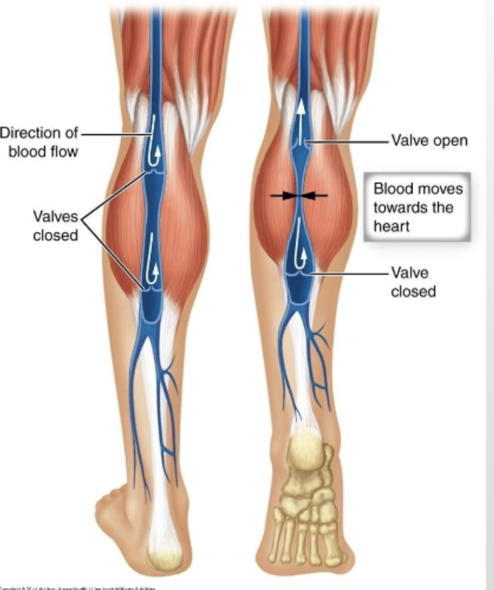
What is the function of the one-way venous valves?
They ensure blood flows only toward the heart.
When blood flows backward, the valve cusps fill with blood and close (shown in the "Closed" diagram), preventing backflow.
How does the skeletal muscle pump assist venous return?
Muscles contract: Squeezes the vein, pushing blood upward (proximal valve opens, distal valve closes).
Muscles relax: The vein expands, and the proximal valve closes to stop backflow while blood refills the vein from below.
How does breathing (the respiratory pump) help blood return to the heart?
Inhalation: Thoracic pressure decreases and abdominal pressure increases. This pressure difference squeezes abdominal veins and pulls blood into the chest.
Exhalation: The pressure reverses, but valves prevent blood from flowing backward.
How does the normal (dilated) state of a vein compare to the venoconstriction state?
Normal/Dilated State (Lower Venous Return):
High venous capacitance (veins hold more blood)
Large vein diameter
Lower internal pressure
Venoconstriction State (Increased Venous Return):
Decreased venous capacitance
Decreased vein diameter
Increased venous pressure
Increased pressure gradient to the heart
What triggers venoconstriction, and what is the end result for the heart?
Trigger: Sympathetic nerves stimulate the smooth muscle in the vein wall (e.g., via Norepinephrine).
Result: The vein narrows (venoconstriction), which increases blood flow back to the heart.
Final effect: This increases venous return, which increases Cardiac Output (thanks to the Frank-Starling Law).
If venous return increases (while End-Systolic Volume stays unchanged), what happens to Stroke Volume according to the formula SV = EDV - ESV?
Increased venous return means more blood enters the heart.
This raises End-Diastolic Volume (EDV).
Because ESV is unchanged, the higher EDV results in an increased Stroke Volume (SV).
(More blood in equals more blood pumped out.)
How do Preload and Afterload differ in terms of definition?
Preload (Think: STRETCH): The volume of blood returning to the heart at the end of diastole. It stretches the muscle fibers just before contraction.
Afterload (Think: SQUEEZE): The pressure the heart (specifically the left ventricle) has to overcome to eject blood out of the heart.

What is Afterload, and what is its main determining factor?
Definition: The amount of resistance the heart must pump against when ejecting blood out of the ventricles.
Main Factor: Vascular Resistance (the pressure in the blood vessels, specifically the arteries, that the heart has to push against to open the valves).
What specific pressure must the ventricles overcome during afterload?
Specific pressure: Aortic pressure (the pressure in the aorta that the ventricles must overcome to push blood out).
How does Hypertension (high blood pressure) affect Afterload, End-Systolic Volume (ESV), and Stroke Volume (SV)?
Afterload: Increases.
End-Systolic Volume (ESV): Higher (more blood is left over because it's harder to pump out).
Stroke Volume (SV): Lower (less blood is ejected per beat).
What is a common example of a condition that increases afterload, and what does the diagram show?
Atherosclerosis (narrowing of the arteries due to plaque buildup).
plaques (fatty deposits) build up in the artery walls (intima/media), narrowing the lumen (the space for blood). This makes it harder for the heart to push blood through, increasing afterload.

According to the formula SV = EDV - ESV, what happens to Stroke Volume (SV) if afterload increases (like in hypertension) while End-Diastolic Volume (EDV) remains unchanged?
Increased afterload makes it harder for the heart to pump blood out.
This causes the End-Systolic Volume (ESV) to increase (more blood is left in the ventricle).
Because EDV is unchanged, the higher ESV means the Stroke Volume (SV) must decrease.
Which step is controlled by Acetylcholine (ACh), and via which receptor?
Step Controlled: It slows down the rate of depolarization in autorhythmic cells (which lowers Heart Rate).
Pathway: Parasympathetic innervation.
Receptor: Muscarinic receptors.
Which steps are controlled by Norepinephrine (NE) and Epinephrine, and via which receptors?
Step Controlled 1 (Heart Rate): Sympathetic innervation and Epinephrine make the rate of depolarization in autorhythmic cells faster.
Step Controlled 2 (Stroke Volume): Sympathetic innervation and Epinephrine influence Contractility (force of contraction in ventricular myocardium).
Receptor: Beta-1 (β1) receptors.
What is Contractility?
The intrinsic strength of the ventricle.
It is independent of loading (preload) conditions (meaning the strength of the squeeze can change even if the amount of blood filling the heart stays the same).
What does the graph show about the effect of Norepinephrine on Stroke Volume?
At the exact same filling volume (Point A on the x-axis), the Norepinephrine curve is higher than the Control curve.
This means Norepinephrine increases contractility, which pumps out more blood (higher Stroke Volume) for any given End-Diastolic Volume.

How do Epinephrine and Norepinephrine trigger a change in cardiac contraction?
Epinephrine and Norepinephrine bind to β1-receptors.
This activates the cAMP second messenger system.
This leads to two outcomes: a more forceful contraction and a shorter duration of contraction.

According to the formula SV = EDV - ESV, what happens to Stroke Volume (SV) when cardiac contractility increases (such as during sympathetic stimulation), assuming End-Diastolic Volume (EDV) remains unchanged?
Increased contractility means the heart squeezes harder.
This causes End-Systolic Volume (ESV) to decrease (less blood is left over in the ventricle).
Because EDV is unchanged, the lower ESV means Stroke Volume (SV) must increase.