Anatomy and Physiology - Urinary System
 Call Kai
 Learn
 Practice Test
 Spaced Repetition
 Match
 Flashcards
 Knowt Play
1/28

There's no tags or description
Looks like no tags are added yet.
29 Terms
Urinary tract
Ureters, urinary bladder, and urethra; they transport urine from kidneys to outside body
Processes that occur as filtrate is converted to urine
Elimination of metabolic wastes
Regulation of ion levels (ex: Na+, K+, Ca2+)
Regulation of acid-base balance: alters levels of H+/hydron (acid) and HCO3-/bicarbonate (base)
Regulation of blood pressure
Elimination of biologically active molecules: hormones, drugs
Formation of calcitriol: increases absorption of calcium from small intestine
Secretes erythropoietin (EPO) in response to low blood oxygen
Stimulates red bone marrow to increase erythrocyte production
Potential to engage in gluconeogenesis to maintain glucose levels
During prolonged fasting or starvation
Kidney structure
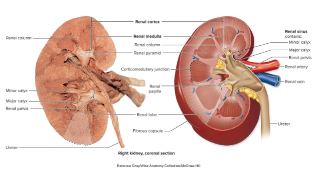
Against the posterior abdominal wall & are retroperitoneal (posterior to the parietal peritoneum)
Only the anterior surface is covered by the parietal peritoneum
Tissue layers (inner → outer): fibrous capsule, perinephric fat, renal fascia, and paranephric fat
Two distinct regions: outer renal cortex and an inner renal medulla
Concave medial border (hilum) where vessels, nerves, and the ureter connect to it

Peritoneum
Serous membranes lining the internal walls of the abdominopelvic cavity and covering most of the organs within the cavity

Nephron
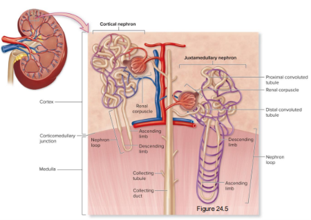
Functional filtration unit of the kidney that mostly resides in the renal cortex; composed of:
Renal corpuscle: includes the glomerulus and glomerular capsule/Bowman’s capsule
Renal tubule: includes the proximal convoluted tube (PCT), nephron loop/loop of Henle, and distal convoluted tubule (DCT)
Not part of the nephron but associated with it are the afferent arteriole, efferent arteriole, collecting tubule, and collecting duct

Glomerulus (renal corpuscle)
Thick tangle of blood capillary loops
Blood enters by afferent arteriole and exits by an efferent arteriole
Primary location of blood filtration

Fluid flow through the renal tubule
First region is the PCT, originating at the renal corpuscle
Bends sharply to form a nephron loop that juts into the renal medulla
Re-enters the renal cortex at the DCT
Drain into a connecting tubule
Multiple collecting tubules empty into larger collecting ducts


Types of nephrons
Functions are filtration and reabsorption
Cortical: located almost completely in the cortex with short nephron loops that barely penetrate into the medulla; 85% of nephrons
Juxtamedullary: lie close to the corticomedullary junction and have relatively long nephron loops that extend deep into the medulla; remaining 15%
Additional functions of urine concentration and electrolyte balance

Juxtaglomerular apparatus
Formed by the physical contact between the afferent arteriole and the adjacent DCT
Regulates urine production and blood pressure
Composed of:
Extraglomerular mesangial cells
Macula densa of DCT
Granular cells of affertent arteriole

Fluid flow through the urinary system
As filtrate: capsular space (between glomerulus and glomerular capsule)
As tubular fluid: PCT
Descending limb of nephron loop
Ascending limb of nephron loop
DCT
Collecting tubules
Collecting ducts
As urine: papillary duct
Minor calyx (part of renal sinus)
Major calyx (part of renal sinus)
Renal pelvis (part of renal sinus)
Ureter
Urinary bladder
Urethra
Filtrate vs. tubular fluid vs. urine
Essentially plasma (water + solutes) minus large solutes (e.g., most proteins)
Filtrate enters the PCT → called tubular fluid
Leaves the collecting ducts → called urine
Process of urine formation
Glomerular filtration: movement of substances from the blood within the glomerulus into the capsular space to form filtrate
Tubular reabsorption: movement of substances from the tubular fluid back into the blood
Tubular secretion: movement of substances from the blood into the tubular fluid
(Tubule is focused on fine-tuning filtrate, ISF, and blood concentrations)
Peritubular capillaries and vasa recta
Tubular reabsorption and secretion occur between the tubules and the peritubular capillaries + vasa recta
Specialized blood vessels in the kidney that play a critical role
in maintaining the medullary osmotic gradient and preventing its washout
Branches from efferent arteriole and merges into venules
Peritubular capillaries: surround the DCT and PCT; provide oxygen and nutrients in the cortex
Vasa recta: surround the nephron loop; provide oxygen and nutrients in the medulla
Filtration membrane
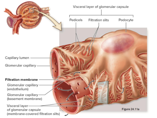
Filtrate is made in renal corpuscle when blood plasma is forced across the filtration membrane under pressure (produced by difference in afferent and efferent arteriole pressures)
3 layers (inner → outer)
Fenestrated endothelium of a glomerular capillary: blocks formed elements
Basement membrane of a glomerular capillary: blocks large proteins
Visceral layer of glomerular capsule (membrane-covered filtration slits): blocks small proteins

Podocytes
Specialized epithelial cells in the kidneys that play a critical role in glomerular filtration
Spaces between the pedicels (footlike processes) form the membrane-covered filtration slits of the filtration membrane