OTR 704 2.1 Head, Neck, Trunk & Spine
 Call Kai
 Learn
Learn Practice Test
 Spaced Repetition
 Match
Match Flashcards
 Knowt Play
1/156

There's no tags or description
Looks like no tags are added yet.
Name | Mastery | Learn | Test | Matching | Spaced | Call with Kai | Chat |
|---|
No analytics yet
Send a link to your students to track their progress
157 Terms

What structure is shown?
Cranium
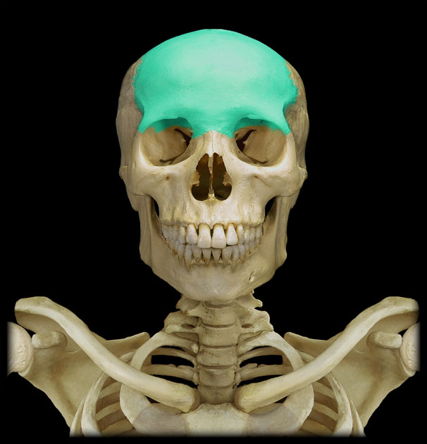
What structure is shown?
Frontal bone
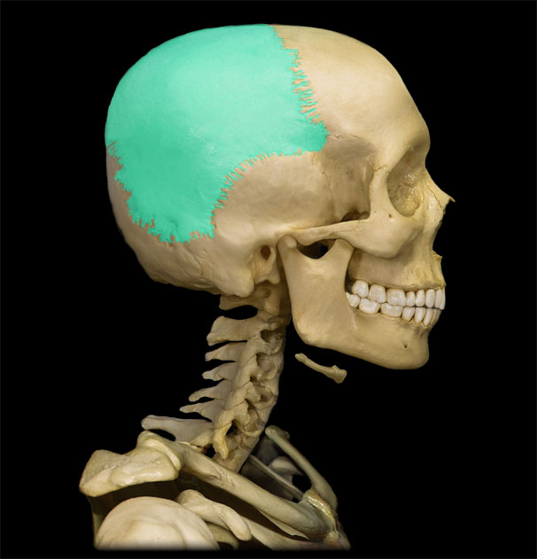
What structure is shown?
Parietal bone

What structure is shown?
Temporal bone

What structure is shown?
Occipital bone
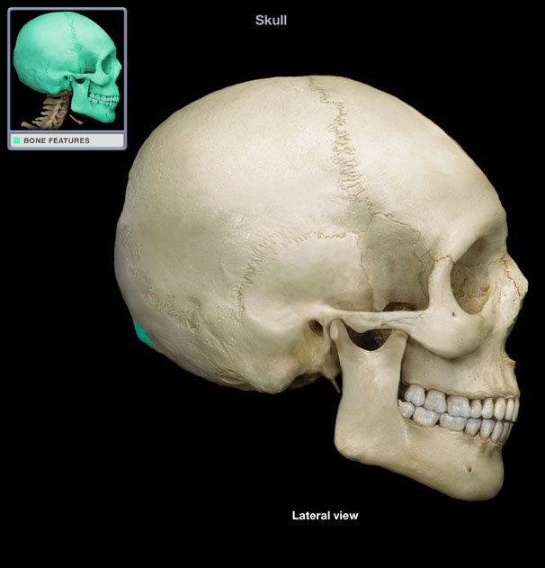
What structure is shown?
External occipital protuberance

What structure is shown?
Mastoid process

What structure is shown?
Mandible
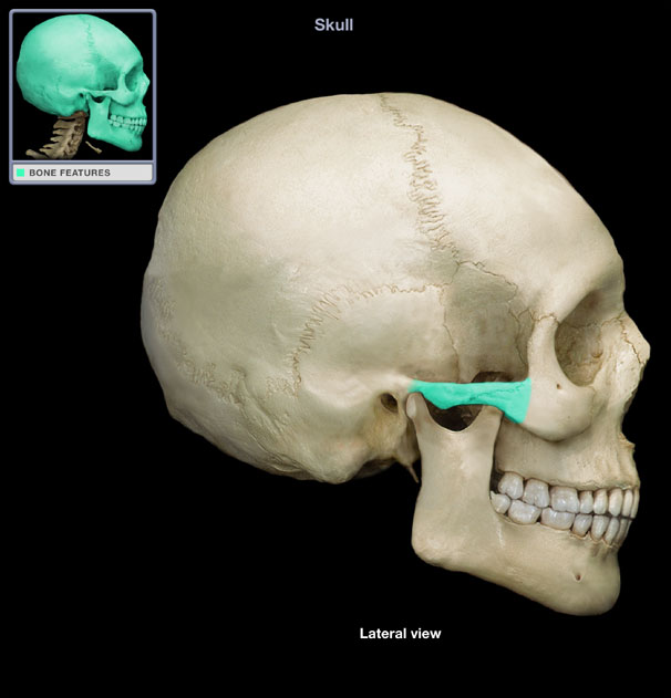
What structure is shown?
Zygomatic arch
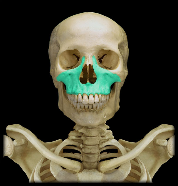
What structure is shown?
Maxilla

What structure is shown?
Orbit

What structure is shown?
Foramen magnum

What structure is shown?
Hyoid bone
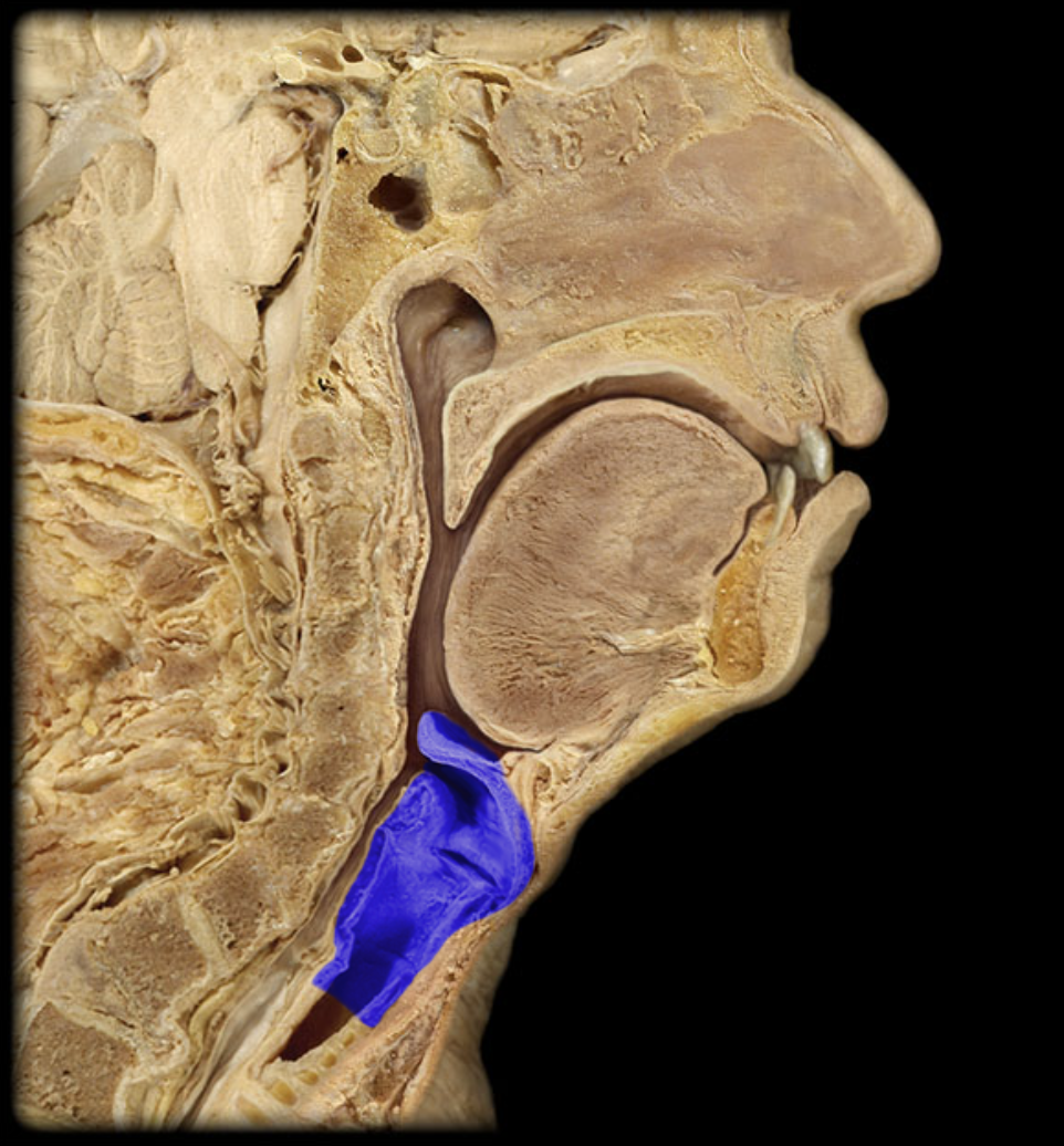
What structure is shown?
Larynx

What structure is shown?
True ribs (1-7)

What structure is shown?
False ribs (8-10)

What structure is shown?
Floating ribs (11-12)

What structure is shown?
Intercostal spaces
Hyoid bone
Suspended, U-shaped bone; point of attachment for many muscles involved in speech and swallowing
“Floating” — not directly attached to any other bones
Elevates to close off the larynx and airway during swallowing
Larynx
“Voice box” formed by several cartilages
Temporomandibular joints (TMJ)
Synovial (modified hinge) joints
Work in tandem for symmetrical functional motions of the mandible — elevation, depression, protraction, retraction
Important for chewing, speaking
Integumentary muscles
Muscles of facial expression
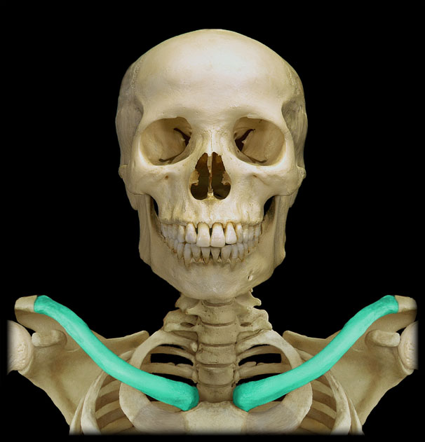
What structure is shown?
Clavicle

What structure is shown?
Sternoclavicular joint
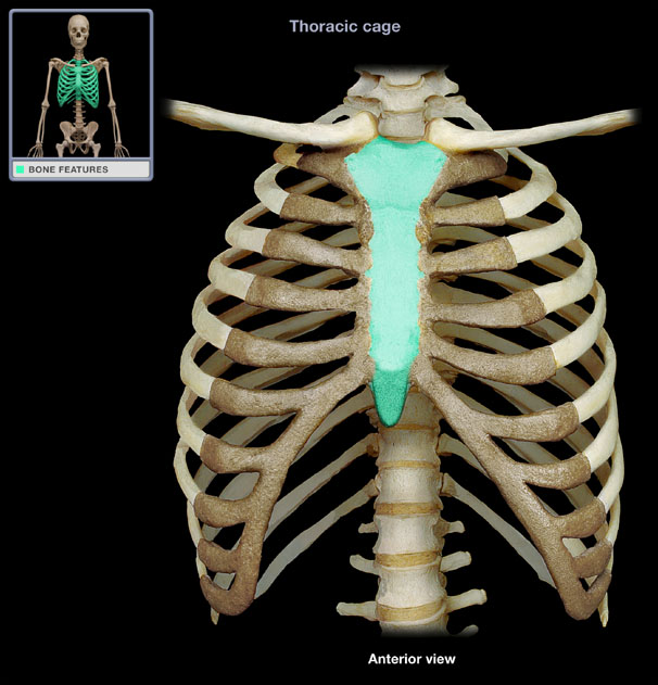
What structure is shown?
Sternum
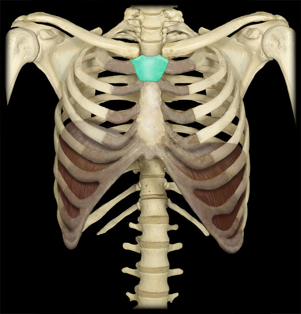
What structure is shown?
Manubrium

What structure is shown?
Sternal body

What structure is shown?
Xiphoid process
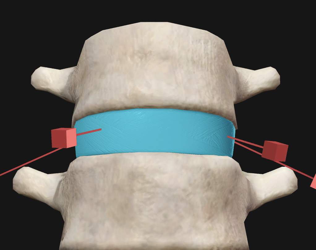
What structure is shown?
Intervertebral disc

What structure is shown?
Coccyx (4 fused/semi-fused vertebrae)

What structure is shown?
Sacrum (5 fused vertebrae)

What structure is shown?
Lumbar vertebrae (5)

What structure is shown?
Thoracic vertebrae (12)
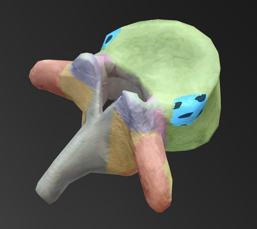
What structure is shown (blue highlight)?
Costal facets

What structure is shown?
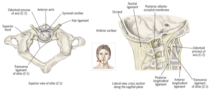
Axis (C2)

What structure is shown?
Dens (odontoid process)

What structure is shown?
Atlas (C1)

What structure is shown?
Cervical vertebrae (7)

What structure is shown?
Vertebral body

What structure is shown?
Spinous process(es)

What structure is shown?
Transverse process(es)
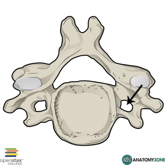
What structure is shown?
Transverse foramen

What structure is shown?
Lamina

What structure is shown?
Pedicle

What structure is shown?
Superior articular facet
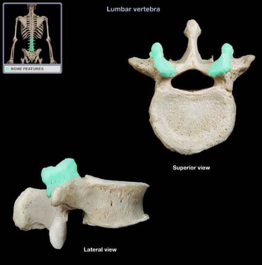
What structure is shown?
Superior articular processes

What structure is shown?
Inferior articular processes
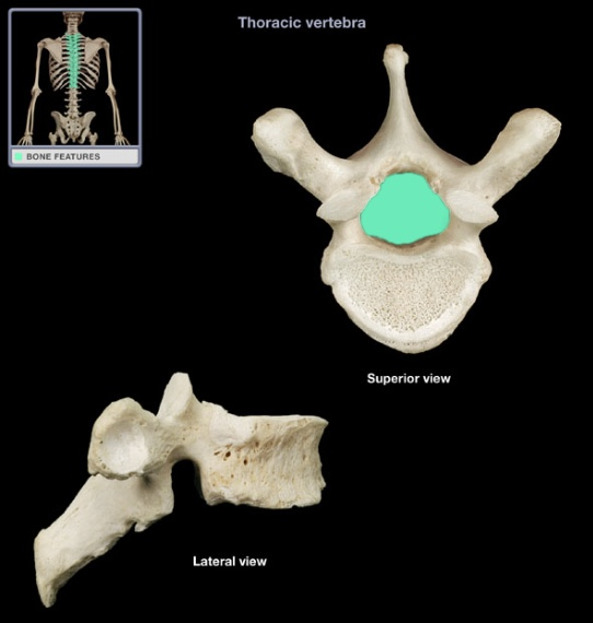
What structure is shown?
Vertebral foramen
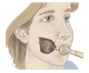
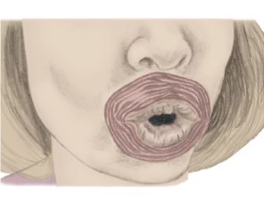
Buccinator action
Contracts cheeks to narrow mouth; directs bolus inside mouth; engages when blowing air out of narrowed mouth (e.g., balloon)
Orbicularis oris action
Seals mouth for swallowing; drinking from a straw; eating from a spoon
Masseter action
Elevates and protrudes mandible (e.g., chewing, speech)
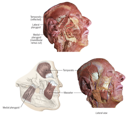
Masseter origin
Zygomatic arch
Masseter insertion
Angle and ramus of mandible
Masseter innervation
CN V (Trigeminal n.)
Temporalis action
Elevates and retracts mandible (e.g., chewing, speech)

Temporalis innervation
CN V (Trigeminal n.)
Pharynx
“Fork in the road” between respiratory and digestive systems; the throat (nasopharynx + oropharynx + laryngopharynx)

Suprahyoids action
Elevate hyoid, tongue, and larynx during swallowing; depress mandible; retracts the mandible (digastric only); engages during verbal communication, swallowing

Infrahyoids action
Depresses hyoid and thyroid cartilage during speech and swallowing

Pre-oral phase of swallowing/feeding
Anything that works to bring food/drink to the mouth
Oral preparatory phase of swallowing/feeding
1st phase — mastication and salivation create a bolus
VOLUNTARY
Oral transit phase of swallowing/feeding
2nd phase — tongue propels bolus toward pharynx
VOLUNTARY
Pharyngeal phase of swallowing/feeding
3rd phase — hyoid/larynx elevates, nasopharynx are oropharynx are sealed, epiglottis covers larynx, and bolus descends
INVOLUNTARY
Esophageal phase of swallowing/feeding
4th phase — esophageal peristalsis advances bolus toward the stomach
INVOLUNTARY
Aspiration
Food or liquid descending beneath vocal folds and entering trachea
Cranial nerves
Twelve pairs of nerves that arise from the brainstem and innervate the head and neck; identified by Roman numeral and name
CNs do not have contralateral control → ipsilateral deficits with cortical injury (exception: CN II & CN III)

CN I
Name: Olfactory
Sensory or motor: Sensory
Function: Olfaction (smell)
CN II
Name: Optic
Sensory or motor: Sensory
Function: Vision
CN III
Name: Oculomotor
Sensory or motor: Motor
Function: Moving eye up, down, medially (superior/inferior/medial rectus muscles); raising eyelid (levator palpebrae muscle); constricting pupil (sphincter pupillae muscle)
CN IV
Name: Trochlear
Sensory or motor: Motor
Function: Moving eye down and medially (superior oblique muscle)
CN V
Name: Trigeminal
Sensory or motor: Both
Function: Mastication (masseter, temporalis, pterygoid muscles); sensation to face and TMJ
CN VI
Name: Abducens
Sensory or motor: Motor
Function: Abducting the eye (lateral rectus muscle)
CN VII
Name: Facial
Sensory or motor: Both
Function: Facial expression; taste (sensory innervation to anterior 2/3 of tongue)
CN VIII
Name: Vestibulocochlear
Sensory or motor: Sensory
Function: Hearing, vestibular sense (head position relative to gravity)
CN IX
Name: Glossopharyngeal
Sensory or motor: Both
Function: Swallowing; taste; salivation
CN X
Name: Vagus
Sensory or motor: Both
Function: Visceral regulation (parasympathetic); speech
CN XI
Name: Spinal accessory
Sensory or motor: Motor
Function: Scapular elevation (trapezius muscle); rotation of head (sternocleidomastoid muscle)
CN XII
Name: Hypoglossal
Sensory or motor: Motor
Function: Movements of tongue
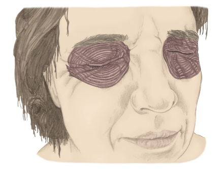
Orbicularis oculi action
Specialized fibers contribute to involuntary closure during blinking or sleeping
Semi-voluntary closure to squint or when in pain
Levator palpebrae action
Elevates eyelid
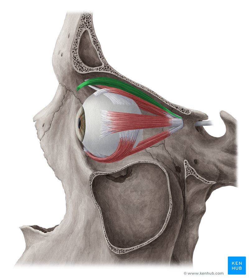
Levator palpebrae innervation
CN III (Oculomotor n.)
Superior rectus action
Elevates (upward movement of) eye

Superior rectus innervation
CN III (Oculomotor n.)
Inferior rectus action
Depresses (downward movement of) eye
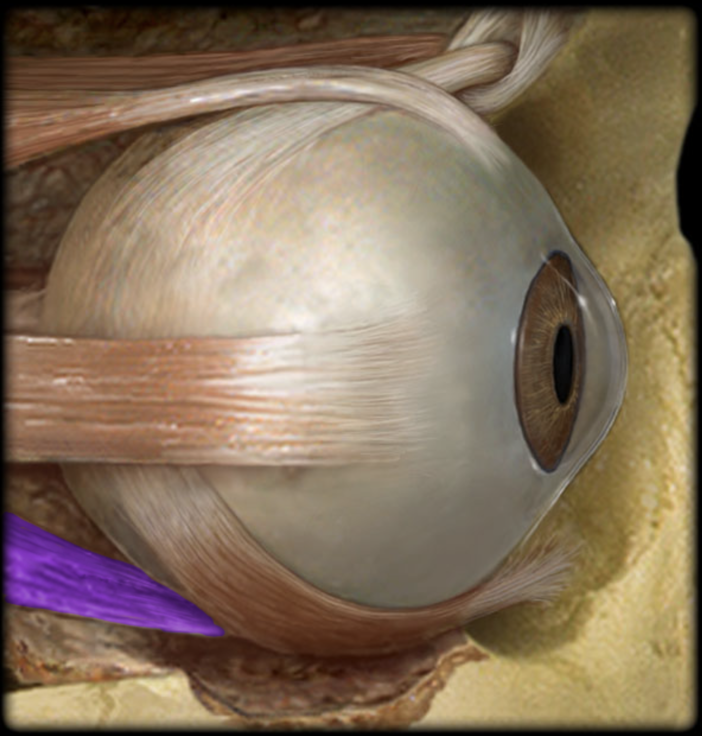
Inferior rectus innervation
CN III (Oculomotor n.)
Medial rectus action
Turns eye medially (inward)

Medial rectus innervation
CN III (Oculomotor n.)
Lateral rectus action
Turns eye laterally (outward/abduction)

Lateral rectus innervation
CN VI (Abducens n.)
Superior oblique action
Downward & medial rotation of the eye

Superior oblique innervation
CN IV (Trochlear n.)
Inferior oblique action
Upward rotation of the eye

Inferior oblique innervation
CN III (Oculomotor n.)
What cranial nerve that controls eye movement is most commonly impacted by traumatic head injury?
CN VI (Abducens n.); located laterally on the side of the head and is more vulnerable to trauma
Core (trunk) components
The back, abdomen, thorax (chest), and pelvis
Bones, connective tissues, muscles, neuromuscular innervation
Osteo components: vertebral column, ribs, sternum

Regions of the vertebral column
Cervical (7)
Thoracic (12)
Lumbar (5)
Sacral (5 fused)
Coccygeal (4 semi-fused vertebrae)

Spinal curves
Lordosis (cervical & lumbar) — anterior
Kyphosis (thoracic & sacral) — posterior
Spinal curve development
Born with a C-spine (single kyphotic curve)
Lordotic curvatures develop through experience
Tummy time → cervical curvature
Creeping (quadruped) → lumbar curvature
Further development of spinal curvature through weight-bearing in sitting & walking

Atlantooccipital joint
Articulation of atlas (C1) and the occiput (skull); allows for capital flexion and extension

Atlantoaxial joint
Articulation of the atlas (C1) and axis (C2); allows for rotation of the head