IPHY 3410 - Chapter 10: Cardiovascular System
 Call Kai
 Learn
 Practice Test
 Spaced Repetition
 Match
 Flashcards
 Knowt Play
1/89

There's no tags or description
Looks like no tags are added yet.
90 Terms
arteries
carries blood AWAY from heart
veins
carry blood TOWARD the heart
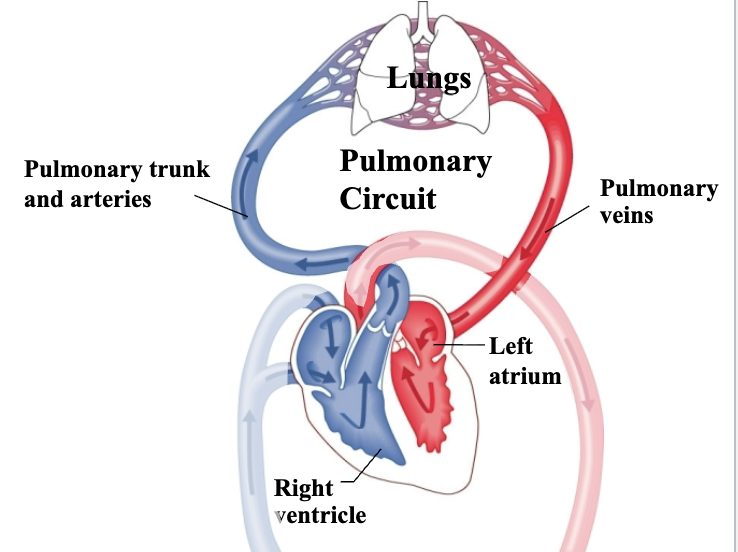
pulmonary circuit
route between heart and lungs that allow blood to pick up oxygen
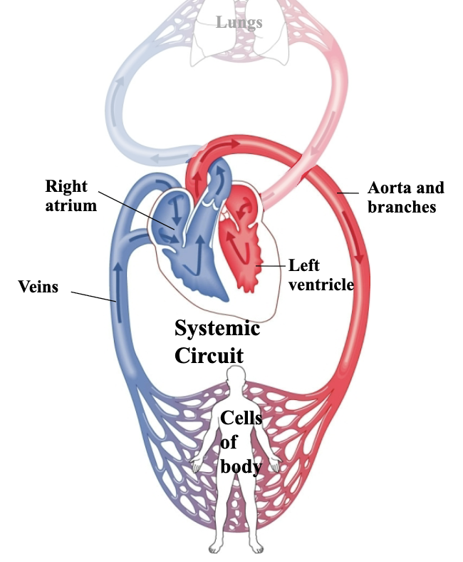
systemic circuit
route between heart & tissues of the body (other than the lungs) that brings oxygen out to cells

transposition of great arteries
aorta and pulmonary trunk are switched. blood leaving RV never get to the lungs, blood leaving LV would travel to lungs
major purpose of pericardium
anchor the heart to surroundings.
layers of pericardium superficial to deep
fibrous pericardium, serous pericardium (parietal, visceral, pericardial cavity), myocardium, endocardium

fibrous pericardium
Touch outer layer of sac that anchors heart and prevents overfilling; dense irreg CT
serous pericardium
serous membrane around heart
parietal layer of serous pericardium
inner layer of sac, secretes serous fluid
visceral layer of serous pericardium
outer layer of heart, secretes serous fluid (=epicardium)
pericardial cavity laayer of serous pericardium
space btwn parietal & visceral; filled w/ serous fluid
myocardium
cardiac muscle tissue
endocardium
simple squamous epith

Superior & Inferior vena cava
large veins that return deoxygenated blood from body

Tricuspid (R. atrioventricular) valve
separates right atrium from right ventricle

Chordae tendineae
Dense reg CT that attaches cusps of tricuspid valve to ventricle wall via papillary muscle

papillary muscle
projection of cardiac muscle

pulmonary semilunar valve
separates right ventricle from pulmonary trunk (artery)

pulmonary veins
return oxygenated blood to heart from lungs

Bicuspid/mitral (left atrioventricular) valve
separates left atrium from left ventricle
aortic semilunar valve purpose
separates left ventricle from aorta
how do tricupsid and bicupsid valves work
open at rest, pressure then pushes fold back to atrium, chordae tendineae pull valves from pushing too far back into atrium
Pulmonary & Aortic Semilunar valves
•Valve cusps resemble pockets
•Blood pushes them open when ventricles contract
•When ventricles relax, blood fills pockets and forces valve
closed
how do semilunar valves work
valves open w blood flow, and fall back down w gravity
heart murmur
Noise in heart caused by blood leaking past closed valve

mitral valve prolapse
most common; weakness in collagen of valve or chordae tendinae
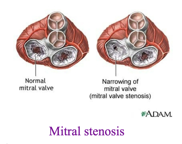
mitral stenosis
narrowed opening between valves, may be caused by calcium deposits or illness
characteristics of cardiac muscle
striated, connected by intercalated discs.
some cells adapted to conduct impulse instead of contracting

fascia adherens
desmosome like connections, provide strength
how are intercalated discs arranged
zig zags


Sinoatrial (SA) node
in RA, initiates an electrical impulse
inherent rhythmicity - pacemaker
Only in RA

internodal pathway
carries impulse away from SA node to AV node

atrioventricular (AV) node
delays impulse before it is passed on to ventricles
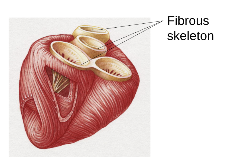
fibrous skeleton
collagen barrier between atria and ventricles that prevent an electrical impulse from passing
pathway of nerve impulse in the heart
SA node (pacemaker) —> internodal pathway —> AV node (bundle of His) —> bundle branches —> purkinje fibers

how do atria vs ventricles contract
atria:top to bottom; ventricles: bottom to top
cardiac cycle - phase 1 (atria contract)
atrial: systolic
ventricles: diastolic
AV valves: open
SL valves: closed
cardiac cycle - phase 2 (ventricles contract)
atrial: diastolic
ventricles: systolic
AV valves: close
SL valves: open
cardiac cycle - phase 3 (all 4 chambers rest)
atrial: diastolic
ventricle: diastolic
AV valves: open
SL valves: closed
systole
contraction (forcing blood out)
diastole
relaxation (filling w blood)
what supplies the cardia muscles w an external blood supply
right and left coronary arteries


artherosclerosis
build up of plaques which causes narrowing of an artery.
most common cause of arteriosclerosis (any hardening of arteries)
arteriosclerosis
ay hardening of arteries
myocardial infarction (heart attack)
result of blood not reaching cardiac muscle tissue (thrombus formed, tissues become ischemic)
thrombus
blood clot
ischemic
restriction in blood supply resulting in O2 and glucose deprivation in tissues
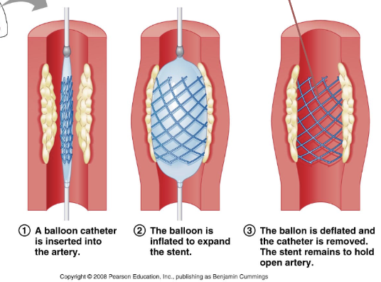
Tx for artherosclerosis
balloon angioplasty w stent
coronary bypass
new artery taken and goes around the old blocked vein, blocked vein isn’t removed, new artery just goes around it.
what do lymph vessels
transport lymph (fluid accumulated in tissues), ONE WAY, same organization as blood vessels

layers of vessel walls? superficial to deep
tunica externa (w/ vaso vasorum), external elastic membrane, tunica media, subendothelial layer, endothelium lumen
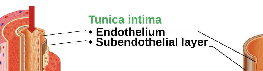
tunica intima
endothelium (simple squamous) and subendothelial layer (loose areolar CT)

tunica media
smooth muscle arranged circularly to allow for vasoconstriction

tunica externa
loose areolar CT

vaso vasorum
blood vessels that run through the layer of tunica externa to supply cells of vessel wall w blood.
how are arterial walls different from vein walls
Thicker and stronger, they have more elastic fibers; they r under higher pressure. Veins also have valves

what part of the wall forms the valves in veins
tunica intima
circle of flow from and back to heart
heart —> elastic artery —> muscular artery —> arteriole —> capillary —> venule —> medium vein —> large vein —> heart

elastic arteries
largest, branches near heart.
high concentration of elastin in tunica media. (regulates BP)
muscular arteries
supply organs and skeletal muscles
tunica media has less elastin and more smooth muscle. (more control)
sheets of elastin support and surround muscle (internal and external elastic membrane)

arterioles
branches of musclar arteries
constrict and dilate quickly
larger: all 3 tunics and elastic lamina
smaller: tunica media and endothelium only
capillary beds
diameter of about one RBC
lack tunica media and externa (thin for diffusion)

metarteriole
passage of blood cells in capillaries from arteriole to venules, always open
precapillary sphincters
smooth muscle cells, regulate RBCs - open/close
capillary characteristics
thin walls
endothelium: simple squamous epithelium
basement membrane: CT
3 types of capillaries
continuous, fenestrated, sinusoidal
continuous capillary
most common type (skeletal muscles, skin, nervous system)
tight junctions (least leaky)

fenestrated capillary
found where there is a high rate of exchange (digestive organs)
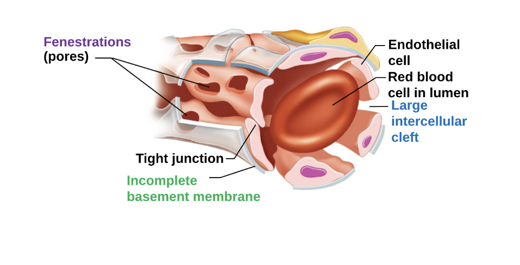
fenestrations
complete basement membrane

intercellular clefts
areas where there is no cell junction
fenestrations
pores thru endothelial cells
sinsoidal capillary (discontinuous)
leakiest
allows passage of large materials
large intercellular cleft and incomplete basement membrane
fewer tight junctions
some locations: spleen, bone marrow, lymph nodes
venules
smallest, drain capillaries
medium veins
drain venuels
large veins
drain large regions back towards heart
varicose veins
most commonly affect veins that r superficial on lower leg
failure of valves allow blood to pool, stretched veins and causes misshapen vessels
formed elements (cellular components)
erythrocytes (RBCs)
buffy coat - leukocytes (WBCs) + platelets
~46% of blood volume
plasma (extracellular matrix)
55% blood volume (mostly water)
contains dissolved solutes: wastes, nutrients, hormones
proteins: albumin and fibrinogen
albumin
bind above solutes
fibrinogen
needed for blood clotting
hemophilia
erythrocyte characteristics
mature red blood cell
anucleate and no organelles
anareobic - use glycolysis for E
97% hemoglobin
bioconcave - large surface area
3-4mo lifespan
hematopoiesis (hemopoiesis)
new RBCs form from stem cells in red bone marrow
Fe needed to make new cells
anemia
lower than normal number of RBCs to carry O2 to body
due to low levels of iron which leads to fewer RBCs
fewer RBCs to transport O2 to cells
also caused by blood loss and menstruation
leukocytes (WBCs)
complete cells, diapedesis (can squeeze thru capillary walls), immune response
platelets
blood clotting, cell fragments of larger megakaryocyte
what kind of antigens does type A blood have
A antigens only

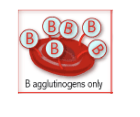
what kind of antigens does type B have
B antigens only
what kind of antigens does type AB have
both A and B antigens

what kind of antigens does type O have
neither A or B

Rh factor
Rh is a protein that is either present (+) or absent (-) on erythrocytes unlike A or B antibodies, Rh antibodies are able to pass thru the pacenta and attack the fetus.
prob is mother is Rh-